. 2023 Mar 30;12(7):1056.
doi: 10.3390/cells12071056.

ABCB1 and ABCG2 Overexpression Mediates Resistance to the Phosphatidylinositol 3-Kinase Inhibitor HS-173 in Cancer Cell Lines

Affiliations

ABCB1 and ABCG2 Overexpression Mediates Resistance to the Phosphatidylinositol 3-Kinase Inhibitor HS-173 in Cancer Cell Lines

Chung-Pu Wu et al. Cells. .

Abstract

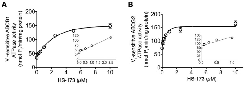
Constitutive activation of the phosphoinositide-3-kinase (PI3K)/Akt signaling pathway is crucial for tumor growth and progression. As such, this pathway has been an enticing target for drug discovery. Although HS-173 is a potent PI3K inhibitor that halts cancer cell proliferation via G2/M cell cycle arrest, the resistance mechanisms to HS-173 have not been investigated. In this study, we investigated the susceptibility of HS-173 to efflux mediated by the multidrug efflux transporters ABCB1 and ABCG2, which are two of the most well-known ATP-binding cassette (ABC) transporters associated with the development of cancer multidrug resistance (MDR). We found that the overexpression of ABCB1 or ABCG2 significantly reduced the efficacy of HS-173 in human cancer cells. Our data show that the intracellular accumulation of HS-173 was substantially reduced by ABCB1 and ABCG2, affecting G2/M arrest and apoptosis induced by HS-173. More importantly, the efficacy of HS-173 in multidrug-resistant cancer cells could be recovered by inhibiting the drug-efflux function of ABCB1 and ABCG2. Taken together, our study has demonstrated that HS-173 is a substrate for both ABCB1 and ABCG2, resulting in decreased intracellular concentration of this drug, which may have implications for its clinical use.

Keywords: ABCB1; ABCG2; HS-173; PI3K; multidrug resistance.


Conflict of interest statement

The authors declare no conflict of interest.

Figures

Figure 1
Figure 1
Dose–response curves for parental and multidrug-resistant cell lines overexpressing ABCB1 or ABCG2 treated with HS-173. The cytotoxicity of HS-173 in (A) the parental human KB-3-1 epidermal cancer cell line (open circles) and its ABCB1-overexpressing multidrug-resistant subline KB-V1 (filled circles); (B) parental human OVCAR-8 ovarian cancer cell line (open circles) and its ABCB1-overexpressing multidrug-resistant subline NCI-ADR-RES (filled circles); (C) parental human S1 colon cancer cell line (open circles) and its ABCG2-overexpressing multidrug-resistant subline S1-MI-80 (filled circles); (D) parental human MCF-7 breast cancer cell line (open circles) and its ABCG2-overexpressing multidrug-resistant subline MCF7-FLV1000 (filled circles); as well as (E) the parental HEK293 cells (open circles) and HEK293 cells transfected with human ABCB1 (MDR19-HEK293, open squares) or human ABCG2 (R482-HEK293, filled circles). Points, mean values from more than three independent experiments; bars, SEM.
Figure 2
Figure 2
Differential effect of HS-173 on G2/M cell cycle arrest in drug-sensitive and multidrug-resistant cancer cells overexpressing ABCB1 or ABCG2. (A) KB-3-1 and KB-V1 cells were treated with DMSO (control), 1 μM tariquidar, 2 μM HS-173, or a combination of HS-173 and tariquidar; (B) S1 and S1-MI-80 cells were treated with DMSO (control), 1 μM Ko143, 2 μM HS-173, or a combination of HS-173 and Ko143 for 24 h before harvesting for cell cycle analysis, as described in the Materials and Methods section. * p < 0.05; ** p < 0.01; *** p < 0.001.
Figure 3
Figure 3
Differential effect of HS-173-induced apoptosis in drug-sensitive cancer cells and multidrug-resistant cancer cells overexpressing ABCB1 or ABCG2. (A) KB-3-1 and KB-V1 cells were treated with DMSO (control), 1 μM tariquidar, 2 μM HS-173, or a combination of HS-173 and tariquidar; (B) S1 and S1-MI-80 cells were treated with DMSO (control), 1 μM Ko143, 2 μM HS-173, or a combination of HS-173 and Ko143 for 48 h, as described in the Materials and Methods section. The cells were later processed using the Annexin V-FITC and PI staining method and analyzed by means of flow cytometry. Representative dot plots (left panel) and quantified values (right panel) are mean values ± SD calculated from more than three independent experiments. * p < 0.05; ** p < 0.01, versus the same treatment with the addition of tariquidar or Ko143.
Figure 4
Figure 4
ABCB1- and ABCG2-mediated transport reduces the intracellular accumulation of HS-173 in human cancer cells. (A) The chemical structure and product ion mass spectra of HS-173 (precursor ion m/z 423.2 in positive mode). The fragment ion m/z 395.1 was selected for quantitative analysis. (B) Quantification of intracellular concentration of HS-173 by HPLC-MS/MS analysis in drug-sensitive OVCAR-8 cells (open bars), ABCB1-overexpressing NCI-ADR-RES (striped bars), drug-sensitive S1 (gray bars), and ABCG2-overexpressing S1-MI-80 (black bars) cells with or without the addition of 10 μM tariquidar or Ko143 as described in the Materials and Methods section. Values are mean values ± SD calculated from more than three independent experiments. ** p < 0.01 and *** p < 0.001 versus the treatment with tariquidar or Ko143.
Figure 5
Figure 5
HS-173 inhibits ABCB1- and ABCG2-mediated drug transport. The accumulation of fluorescent calcein in the KB-3-1 cell line (A, left panel) and its ABCB1-overexpressing KB-V1 line (A, right panel); OVCAR-8 cell line (B, left panel) and its ABCB1-overexpressing NCI-ADR-RES (B, right panel) cells; HEK293 cells stably transfected with the pcDNA3.1 empty vector (C, left panel) and HEK293 cells stably transfected with human ABCB1 (C, right panel), or fluorescent pheophorbide A (PhA) in the S1 cell line (D, left panel) and its ABCG2-overexpressing S1-MI-80 line (D, right panel); MCF-7 cell line (E, left panel) and its ABCG2-overexpressing MCF7-FLV1000 line (E, right panel); HEK293 cells stably transfected with the pcDNA3.1 empty vector (F, left panel) and HEK293 cells stably transfected with human ABCG2 (F, right panel), measured without (solid lines) or with the addition of 40 μM HS-173 (shaded, solid lines) or 1 μM tariquidar or Ko143 (dotted lines) and analyzed immediately by means of flow cytometry, as described in the Materials and Methods section. Representative histograms of three independent experiments are shown.
Figure 6
Figure 6
HS-173 stimulates the ATPase activity of ABCB1 and ABCG2. The effect of HS-173 (0–10 µM) on the vanadate (Vi)-sensitive ATP hydrolysis of (A) ABCB1 and (B) ABCG2, determined by endpoint Pi assay, as described previously [37,57]. Points, mean from a minimum of three independent experiments; bars, SEM.
Figure 7
Figure 7
The lowest energy poses for the docking binding of HS-173 within the drug-binding pocket of (A) ABCB1 (PDB:6QEX) and (B) ABCG2 (PDB: 6VXH) were predicted using BIOVIA Discovery Studio 4.0 software as described in the Materials and Methods section. The molecular model of HS-173 is presented in (yellow) stick representation, and the atoms for interacting amino acid residues are colored: carbon (gray), oxygen (red), nitrogen (blue), hydrogen (light gray), and sulfur (yellow). Dashed lines represent the proposed interactions. The residues from monomer 2 of ABG2 are indicated with prime symbols.

References

    1. Liu P., Cheng H., Roberts T.M., Zhao J.J. Targeting the phosphoinositide 3-kinase pathway in cancer. Nat. Rev. 2009;8:627–644. doi: 10.1038/nrd2926. - DOI - PMC - PubMed
    1. Alzahrani A.S. PI3K/Akt/mTOR inhibitors in cancer: At the bench and bedside. Semin. Cancer Biol. 2019;59:125–132. doi: 10.1016/j.semcancer.2019.07.009. - DOI - PubMed
    1. Bleau A.M., Hambardzumyan D., Ozawa T., Fomchenko E.I., Huse J.T., Brennan C.W., Holland E.C. PTEN/PI3K/Akt pathway regulates the side population phenotype and ABCG2 activity in glioma tumor stem-like cells. Cell Stem Cell. 2009;4:226–235. doi: 10.1016/j.stem.2009.01.007. - DOI - PMC - PubMed
    1. Burris H.A., 3rd Overcoming acquired resistance to anticancer therapy: Focus on the PI3K/AKT/mTOR pathway. Cancer Chemother. Pharmacol. 2013;71:829–842. doi: 10.1007/s00280-012-2043-3. - DOI - PubMed
    1. Greenwell I.B., Flowers C.R., Blum K.A., Cohen J.B. Clinical use of PI3K inhibitors in B-cell lymphoid malignancies: Today and tomorrow. Expert Rev. Anticancer Ther. 2017;17:271–279. doi: 10.1080/14737140.2017.1285702. - DOI - PubMed

Publication types

MeSH terms

Substances