. 2023 Mar 31;12(7):1059.
doi: 10.3390/cells12071059.

Primary Cilium Identifies a Quiescent Cell Population in the Human Intestinal Crypt

Affiliations

Primary Cilium Identifies a Quiescent Cell Population in the Human Intestinal Crypt

Blanche Sénicourt et al. Cells. .

Abstract

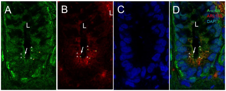
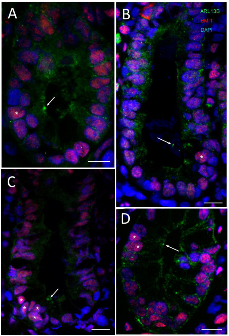
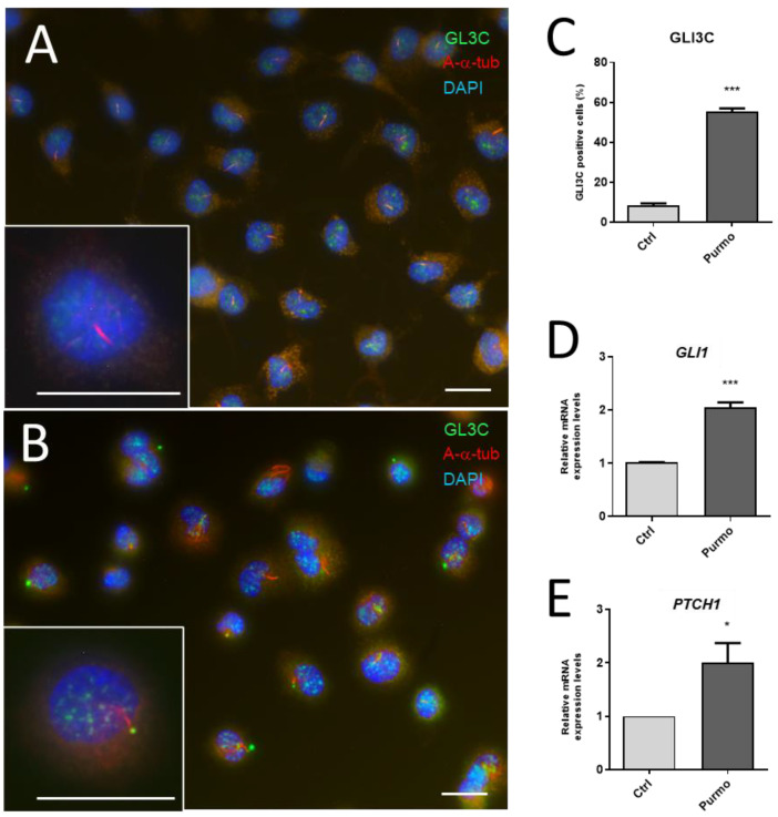
Primary cilia are sensory antennae located at the cell surface which mediate a variety of extracellular signals involved in development, tissue homeostasis, stem cells and cancer. Primary cilia are found in an extensive array of vertebrae cells but can only be generated when cells become quiescent. The small intestinal epithelium is a rapidly self-renewing tissue organized into a functional unit called the crypt-villus axis, containing progenitor and differentiated cells, respectively. Terminally differentiated villus cells are notoriously devoid of primary cilia. We sought to determine if intestinal crypts contain a quiescent cell population that could be identified by the presence of primary cilia. Here we show that primary cilia are detected in a subset of cells located deep in the crypts slightly above a Paneth cell population. Using a normal epithelial proliferative crypt cell model, we show that primary cilia assembly and activity correlate with a quiescent state. These results provide further evidence for the existence of a quiescent cell population in the human small intestine and suggest the potential for new modes of regulation in stem cell dynamics.

Keywords: BMI1; GLI; HIEC-6 cell line; Hedgehog pathway; intestinal epithelial cells; patched; primary cilium; stem cells; tubulin.


Conflict of interest statement

The authors declare no conflict of interest. The funders had no role in the design of the study; in the collection, analyses, or interpretation of data; in the writing of the manuscript; or in the decision to publish the results.

Figures

Figure 1
Figure 1
Primary cilia in the human small intestine. (A) Schematic of the crypt–villus axis showing the crypt region depicted in B and C. (B) Representative immunodetection of primary cilia using anti-acetylated-α-tubulin (A-α-tub; arrow, green staining) in the epithelial cells (E-cadherin, E-cad; red staining) of a crypt in the human adult small intestine (L, lumen). Nuclei were stained with DAPI (blue staining). Smaller structures that could be related to either midbody remnants [64] or centrosomes (see below) were also detected (arrowheads). (C) 3D reconstruction of double immunostaining of anti-α-acetylated-tubulin (green), showing primary cilia (arrow) and smaller structures (arrowheads) as well as anti-phospholipase A2 (red), a Paneth cell marker (L, lumen; P, Paneth cell). Nuclei were stained with DAPI (blue) and are out of focus because they are present on a lower plane of focus. Scale bars are equal to 10 µm. (B’) Higher magnification of the primary cilium seen in B (arrow).
Figure 2
Figure 2
Primary cilia in the lower half of the crypts of the human small intestine. Representative confocal imaging for the detection of primary cilia using anti-acetylated-α-tubulin (A-α-tub; green staining, arrow in (A,D)) and anti-ARL13B antibody (red staining, arrow in (B,D)) in epithelial cells of the human adult small intestine (L, lumen of the crypt). Nuclei were stained with DAPI (blue) (C,D). Note that anti-acetylated-α-tubulin and anti-ARL13B stained smaller dots that were co-stained (white arrowheads) or not stained (green and orange arrowheads) in (A,B,D). Scale bar is equal to 10 µm.
Figure 3
Figure 3
Primary cilia in the lower part of the crypts of the human small intestine. Representative confocal imaging for the detection of primary cilia using anti-glutamylated tubulin (Pglu-tub; green staining, arrow in (A,D)) and anti-ARL13B antibody (red staining, arrow in (B,D)) in epithelial cells of the human adult small intestine (L, lumen of the crypt). (A’,B’,D’) are higher magnifications of the (A,B,D) panels. Nuclei were stained with DAPI (C,D). As observed above, the 17711-1-AP antibody (B,D) identified dots that were only weakly stained or not stained with the anti-Pglu-tub antibody. Scale bar is equal to 10 µm.
Figure 4
Figure 4
Primary cilia in BMI1-positive cells of the crypts of the human small intestine. (A) Representative immunodetection of primary cilia using the anti-ARL13b 17711-1-AP antibody (arrow, red staining) and positive BMI1 nuclei (green staining, stars) in epithelial crypt cells of the human adult small intestine (L, lumen of the crypts). Nuclei were stained with DAPI (blue). The square delimits the portion of the lower crypt shown in panels (BE) and the rectangle delimits the portion of the luminal crypt epithelium shown in panel (F). (BE) Higher magnification of the lower crypt region showing a cluster of positive 17711-1-AP structures in the luminal aspect of lower crypt cells (arrows in B,E) and positive nuclei for BMI1 staining (stars in C,E). Arrowheads in the (A,B,E,F) panels identify smaller 17711-1-AP-stained dots that do not seem to be related to the primary cilium. (G,H) Another region showing the expression of 17711-1-AP-stained structures (arrows) in cells displaying positive BMI-1-stained nuclei (stars). Nuclei were stained with DAPI (A,D,E,I). Scale bar is equal to 10 µm.
Figure 5
Figure 5
Primary cilia in the lower third of a crypt of the human small intestine. A, Representative immunodetection of primary cilia using the anti-ARL13b 90413h antibody (green staining) (arrow) in one of the crypts (C). Nuclei were stained with DAPI (blue staining). Scale bar is equal to 10 µm.
Figure 6
Figure 6
Primary cilia and centrosomes in the lower third of the crypts of the human small intestine. (AE) Representative immunodetection of primary cilia using the anti-ARL13b 90413h antibody (green staining, arrow) and of centrosomes using an anti-pericentrin antibody (red staining) in various crypts. In most cases of positive crypts, the 90413h antibody stained one predominant dot (AE) that was localized adjacent to a positive pericentrin-labeled dot, as in (BE). (E’) Higher magnification of the section delimited by the square in (E) showing adjacent dots stained by ARL13B and pericentrin. (F,G) are high magnifications of the stroma around the crypts showing a similar distribution of ARL13B and pericentrin in ciliated fibroblasts. Nuclei were stained with DAPI (blue staining). Scale bars are equal to 10 µm.
Figure 7
Figure 7
Primary cilia and midbody remnants in the lower third of the crypts of the human small intestine. (AE) Representative immunodetection of primary cilia using anti-ARL13b 90413h antibody (green staining, arrow) and of midbody remnants using an anti-MKLP1 antibody (red staining) in various crypts. In most cases of positive crypts, the 90413h antibody stained one predominant dot (AF) that was localized adjacent to a positive MKLP1-labeled dot. (A’,B’) Higher magnification of the section delimited by the squares in (A,B) showing adjacent dots stained by ARL13B and MKLP1. Nuclei were stained with DAPI (blue staining). Scale bars are equal to 10 µm.
Figure 8
Figure 8
Primary cilia and midbody remnants in the lower third of the crypts of the human small intestine. Representative immunodetection of primary cilia using anti-acetylated tubulin antibody (green staining) and midbody remnants using an anti-MKLP1 antibody (red staining) in the lower portion of a crypt. The 90413h antibody stained one predominant structure that was localized adjacent to a positive MKLP1-labeled dot. Insert in the upper right corner, higher magnification of the section delimited by the square showing the co-staining. Nuclei were stained with DAPI (blue staining). Scale bar is equal to 10 µm.
Figure 9
Figure 9
Primary cilia in association with BMI1-positive cells of the crypts of the human small intestine. (A) Representative immunodetection of primary cilia using the anti-ARL13b 90413h antibody (arrows, green staining) and positive BMI1 nuclei (red staining, stars) in epithelial crypt cells of the human adult small intestine. In some instances, single nuclei staining was observed to be more intensive for BMI1 in relation to the detection of cilia (A,B) while in other cases, a cluster of BMI1-positive nuclei coinciding with ARL13b-positive structures was noted (C,D). Nuclei were stained with DAPI (blue). Scale bars are equal to 10 µm.
Figure 10
Figure 10
Constitutive expression of BMI1 and assembly of the primary cilia in HIEC-6 cells. (A,B): Representative immunodetection of BMI1 (A) and DAPI nuclear co-staining (B) in newly confluent HIEC cells. (C): BMI1 transcript expression in subconfluent (SC) and 5-, 10- and 15-day post-confluent (5PC, 10PC and 15PC) HIEC-6 cells. (D): Immunodetection of acetylated-α-tubulin (A-α-tub) in newly confluent HIEC-6 (green) cells. Scale bars are equal to 25 µm.
Figure 11
Figure 11
Expression of the primary cilia in quiescent HIEC cells. (A,B): Representative indirect immunofluorescence for the detection of ARL13b with 17711-1-AP (green staining) and polyglutamylated tubulin (Pglu-tub; red) in subconfluent (A) and postconfluent (B) HIEC cells. (C). Representative experiment showing primary cilium (PC) and BrdU counts in percentage of total DAPI stained cells in synchronized cells at 24 h intervals after passage. PC and BrdU counts were performed in separate dishes. The experiment was repeated three times. Bars are equal to 10 µm.
Figure 12
Figure 12
Accumulation of GLI1 expression in quiescent HIEC cells. (AD): Representative indirect immunofluorescence for the detection of GLI1 (A,D, red staining) and acetylated-α-tubulin (A-α-tub; B,D, green staining) in post-confluent HIEC cells. Nuclei were stained with DAPI (C,D). Scale bar is equal to 10 µm. (E): GLI1 transcript expression in subconfluent (SC) and 5-, 10- and 15-day post-confluent (5PC, 10PC and 15PC) HIEC-6 cells. **, p < 0.01; ***, p < 0.0005.
Figure 13
Figure 13
Stimulation of the HH pathway in response to purmorphamine in HIEC cells. Quiescent post-confluent HIEC cells were untreated (A) or treated with 2µM purmorphamine (Purmo) (B) for 24 h and analyzed for the HH activity marker GLI3C (AC) and expression of downstream target genes GLI1 (D) and PTCH1 (E). Primary cilia were detected using an anti-acetylated-α-tubulin (A-α-tub; red staining) in most post-confluent HIEC cells (A,B). Accumulation of GLI3C expression at the tip of primary cilia with an anti-GLI3C (green staining) was found in ~10% of control cells (A,C, Ctrl) while it was detected in more than 50% of the Purmo-treated cells (B,C, Purmo). Bars in (A,B) are equal to 10 µm. (D,E): Expression of GLI1 and PTC1 transcripts was also significantly increased in Purmo-treated cells. *, p < 0.05; ***, p < 0.001.
Figure 14
Figure 14
Quiescence of post-confluent HIEC cells is reversible and is regulated by the HH pathway. (A): In post-confluent HIEC-6 cells, the GLI1 inhibitor GANT-61 (GT) reduced the expression of both GLI1 and PTCH1 transcripts, the main HH downstream target genes, after 48 h treatment. (B): In contrast to sub-confluent (SC) cells, HIEC-6 cells that maintained up to 30 days of post-confluent (30PC) culture did not synthesize DNA as evaluated by the lack of BrdU staining. However, BrdU staining was restored to basal levels after 3 passages. (C): 20+ day-post-confluent HIEC-6 cells were treated for 48 h with GT before being passed and allowed to recover for 48 h then processed for BrdU staining and cell counting. *, p < 0.05; ***, p < 0.001.
Figure 15
Figure 15
Schematic illustration of the findings of the current study. Primary cilium (PC)-bearing cells (pink) were found in the lower crypt just above the stem/Paneth cell zone at a frequency estimated at one cell per crypt in the human small intestine and corresponding to the +4 reserve stem cells (RSC) described previously. HIEC-6 cells were then used to further investigate the expression of the PC and signaling. HIEC-6 cells were previously found to be unique in their abilities to undertake a differentiation program under the influence of pro-differentiation transcription factors, such as CDX2 and HNF1α [51,83], as well as to be induced to adopt a primordial stem cell (PSC) phenotype upon activation of the WNT pathway [38]. Herein, we showed that post-confluent HIEC-6 cells express a PC when becoming quiescent and that this PC mediates the activation of the HH pathway, which in turn appears to regulate the cell cycle.

Similar articles

Cited by

References

    1. Eguether T., Hahne M. Mixed signals from the cell’s antennae: Primary cilia in cancer. EMBO Rep. 2018;19:e46589. doi: 10.15252/embr.201846589. - DOI - PMC - PubMed
    1. Wheatley D.N. Primary cilia: Turning points in establishing their ubiquity, sensory role and the pathological consequences of dysfunction. J. Cell Commun. Signal. 2021;15:291–297. doi: 10.1007/s12079-021-00615-5. - DOI - PMC - PubMed
    1. Wheway G., Nazlamova L., Hancock J.T. Signaling through the Primary Cilium. Front. Cell Dev. Biol. 2018;6:8. doi: 10.3389/fcell.2018.00008. - DOI - PMC - PubMed
    1. Yanardag S., Pugacheva E.N. Primary Cilium Is Involved in Stem Cell Differentiation and Renewal through the Regulation of Multiple Signaling Pathways. Cells. 2021;10:1428. doi: 10.3390/cells10061428. - DOI - PMC - PubMed
    1. Praetorius H.A., Spring K.R. Bending the MDCK cell primary cilium increases intracellular calcium. J. Membr. Biol. 2001;184:71–79. doi: 10.1007/s00232-001-0075-4. - DOI - PubMed

Publication types

LinkOut - more resources