. 2023 Apr 3;12(7):1078.
doi: 10.3390/cells12071078.

Development of Cyclic Peptides Targeting the Epidermal Growth Factor Receptor in Mesenchymal Triple-Negative Breast Cancer Subtype

Affiliations

Development of Cyclic Peptides Targeting the Epidermal Growth Factor Receptor in Mesenchymal Triple-Negative Breast Cancer Subtype

Nancy Nisticò et al. Cells. .

Abstract

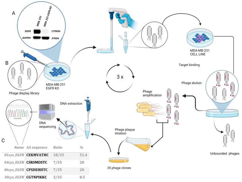
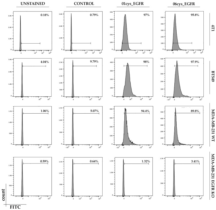
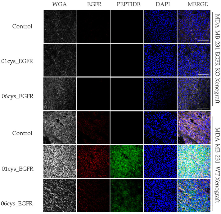
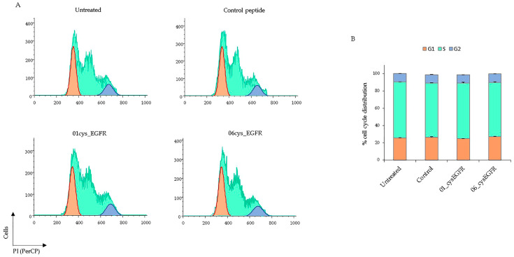
Triple-negative breast cancer (TNBC) is an aggressive malignancy characterized by the lack of expression of estrogen and progesterone receptors and amplification of human epidermal growth factor receptor 2 (HER2). Being the Epidermal Growth Factor Receptor (EGFR) highly expressed in mesenchymal TNBC and correlated with aggressive growth behavior, it represents an ideal target for anticancer drugs. Here, we have applied the phage display for selecting two highly specific peptide ligands for targeting the EGFR overexpressed in MDA-MB-231 cells, a human TNBC cell line. Molecular docking predicted the peptide-binding affinities and sites in the extracellular domain of EGFR. The binding of the FITC-conjugated peptides to human and murine TNBC cells was validated by flow cytometry. Confocal microscopy confirmed the peptide binding specificity to EGFR-positive MDA-MB-231 tumor xenograft tissues and their co-localization with the membrane EGFR. Further, the peptide stimulation did not affect the cell cycle of TNBC cells, which is of interest for their utility for tumor targeting. Our data indicate that these novel peptides are highly specific ligands for the EGFR overexpressed in TNBC cells, and thus they could be used in conjugation with nanoparticles for tumor-targeted delivery of anticancer drugs.

Keywords: EGFR; breast cancer biomarkers; peptide; phage display; triple-negative breast cancer; tumor targeting.


Conflict of interest statement

The authors declare no conflict of interest.

Figures

Figure 1
Figure 1
Selection and identification of peptide ligands for EGFR in the TNBC cell line MDA-MB231. Schematic representation of phage display screening: (A) Protein expression levels of EGFR in MDA-MB-231 wild-type and EGFR KO cells were verified by Western blot analysis; GAPDH expression was included as loading control. (B) Non-specifically bound phages were eliminated by incubation of the library with MDA-MB-231 EGFR KO cells. Unbound phages were then incubated with MDA-MB-231 overexpressing EGFR to select phages with high affinity binding. Three cycles of selection and amplification were performed with increasing stringency conditions. The genomic DNA of selected phage clones was extracted and sequenced to determine the peptide insert required for EGFR binding. (C) Peptide sequence and occurrence frequency of selected phage clones.
Figure 2
Figure 2
Three-dimensional front view of EGFR. Domain I (1–165 amino acid residues), II (166–310 amino acid residues), III (311–79 amino acid residues), and IV (480–614 amino acid residues) are colored in grey, green, orange and blue, respectively. The cavity region between Domain I and III in the EC region is highlighted in the lateral popup and colored in grey dots (PDB code: 3NJP).
Figure 3
Figure 3
Principal amino acid (aa) interactions involved in the binding site of EGFR between 01_cys EGFR (A) and 06_cys EGFR (B) are labeled and highlighted in the stick model. On the right of the figure, the chemical pharmacophore features are explained. The EGFR cavity is shown in solid style.
Figure 4
Figure 4
Expression of the EGFR protein in Ramos cells, murine 4T1, human BT-549 TNBC cell lines, and thyroid cancer cell lines K1 and Cal62. Total cell extracts were separated by SDS-PAGE and analyzed by Western blotting with anti-EGFR and anti-GAPDH antibodies.
Figure 5
Figure 5
Binding assay of peptides to TNBC cells. Cells were incubated with 01cys_EGFR, 06cys_EGFR peptides, or the irrelevant peptide as control and then analyzed by flow cytometry. Percentage of binding efficiency of peptides is shown in each panel. The TNBC cell lines were: MDA-MB-231, BT-549, and 4T1. The cell line MDA-MB-231 EGFR-KO was included as a negative control.
Figure 6
Figure 6
Immunostaining of MDA-MB-231 tumor xenografts with 01_cysEGFR and 06_cysEGFR peptides. Representative confocal images were acquired from MDA-MB-231 tumor xenograft tissue sections stained with anti-EGFR (red), the peptides irrelevant control, 01_cysEGFR and 06_cysEGFR (green), anti-WGA (white) and DAPI (blue). The merge of these channels confirmed the co-localization of EGFR and peptides on the cell membrane. Images were acquired at magnification 40× using a confocal microscope (Leica TC-SP2 Confocal System, Leica Microsystem Srl, Milan, Italy). Scale bar = 100 μm. Representative images of multiple serial sections are shown.
Figure 7
Figure 7
(A) The proportion of BT549 cells untreated or treated with 25 μM of control, 01cys_EGFR, and 06cys_EGFR peptides in G1, S, and G2 phases were determined by flow cytometry and quantified by Flow Jo software using Watson Pragmatic method. (B) Histograms showed the percentage of cell cycle phase distribution of PI-stained TNBC cells. Data are collected from two different experiments and presented as means ± SD. Paired t-test indicates no statistical significance between peptide-stimulated and unstimulated cells.

Similar articles

Cited by

References

    1. Yin L., Duan J.-J., Bian X.-W., Yu S.-C. Triple-negative breast cancer molecular subtyping and treatment progress. Breast Cancer Res. 2020;22:61. doi: 10.1186/s13058-020-01296-5. - DOI - PMC - PubMed
    1. Oakman C., Viale G., Di Leo A. Management of triple negative breast cancer. Breast. 2010;19:312–321. doi: 10.1016/j.breast.2010.03.026. - DOI - PubMed
    1. Penault-Llorca F., Viale G. Phathological and molecular diagnosis of triple negative breast cancer: A clinical perspective. Ann. Oncol. 2012;23:vi19–vi22. doi: 10.1093/annonc/mds190. - DOI - PubMed
    1. Dass S., Tan K., Rajan R.S., Mokhtar N., Adzmi E.M., Rahman W.W.A., Din T.T., Balakrishnan V. Triple Negative Breast Cancer: A Review of Present and Future Diagnostic Modalities. Medicina. 2021;57:62. doi: 10.3390/medicina57010062. - DOI - PMC - PubMed
    1. Gupta G.K., Collier A.L., Lee D., Hoefer R.A., Zheleva V., Van Reesema L.L.S., Tang-Tan A.M., Guye M.L., Chang D.Z., Winston J.S., et al. Perspectives on Triple-Negative Breast Cancer: Current Treatment Strategies, Unmet Needs, and Potential Targets for Future Therapies. Cancers. 2020;12:2392. doi: 10.3390/cancers12092392. - DOI - PMC - PubMed

Publication types