. 2023 Apr 6;12(7):1098.
doi: 10.3390/cells12071098.

Glucagon Promotes Gluconeogenesis through the GCGR/PKA/CREB/PGC-1α Pathway in Hepatocytes of the Japanese Flounder Paralichthys olivaceus

Affiliations

Glucagon Promotes Gluconeogenesis through the GCGR/PKA/CREB/PGC-1α Pathway in Hepatocytes of the Japanese Flounder Paralichthys olivaceus

Mengxi Yang et al. Cells. .

Abstract

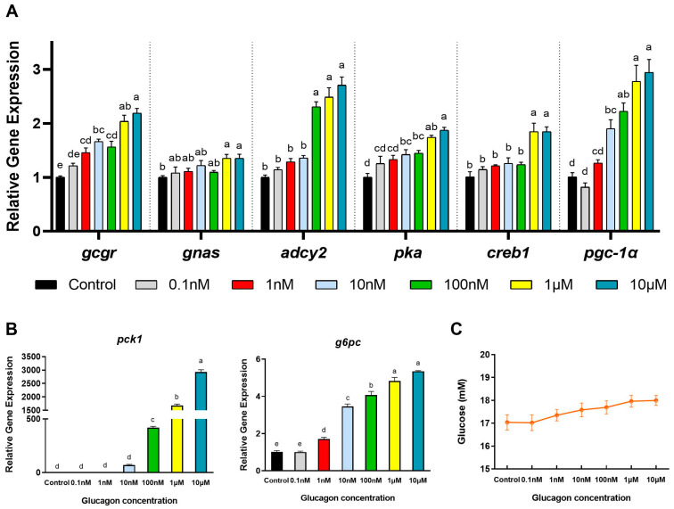
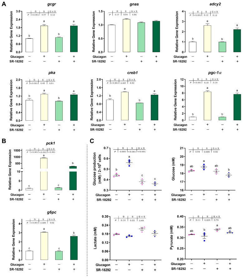
In order to investigate the mechanism of glucagon regulation of gluconeogenesis, primary hepatocytes of the Japanese flounder (Paralichthys olivaceus) were incubated with synthesized glucagon, and methods based on inhibitors and gene overexpression were employed. The results indicated that glucagon promoted glucose production and increased the mRNA levels of glucagon receptor (gcgr), guanine nucleotide-binding protein Gs α subunit (gnas), adenylate cyclase 2 (adcy2), protein kinase A (pka), cAMP response element-binding protein 1 (creb1), peroxisome proliferator-activated receptor-γ coactivator 1α (pgc-1α), phosphoenolpyruvate carboxykinase 1 (pck1), and glucose-6-phosphatase (g6pc) in the hepatocytes. An inhibitor of GCGR decreased the mRNA expression of gcgr, gnas, adcy2, pka, creb1, pgc-1α, pck1, g6pc, the protein expression of phosphorylated CREB and PGC-1α, and glucose production. The overexpression of gcgr caused the opposite results. An inhibitor of PKA decreased the mRNA expression of pgc-1α, pck1, g6pc, the protein expression of phosphorylated-CREB, and glucose production in hepatocytes. A CREB-targeted inhibitor significantly decreased the stimulation by glucagon of the mRNA expression of creb1, pgc-1α, and gluconeogenic genes, and glucose production decreased accordingly. After incubating the hepatocytes with an inhibitor of PGC-1α, the glucagon-activated mRNA expression of pck1 and g6pc was significantly down-regulated. Together, these results demonstrate that glucagon promotes gluconeogenesis through the GCGR/PKA/CREB/PGC-1α pathway in the Japanese flounder.

Keywords: Paralichthys olivaceus; glucagon; gluconeogenesis; glucose; signaling pathway.


Conflict of interest statement

The authors declare no conflict of interest.

Figures

Figure 1
Figure 1
Gene cloning, functional analysis, and tissue distribution of GCG. (A), Nucleotide sequence, deduced amino acid sequence, and predicted domains of the GCG gene. The signal peptide is marked with a black underline; the GLUCA domain is marked with a black box; The asterisk (*) indicatesthe stop codon. (B), Comparison of the amino acid sequences of GCG. Comparison of the deduced amino acid (aa) sequence of GCG from Japanese flounder with those from other fish, mouse, and human. The aa sequences were aligned using ClustalW, and similarity shading was based on a 75% identity threshold. Identical residues are shaded dark, and similar residues are shaded light. Accession numbers: Hippoglossus stenolepis (XP_035030856.1); Scophthalmus maximus (XP_035459699.1); Mastacembelus armatus (XP_026151650.1); Sparus aurata (XP_030284999.1); Larimichthys crocea (XP_010730517.1); Danio rerio (NP_001229699.1); Mus musculus (AAH12975.1); Homo sapiens (AAH05278.1). (C), NJ phylogenetic tree based on amino acid sequences. The phylogenetic tree is based on these sequences: Ceratotherium simum simum (XP_004428320.1); Rhinolophus ferrumequinum (KAF6361488.1); Leptonychotes weddellii (XP_006747175.1); Tursiops truncates (XP_033716238.1); Erinaceus europaeus (XP_007523834.1); Oryctolagus cuniculus (XP_008256891.1); Hylobates moloch (XP_032611454.1); Homo sapiens (AAH05278.1); Mus musculus (AAH12975.1); Gallus gallus (CAA68827.1); Taeniopygia guttata (XP_002197220.1); Alligator sinensis (AFV96267.1); Gopherus evgoodei (XP_030436009.1); Nanorana parkeri (XP_018412196.1); Rhinatrema bivittatum (XP_029460758.1); Labeo rohita (RXN30499.1); Chanos chanos (XP_030642916.1); Danio rerio (NP_001229699.1); Oreochromis aureus (XP_031599195.1); Larimichthys crocea (XP_010730517.1); Sparus aurata (XP_030284999.1); Mastacembelus armatus (XP_026151650.1); Scophthalmus maximus (XP_035459699.1); Paralichthys olivaceus (MW727222); Hippoglossus stenolepis (XP_035030856.1); The red words represent Japanese flounder. (D), Schematic diagram of the three-dimensional structure of the GCG protein from Japanese flounder and human. (a) GCG in Japanese flounder; (b) GCG in human. (E), Tissues distribution of GCG in Japanese flounder. The values represent the means ± S.E., n = 3, one-way ANOVA with Tukey post-hoc test. Values with different letters mean statistical differences (p < 0.05).
Figure 2
Figure 2
Time-dependent effects of glucagon treatment on gene expression in hepatocytes and glucose concentration in the medium. (A), qPCR analysis of the mRNA levels of glucagon pathway-related genes (gcgr; gnas; adcy2; pka; creb1; pgc-1α) in hepatocytes at different times in response to glucagon treatment (1 μM) or DMSO. (B), qPCR analysis of the mRNA levels of gluconeogenic genes (pck1 and g6pc) in hepatocytes at different times in response to glucagon treatment (1 μM) or DMSO. (C), Glucose concentration in the medium at different times in response to glucagon treatment (1 μM) or DMSO. All data are expressed as mean ± SE, n = 3, two-way ANOVA with Tukey post-hoc test. Values with different letters mean statistical differences (p < 0.05). T: time points; I: reagents for incubation; T × I: interaction between time and reagents.
Figure 3
Figure 3
Dose-dependent effects of glucagon treatment on the mRNA levels of genes in hepatocytes and glucose concentration in the medium. (A), qPCR analysis of the mRNA levels of glucagon pathway-related genes (gcgr; gnas; adcy2; pka; creb1; pgc-1α) in hepatocytes in response to glucagon treatment at different doses or DMSO (24 h). (B), qPCR analysis of the mRNA levels of gluconeogenic genes (pck1 and g6pc) in hepatocytes in response to glucagon treatment at different doses or DMSO (24 h). (C), Glucose concentration in the medium in response to glucagon treatment at different doses or DMSO (24 h). All data are expressed as mean ± SE, n = 3, one-way ANOVA with Tukey post-hoc test. Values with different letters mean statistical differences (p < 0.05).
Figure 4
Figure 4
Adomeglivant inhibits the glucagon pathway in hepatocytes by targeting GCGR. (A), qPCR analysis of the mRNA levels of glucagon pathway-related genes (gcgr; gnas; adcy2; pka; creb1; pgc-1α) following glucagon stimulation and treatment with adomeglivant (50 μM) in hepatocytes. (B), qPCR analysis of the mRNA levels of gluconeogenic genes (pck1 and g6pc) following glucagon stimulation and treatment with adomeglivant (50 μM) in hepatocytes. (C), Western blot analysis of p-CREB (Ser133) and PGC-1α following glucagon stimulation and treatment with adomeglivant (50 μM) in hepatocytes. (D), Glucose production in hepatocytes, glucose, lactate, and pyruvate concentrations in the medium following glucagon stimulation and treatment with adomeglivant (50 μM). All data are expressed as mean ± SE, n = 3, two-way ANOVA with Tukey post-hoc test. Values with different letters mean statistical differences (p < 0.05). G: glucagon treatments; A: adomeglivant treatments; G × A: interaction between glucagon and adomeglivant treatments.
Figure 5
Figure 5
Overexpression of gcgr activates the glucagon pathway in hepatocytes. (A), Images of hepatocytes 48 h after transfection; bar = 100 µm. The cells were treated with PBS and transfected with the pcDNA3.1-EGFP plasmid and the pcDNA3.1-GCGR-EGFP plasmid. (B), qPCR analysis of the mRNA levels of glucagon pathway -elated genes (gcgr; gnas; adcy2; pka; creb1; pgc-1α) in hepatocytes treated with PBS and transfected with the pcDNA3.1-EGFP plasmid and the pcDNA3.1-GCGR-EGFP plasmid. (C), qPCR analysis of the mRNA levels of gluconeogenic genes (pck1 and g6pc) in hepatocytes treated with PBS and transfected with the pcDNA3.1-EGFP plasmid and the pcDNA3.1-GCGR-EGFP plasmid. (D), Western blot analysis of GCGR and PCK1 in hepatocytes treated with PBS and transfected with the pcDNA3.1-EGFP plasmid and the pcDNA3.1-GCGR-EGFP plasmid. (E), Glucose production in hepatocytes and glucose, lactate, and pyruvate concentrations in the medium following treatment with PBS and transfection with the pcDNA3.1-EGFP plasmid and the pcDNA3.1-GCGR-EGFP plasmid. All data are expressed as mean ± SE, n = 3, two-tailed t-test. * p < 0.05, ** p < 0.01, *** p < 0.001 versus PBS control.
Figure 5
Figure 5
Overexpression of gcgr activates the glucagon pathway in hepatocytes. (A), Images of hepatocytes 48 h after transfection; bar = 100 µm. The cells were treated with PBS and transfected with the pcDNA3.1-EGFP plasmid and the pcDNA3.1-GCGR-EGFP plasmid. (B), qPCR analysis of the mRNA levels of glucagon pathway -elated genes (gcgr; gnas; adcy2; pka; creb1; pgc-1α) in hepatocytes treated with PBS and transfected with the pcDNA3.1-EGFP plasmid and the pcDNA3.1-GCGR-EGFP plasmid. (C), qPCR analysis of the mRNA levels of gluconeogenic genes (pck1 and g6pc) in hepatocytes treated with PBS and transfected with the pcDNA3.1-EGFP plasmid and the pcDNA3.1-GCGR-EGFP plasmid. (D), Western blot analysis of GCGR and PCK1 in hepatocytes treated with PBS and transfected with the pcDNA3.1-EGFP plasmid and the pcDNA3.1-GCGR-EGFP plasmid. (E), Glucose production in hepatocytes and glucose, lactate, and pyruvate concentrations in the medium following treatment with PBS and transfection with the pcDNA3.1-EGFP plasmid and the pcDNA3.1-GCGR-EGFP plasmid. All data are expressed as mean ± SE, n = 3, two-tailed t-test. * p < 0.05, ** p < 0.01, *** p < 0.001 versus PBS control.
Figure 6
Figure 6
H89 inhibits the glucagon pathway in hepatocytes by targeting PKA. (A), qPCR analysis of the mRNA levels of glucagon pathway-related genes (gcgr; gnas; adcy2; pgc-1α) following glucagon stimulation and treatment with H89 (20 μM) in hepatocytes. (B), qPCR analysis of the mRNA levels of gluconeogenic genes (pck1 and g6pc) following glucagon stimulation and treatment with H89 (20 μM) in hepatocytes. (C), Western blot analysis of a phospho-PKA substrate treated with H89 (20 μM) in hepatocytes. (D), Western blot analysis of p-CREB (Ser133) following glucagon stimulation and treatment with H89 (20 μM) in hepatocytes. (E), Glucose production in hepatocytes and glucose, lactate, and pyruvate concentrations in the medium following glucagon stimulation and treatment with H89 (20 μM). All data are expressed as mean ± SE, n = 3. For (A,B,D,E), p values were determined by two-way ANOVA with Tukey post-hoc test. Values with different letters mean statistical differences (p < 0.05). G: glucagon treatments; H: H89 treatments; G × H: interaction between glucagon and H89 treatments. For C, p values were determined by two-tailed t-test. ** p < 0.01.
Figure 7
Figure 7
The compound 666-15 inhibits the glucagon pathway in hepatocytes by targeting CREB. (A), qPCR analysis of the mRNA levels of glucagon pathway-related genes (gcgr; gnas; adcy2; pka; creb1; pgc-1α) following glucagon stimulation and treatment with 666-15 (40 μM) in hepatocytes. (B), qPCR analysis of the mRNA levels of gluconeogenic genes (pck1 and g6pc) following glucagon stimulation and treatment with 666-15 (40 μM) in hepatocytes. (C), Western blot analysis of p-CREB (Ser133) and PGC-1α following glucagon stimulation and treatment with 666-15 (40 μM) in hepatocytes. (D), Glucose production in hepatocytes and glucose, lactate, and pyruvate concentrations in the medium following glucagon stimulation and treatment with 666-15 (40 μM). All data are expressed as mean ± SE, n = 3, two-way ANOVA with Tukey post-hoc test. Values with different letters mean statistical differences (p < 0.05). G: glucagon treatments; C: 666-15 treatments; G × C: interaction between glucagon and 666-15 treatments.
Figure 8
Figure 8
SR-18292 inhibits the glucagon pathway in hepatocytes by targeting PGC-1α. (A), qPCR analysis of the mRNA levels of glucagon pathway-related genes (gcgr; gnas; adcy2; pka; creb1; pgc-1α) following glucagon stimulation and treatment with SR-18292 (20 μM) in hepatocytes. (B), qPCR analysis of the mRNA levels of gluconeogenic genes (pck1 and g6pc) following glucagon stimulation and treatment with SR-18292 (20 μM) in hepatocytes. (C), Glucose production in hepatocytes and glucose, lactate, and pyruvate concentrations in the medium following glucagon stimulation and treatment with SR-18292 (20 μM). All data are expressed as mean ± SE, n = 3, two-way ANOVA with Tukey post-hoc test. Values with different letters mean statistical differences (p < 0.05). G: glucagon treatments; S: SR-18292 treatments; G×S: interaction between glucagon and SR-18292 treatments.
Figure 9
Figure 9
Glucagon signaling pathway in the Japanese flounder. Gs, guanine nucleotide-binding protein Gs; GDP, guanine dinucleotide phosphate; GTP, guanine trinucleotide phosphate; ADCY, adenylate cyclase; ATP, adenosine triphosphate; cAMP, cyclic adenosine monophosphate; PKA, protein kinase A; CREB, cAMP response element-binding protein; PGC-1α, peroxisome proliferator-activated receptor-γ coactivator 1-α; PCK1, phosphoenolpyruvate carboxykinase 1; G6PC, glucose-6-phosphatase.

Similar articles

Cited by

References

    1. Park M.K. Glucagon. In: Takei Y., Ando H., Tsutsui K., editors. Handbook of Hormones. Academic Press; Oxford, UK: 2016. pp. 129–131.
    1. Herzig S., Long F.X., Jhala U.S., Hedrick S., Quinn R., Bauer A., Rudolph D., Schutz G., Yoon C., Puigserver P., et al. CREB regulates hepatic gluconeogenesis through the coactivator PGC-1. Nature. 2001;413:179–183. doi: 10.1038/35093131. - DOI - PubMed
    1. Jiang G., Zhang B.B. Glucagon and regulation of glucose metabolism. Am. J. Physiol. Endocrinol. Metab. 2003;284:E671–E678. doi: 10.1152/ajpendo.00492.2002. - DOI - PubMed
    1. Forbes J.M., Cooper M.E. Mechanisms of diabetic complications. Physiol. Rev. 2013;93:137–188. doi: 10.1152/physrev.00045.2011. - DOI - PubMed
    1. Richardson L.C., Pollack L.A. Therapy insight: Influence of type 2 diabetes on the development, treatment and outcomes of cancer. Nat. Clin. Pract. Oncol. 2005;2:48–53. doi: 10.1038/ncponc0062. - DOI - PubMed

Publication types