

Sternal Abnormalities on Thoracic Radiographs of Dogs and Cats
- PMID: 37048489
- PMCID: PMC10093590
- DOI: 10.3390/ani13071233
Sternal Abnormalities on Thoracic Radiographs of Dogs and Cats
Abstract
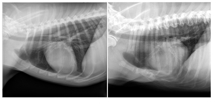
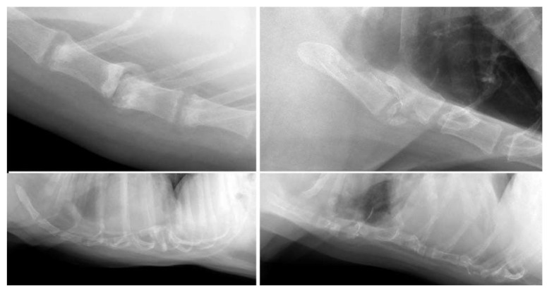
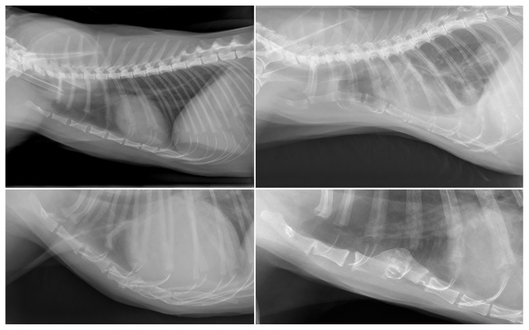
Evaluation of the sternum is part of the routine examination of small animal thoracic radiographs. However, descriptions on frequency and type of abnormalities are lacking. This retrospective observational study aimed to describe abnormal radiographic findings of the sternum in a cross-section of client-owned dogs and cats undergoing thoracic radiography between 1 January 2019 and 1 January 2021 for reasons unrelated to diseases of the sternum. The study population consisted of 777 dogs (mean age, 7.3 ± 3.9 years) and 183 cats (mean age, 7.3 ± 5.1 years). Sternal abnormalities were observed in 189/777 (24%) dogs and 53/183 (29%) cats, mostly around the intersternebral cartilages, accounting for 32/80 (40%) dogs and 20/35 (57%) cats. This was followed by an abnormal number of sternal segments (8% dogs, range 3-9 sternebrae; 15% cats, range 7-9 sternebra). Pectus excavatum was observed in 6/777 (0.8%) dogs and 6/183 (3%) cats, and pectus carinatum in 18/777 (2%) dogs and 2/183 (1%) cats. Post-traumatic changes, such as subluxation, were observed in nine dogs (1.1%) and three cats (1.6%). Presumed prostatic carcinoma metastasis and malignant lymphoma were observed in two dogs (0.2%). Incidental radiographic sternal abnormalities are common in cats and dogs but mostly of unknown clinical relevance.
Keywords: companion animals; dislocation; malformation; osteoarthrosis; pectus carinatum; pectus excavatum; vacuum phenomenon.
Conflict of interest statement
The authors declare no conflict of interest.
Figures
Similar articles
-
Successful Medical Management of an Acute Traumatic Sternal Luxation in a Cat.J Am Anim Hosp Assoc. 2023 May 1;59(3):142-144. doi: 10.5326/JAAHA-MS-7291. J Am Anim Hosp Assoc. 2023. PMID: 37167253
-
Reactive pectus carinatum in patients treated for pectus excavatum.J Pediatr Surg. 2008 Aug;43(8):1468-73. doi: 10.1016/j.jpedsurg.2007.11.019. J Pediatr Surg. 2008. PMID: 18675637
-
Surgical correction of pectus excavatum and carinatum.J Cardiovasc Surg (Torino). 1999 Oct;40(5):725-31. J Cardiovasc Surg (Torino). 1999. PMID: 10597012 Review.
-
Correlation between clinical severity and type and degree of pectus excavatum in twelve brachycephalic dogs.J Vet Med Sci. 2018 May 18;80(5):766-771. doi: 10.1292/jvms.17-0518. Epub 2018 Mar 29. J Vet Med Sci. 2018. PMID: 29593167 Free PMC article.
-
Surgical repair of pectus excavatum and carinatum.Semin Thorac Cardiovasc Surg. 2009 Spring;21(1):64-75. doi: 10.1053/j.semtcvs.2009.03.002. Semin Thorac Cardiovasc Surg. 2009. PMID: 19632565 Review.
Cited by
-
Sternal Dislocation and Associated Lung Lobe Hernia in a Cat.Case Rep Vet Med. 2024 May 14;2024:3719641. doi: 10.1155/2024/3719641. eCollection 2024. Case Rep Vet Med. 2024. PMID: 38774560 Free PMC article.
References
-
- Lahunta A.d., Evans H.E., Hermanson J.W. Miller and Evans’ Anatomy of the Dog. 5th ed. Elsevier; St. Louis, MO, USA: 2019.
-
- Singh B. Dyce, Sack, and Wensing’s Textbook of Veterinary Anatomy. 5th ed. Elsevier; St. Louis, MO, USA: 2018. pp. 38–39.
-
- Hudson L., Hamilton W. Atlas of Feline Anatomy For Veterinarians. Teton NewMedia; Jackson, MS, USA: 2017. p. 31.
-
- König H.E., Liebich H.G. Veterinary Anatomy of Domestic Animals. 7th ed. Thieme; Stuttgart, Germany: 2020. pp. 125–128.
-
- Schwarz T., Johnson V. BSAVA Manual of Canine and Feline Thoracic Imaging. BSAVA; Quedgeley, UK: 2008. p. 340.
LinkOut - more resources
Full Text Sources
Miscellaneous
