Review
. 2023 Mar 24;12(7):2487.
doi: 10.3390/jcm12072487.

The Dark Side of Ultrasound Imaging in Parathyroid Disease

Affiliations
Review

The Dark Side of Ultrasound Imaging in Parathyroid Disease

Roberta Centello et al. J Clin Med. .

Abstract

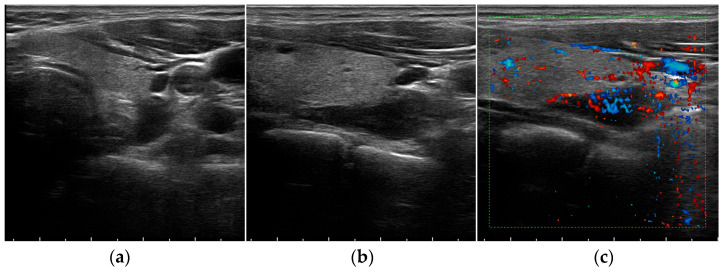
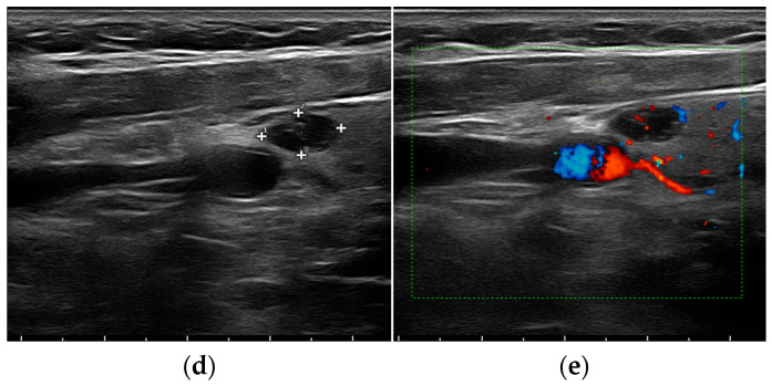
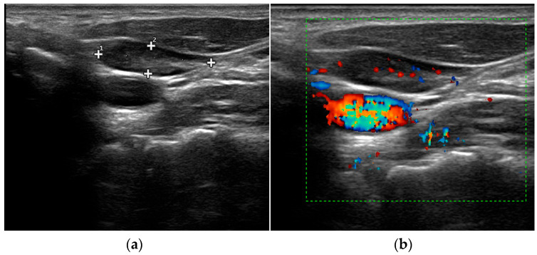
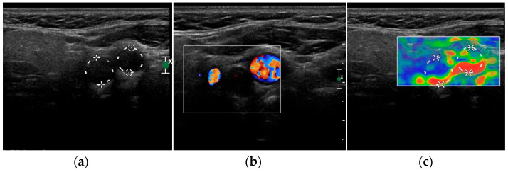
The diagnosis of parathyroid diseases by imaging still has some intrinsic technical limitations due to the differential diagnosis of different structures of the neck that mimic the parathyroid glands. In this view, ultrasound (US) is an established, low-cost, and non-invasive imaging technique that still represents the first-line approach for evaluating patients with parathyroid disease. The objective of this article is to provide a comprehensive review of the applications of USs in clinical practice, discussing the histopathological and US characteristics of the parathyroid glands in normal and pathological conditions, the advantages of preoperative imaging, and novel updates on the most useful and currently available multiparameter US techniques.

Keywords: hyperparathyroidism; parathyroid; ultrasound.


Conflict of interest statement

The authors declare no conflict of interest.

Figures

Figure 1
Figure 1
Grey-scale (a,b,d) and Color Doppler (c,e) aspect of a parathyroid adenoma.
Figure 1
Figure 1
Grey-scale (a,b,d) and Color Doppler (c,e) aspect of a parathyroid adenoma.
Figure 2
Figure 2
Grey-scale (a) and elastography (b) aspect of a hyperplastic parathyroid gland (arrow).
Figure 3
Figure 3
Grey-scale (a) and Color Doppler (b) aspect of cervical lymph node.
Figure 4
Figure 4
Grey-scale (a), Color Doppler (b) and elastography (c) aspect of a carotid body paraganglioma.
Figure 5
Figure 5
Grey-scale (a), Color Doppler (b) and elastography (c) aspect of a infected second branchial cleft cyst.

References

    1. McQueen A.S., Bhatia K.S. Head and neck ultrasound: Technical advances, novel applications and the role of elastography. Clin. Radiol. 2018;73:81–93. doi: 10.1016/j.crad.2017.08.003. - DOI - PubMed
    1. Ovčariček P.P., Giovanella L., Gasset I.C., Hindié E., Huellner M.W., Luster M., Piccardo A., Weber T., Talbot J.-N., Verburg F.A. The EANM practice guidelines for parathyroid imaging. Eur. J. Nucl. Med. Mol. Imaging. 2021;48:2801–2822. doi: 10.1007/s00259-021-05334-y. - DOI - PMC - PubMed
    1. American Institute of Ultrasound in Medicine. AIUM Practice Guideline for the performance of thyroid and parathyroid ultrasound examination. J. Ultrasound. Med. 2003;22:1126–1130. - PubMed
    1. Korwar V., Chang F.Y., Teasdale E., Suchett-Kaye I., Edwards A., Morgan J. Stepwise Approach for Parathyroid Localisation in Primary Hyperparathyroidism. World J. Surg. 2020;44:803–809. doi: 10.1007/s00268-019-05269-4. - DOI - PubMed
    1. Yeh M.W., Barraclough B.M., Sidhu S.B., Sywak M.S., Barraclough B.H., Delbridge L.W. Two hundred consecutive parathyroid ultrasound studies by a single clinician: The impact of experience. Endocr. Pract. 2006;12:257–263. doi: 10.4158/EP.12.3.257. - DOI - PubMed

LinkOut - more resources