Review
. 2023 Mar 27;12(7):2526.
doi: 10.3390/jcm12072526.

Clinical Usefulness of Right Ventricle-Pulmonary Artery Coupling in Cardiovascular Disease

Affiliations
Review

Clinical Usefulness of Right Ventricle-Pulmonary Artery Coupling in Cardiovascular Disease

Qing He et al. J Clin Med. .

Abstract

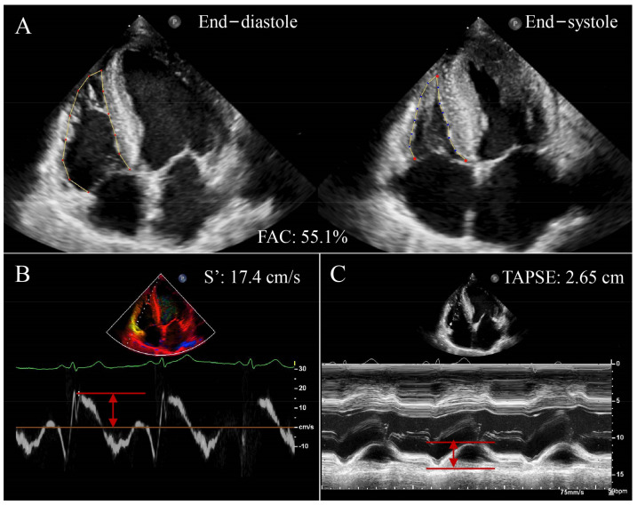
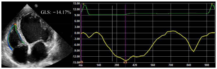
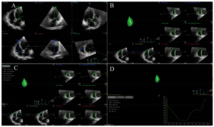
Right ventricular-pulmonary artery coupling (RV-PA coupling) refers to the relationship between RV contractility and RV afterload. Normal RV-PA coupling is maintained only when RV function and pulmonary vascular resistance are appropriately matched. RV-PA uncoupling occurs when RV contractility cannot increase to match RV afterload, resulting in RV dysfunction and right heart failure. RV-PA coupling plays an important role in the pathophysiology and progression of cardiovascular diseases. Therefore, early and accurate evaluation of RV-PA coupling is of great significance for a patient's condition assessment, clinical decision making, risk stratification, and prognosis judgment. RV-PA coupling can be assessed by using invasive or noninvasive approaches. The aim of this review was to summarize the pathological mechanism and evaluation methods of RV-PA coupling, the advantages and disadvantages of each method, and the application value of RV-PA coupling in various cardiovascular diseases.

Keywords: heart failure; hypertension; pulmonary arterial hypertension; right ventricular–pulmonary artery coupling; valvular heart disease.


Conflict of interest statement

The authors declare no conflict of interest.

Figures

Figure 1
Figure 1
Conventional RV function parameters using two-dimensional echocardiography in a patient with HFrEF. (A) RVFAC; (B) S’; (C) TAPSE; RV, right ventricular; HFrEF, heart failure with reduced ejection fraction; RVFAC, right ventricular fractional area change; S’, tricuspid annular systolic velocity;TAPSE, tricuspid annular plane systolic excursion. Red arrows represent measurements for parameters.
Figure 2
Figure 2
RVGLS in a patient with HFrEF. RVGLS, right ventricular global longitudinal strain.
Figure 3
Figure 3
RVEF using 3 DE in a patient with HFrEF. (A) Setting reference points; (B) RV endocardial border identification and tracking at end-diastole; (C) RV endocardial border identification and tracking at end-systole; (D) RVEF is automatically generated. RVEF, right ventricular ejection fraction; 3 DE, three-dimensional echocardiography.

Similar articles

Cited by

References

    1. Jone P.N., Ivy D.D. Comprehensive Noninvasive Evaluation of Right Ventricle-Pulmonary Circulation Axis in Pediatric Patients with Pulmonary Hypertension. Curr. Treat. Options Cardiovasc. Med. 2019;21:6. doi: 10.1007/s11936-019-0710-y. - DOI - PubMed
    1. Kubba S., Davila C.D., Forfia P.R. Methods for Evaluating Right Ventricular Function and Ventricular–Arterial Coupling. Prog. Cardiovasc. Dis. 2016;59:42–51. doi: 10.1016/j.pcad.2016.06.001. - DOI - PubMed
    1. Monge García M.I., Santos A. Understanding ventriculo-arterial coupling. Ann. Transl. Med. 2020;8:795. doi: 10.21037/atm.2020.04.10. - DOI - PMC - PubMed
    1. Naeije R., Brimioulle S., Dewachter L. Biomechanics of the right ventricle in health and disease (2013 grover conference series) Pulm. Circ. 2014;4:395–406. doi: 10.1086/677354. - DOI - PMC - PubMed
    1. Amsallem M., Mercier O., Kobayashi Y., Moneghetti K., Haddad F. Forgotten No More: A Focused Update on the Right Ventricle in Cardiovascular Disease. JACC Heart Fail. 2018;6:891–903. doi: 10.1016/j.jchf.2018.05.022. - DOI - PubMed

LinkOut - more resources