. 2023 Mar 28;12(7):2556.
doi: 10.3390/jcm12072556.

It Is Always the Same-A Complication Classification following Angular Stable Plating of Proximal Humeral Fractures

Affiliations

It Is Always the Same-A Complication Classification following Angular Stable Plating of Proximal Humeral Fractures

Georg Siebenbürger et al. J Clin Med. .

Abstract

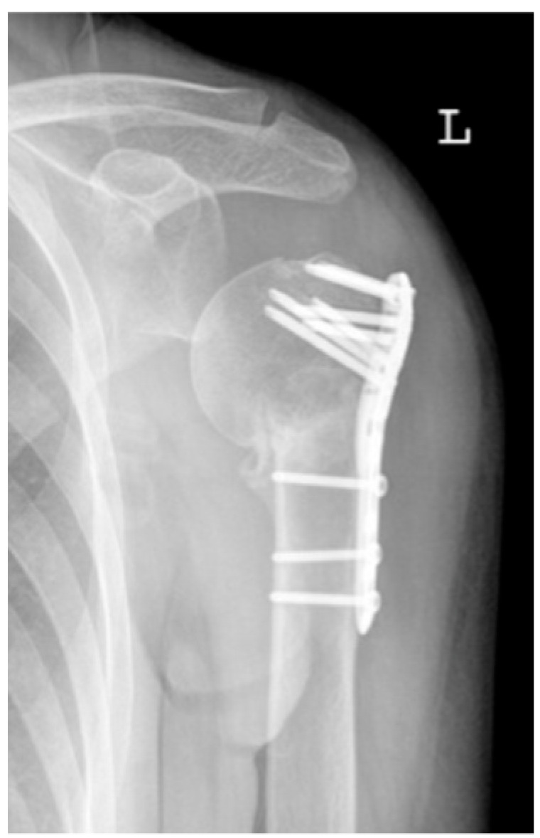
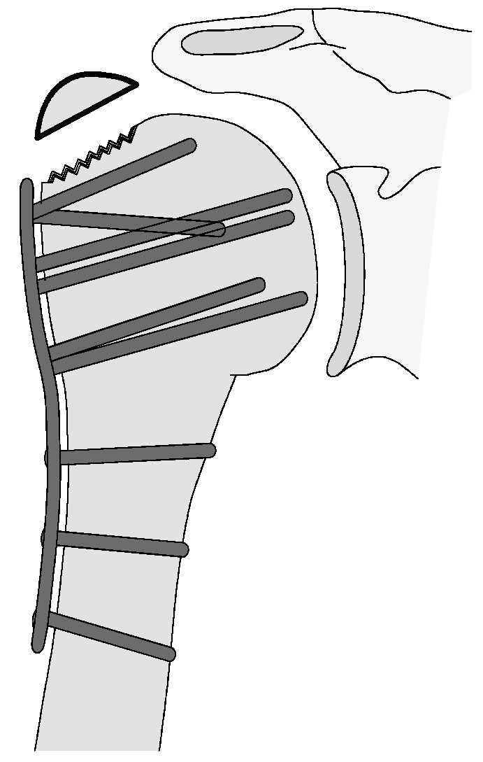
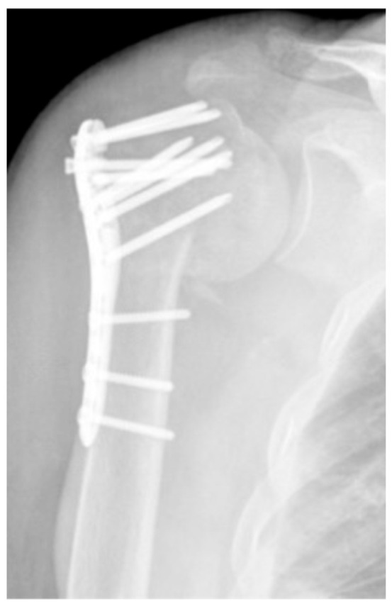
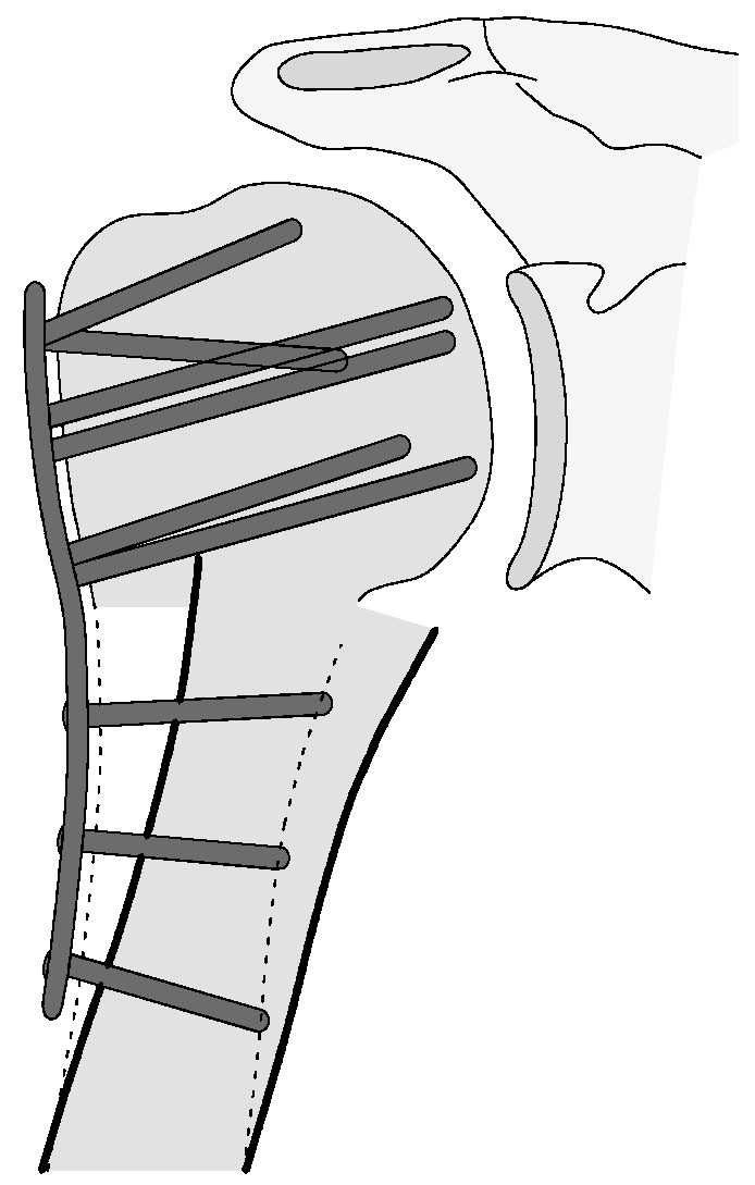
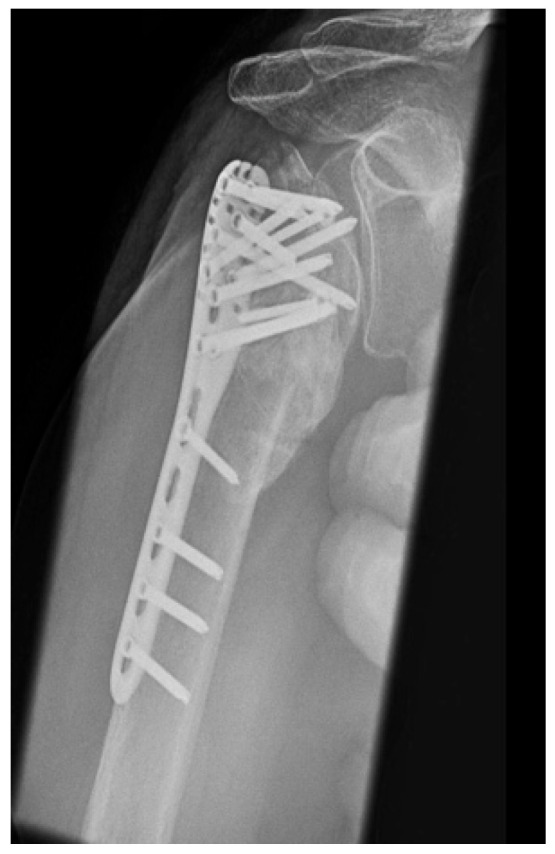
The aim of this study was to create a novel complication classification for osteosynthesis-related complications following angular stable plating of the proximal humerus subsuming the influence of these complications on clinical outcome in relation to fracture morphology and consequent revision strategies. A total of 1047 proximal humerus fractures with overall 193 osteosynthesis-associated complications (24.5%) were included. The following complication types could be clarified: complication Type 1 is defined by mild varus (<20°) or valgus displacement of the humeral head without resulting in a screw cutout through the humeral head cortex. Type 2a is defined by varus displacement (<20°) of the humeral head associated with screw cutout through the humeral head cortex. Type 2b complication is limited to displacement of the greater tuberosity, lesser tuberosity, or both tuberosities. Complication Type 2c is defined by severe varus dislocation (>20°) of the humeral head with screw cutout at the humeral head cortex. Complication Type 3 describes a displacement of the angular stable plate in the humeral shaft region with associated shaft-sided screw cutout, while the position of the humeral head remains static. Complication Type 4 is characterized by the occurrence of AVN with or without glenoidal affection (4a/b). Clinical outcome according to the constant score was mainly affected by type 2-4, leading to a deteriorated result. Depending on the type of complication, specific revision strategies can be considered. Additionally, more complex fracture patterns fostered the incidence of complications.

Keywords: angular stable plating locking plate osteosynthesis; complication classification; complications; constant score; functional outcome; mid-term; proximal humeral fracture.


Conflict of interest statement

The authors declare no conflict of interest.

Figures

Figure 3
Figure 3
Absolute fracture distribution of the total collective, based on the Neer classification, total number n = 1047 (X-axis: Neer classification, Y-axis: number).
Figure 4
Figure 4
Absolute fracture distribution of the total collective, based on the AO-classification, total number n = 1047 (X-axis: AO-classification, Y-axis: number).
Figure 1
Figure 1
Flow chart of patients included at 4 years of follow-up.
Figure 2
Figure 2
Calculation of the humeral head–shaft angle in the a.p. radiographic path by drawing three lines and the intersection of the line along the long axis of the humeral shaft with the perpendicular to the line of inferior + superior articular surface [22,23].
Figure 5
Figure 5
Complication Type 1. a.p. radiograph of the left shoulder after angular stable plate osteosynthesis illustrates mild varus displacement of the humeral head. The humeral head cortex remains intact, so there is no screw cutout.
Figure 6
Figure 6
Complication Type 1. Middle: measurement of varus displacement on an a.p. radiograph of a right shoulder after angular stable plate osteosynthesis. The varus displacement is 16° relative to the anatomic CCD angle of 135°. No screw cutout results.
Figure 7
Figure 7
Complication Type 1. sample image. Mild varus displacement of the humeral head after angular stable plate osteosynthesis is shown by the transition of the dashed drawing (initially correct humeral head position) to the prominent black drawing.
Figure 8
Figure 8
Complication Type 2a. a.p. radiograph of the right shoulder after angular stable plate osteosynthesis shows moderate varus displacement of the humeral head. The most cranial screw breaks through the humeral head cortex (screw cutout).
Figure 9
Figure 9
Complication Type 2a. CCD-relative varus displacement of 19°.
Figure 10
Figure 10
Complication Type 2a. Pattern image. Moderate grade varus displacement of the humeral head after angular stable plate osteosynthesis is illustrated by the transition of the dashed drawing (initially correct humeral head position) to the prominent black drawing with screw cutout (double).
Figure 11
Figure 11
Complication Type 2b. Y-view radiograph of the left shoulder after angular stable plate osteosynthesis illustrates displacement of the greater tuberosity.
Figure 12
Figure 12
Complication Type 2b. Sample image. Following angular stable plate osteosynthesis, the displacement of the greater tuberosity is illustrated by the separation of the prominent black drawing from the serrated drawing (initial regular position).
Figure 13
Figure 13
Complication Type 2c. a.p. radiograph of the right shoulder after angular stable plate osteosynthesis shows severe varus displacement of the humeral head. The most cranial screws penetrate the humeral head cortex, resulting in a screw cutout. The humeral head sintering appears unstable.
Figure 14
Figure 14
Complication Type 2c. Measurement of varus displacement on an a.p. radiograph of a left shoulder after angular stable plate osteosynthesis with same-sided complication. The cranial screws penetrate the cortical bone. Varus displacement is 27° relative to the anatomic CCD angle of 135°.
Figure 15
Figure 15
Complication Type 2c. Pattern image. Severe varus displacement and instability of the humeral head after angle-stable plate osteosynthesis is shown by the transition of the dashed drawing (initially correct humeral head position) to the prominent black drawing. This is followed by a cutout of four screws.
Figure 16
Figure 16
Complication Type 3. a.p. radiograph of the right shoulder after angular stable plate osteosynthesis shows a displacement of the humeral shaft area, so the fixation of the plate osteosynthesis is no longer guaranteed. In contrast, the humeral head position is regular.
Figure 17
Figure 17
Complication Type 3. Pattern image. The displacement of the humeral shaft after angle-stable plate osteosynthesis is shown by the transition of the dashed drawing (initially correct humeral shaft position) to the prominent black drawing.
Figure 18
Figure 18
Complication Type 4a. a.p. radiograph of the right shoulder after angular stable plate osteosynthesis shows necrosis of the humeral head. The glenoid articular surface is still intact.
Figure 19
Figure 19
Complication Type 4a. Sample image. Humeral head necrosis after angle-stable plate osteosynthesis is depicted by the transition of the dashed drawing (regular humeral head anatomy) to the inhomogeneous, prominent black drawing. A cutout of the most cranial screw results, but this does not cause damage to the glenoid articular surface.
Figure 20
Figure 20
Complication Type 4b. a.p. radiograph of the left shoulder after angular stable plate osteosynthesis illustrates necrosis of the humeral head, implying cutout of multiple screws. This has resulted in arrosion of the glenoid articular surface.
Figure 21
Figure 21
Complication Type 4b. Sample image. Humeral head necrosis after angular stable plate osteosynthesis is illustrated by the transition of the dashed drawing (regular humeral head anatomy) to the inhomogeneous, prominent black drawing. A multiple screw cutout results. The damage to the glenoid articular surface based on this is marked by the star symbols on the cavitas glenoidalis.
Figure 22
Figure 22
Comparison CS of collective without complications and complication types (X-axis: study collective, Y-axis: median, IQR, extreme values, outliers).
Figure 23
Figure 23
Comparison nCS of collective without complications and complication types (X-axis: study collective, Y-axis: median, IQR, extreme values, outliers).
Figure 24
Figure 24
Comparison %CS of collective without complications and complication types (X-axis: study collective, Y-axis: median, IQR, o= extreme values, * = outliers).
Figure 25
Figure 25
Comparison of the number with complications (dark) and without complications (light) in relation to the respective fracture type, based on the Neer classification (X-axis: Neer classification, Y-axis: number).
Figure 26
Figure 26
Comparison of the number with complications (dark) and without complications (light) in relation to the respective fracture type, based on the AO classification (X-axis: AO classification, Y-axis: number).

References

    1. Baker H.P., Gutbrod J., Strelzow J.A., Maassen N.H., Shi L. Management of Proximal Humerus Fractures in Adults-A Scoping Review. J. Clin. Med. 2022;11:6140. doi: 10.3390/jcm11206140. - DOI - PMC - PubMed
    1. Rupp M., Walter N., Pfeifer C., Lang S., Kerschbaum M., Krutsch W., Baumann F., Alt V. The Incidence of Fractures Among the Adult Population of Germany—An Analysis from 2009 through 2019. Dtsch. Arztebl. Int. 2021;118:665–669. doi: 10.3238/arztebl.m2021.0238. - DOI - PMC - PubMed
    1. Burkhart K.J., Dietz S.O., Bastian L., Thelen U., Hoffmann R., Müller L.P. The treatment of proximal humeral fracture in adults. Dtsch. Arztebl. Int. 2013;110:591–597. doi: 10.3238/arztebl.2013.0591. - DOI - PMC - PubMed
    1. Porschke F., Bockmeyer J., Nolte P.C., Studier-Fischer S., Guehring T., Schnetzke M. More Adverse Events after Osteosyntheses Compared to Arthroplasty in Geriatric Proximal Humeral Fractures Involving Anatomical Neck. J. Clin. Med. 2021;10:979. doi: 10.3390/jcm10050979. - DOI - PMC - PubMed
    1. Laux C.J., Grubhofer F., Werner C.M.L., Simmen H.P., Osterhoff G. Current concepts in locking plate fixation of proximal humerus fractures. J. Orthop. Surg. Res. 2017;12:137. doi: 10.1186/s13018-017-0639-3. - DOI - PMC - PubMed

LinkOut - more resources