. 2023 Mar 29;16(7):2739.
doi: 10.3390/ma16072739.

Plasma-Polymerised Antibacterial Coating of Ovine Tendon Collagen Type I (OTC) Crosslinked with Genipin (GNP) and Dehydrothermal-Crosslinked (DHT) as a Cutaneous Substitute for Wound Healing

Affiliations

Plasma-Polymerised Antibacterial Coating of Ovine Tendon Collagen Type I (OTC) Crosslinked with Genipin (GNP) and Dehydrothermal-Crosslinked (DHT) as a Cutaneous Substitute for Wound Healing

Ibrahim N Amirrah et al. Materials (Basel). .

Abstract

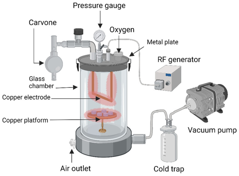
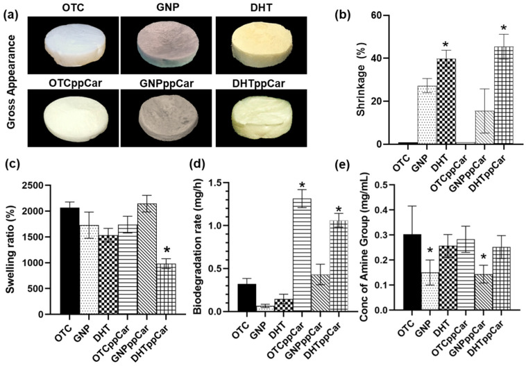
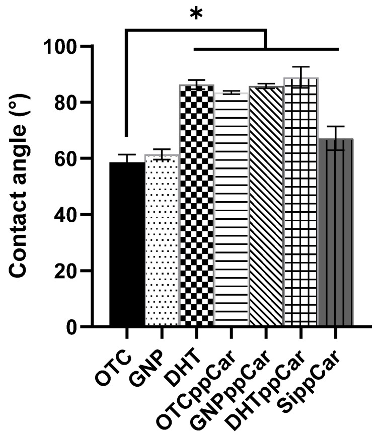
Tissue engineering products have grown in popularity as a therapeutic approach for chronic wounds and burns. However, some drawbacks include additional steps and a lack of antibacterial capacities, both of which need to be addressed to treat wounds effectively. This study aimed to develop an acellular, ready-to-use ovine tendon collagen type I (OTC-I) bioscaffold with an antibacterial coating for the immediate treatment of skin wounds and to prevent infection post-implantation. Two types of crosslinkers, 0.1% genipin (GNP) and dehydrothermal treatment (DHT), were explored to optimise the material strength and biodegradability compared with a non-crosslinked (OTC) control. Carvone plasma polymerisation (ppCar) was conducted to deposit an antibacterial protective coating. Various parameters were performed to investigate the physicochemical properties, mechanical properties, microstructures, biodegradability, thermal stability, surface wettability, antibacterial activity and biocompatibility of the scaffolds on human skin cells between the different crosslinkers, with and without plasma polymerisation. GNP is a better crosslinker than DHT because it demonstrated better physicochemical properties (27.33 ± 5.69% vs. 43 ± 7.64% shrinkage), mechanical properties (0.15 ± 0.15 MPa vs. 0.07 ± 0.08 MPa), swelling (2453 ± 419.2% vs. 1535 ± 392.9%), biodegradation (0.06 ± 0.06 mg/h vs. 0.15 ± 0.16 mg/h), microstructure and biocompatibility. Similarly, its ppCar counterpart, GNPppCar, presents promising results as a biomaterial with enhanced antibacterial properties. Plasma-polymerised carvone on a crosslinked collagen scaffold could also support human skin cell proliferation and viability while preventing infection. Thus, GNPppCar has potential for the rapid treatment of healing wounds.

Keywords: antibacterial; biomaterial; carvone; collagen; dehydrothermal treatment; genipin; plasma polymerisation; wound healing.


Conflict of interest statement

The authors declare no conflict of interest.

Figures

Figure 1
Figure 1
Schematic illustration of the plasma polymerisation mechanism of the collagen scaffolds and the plasma reactor equipment.
Figure 2
Figure 2
(a) Cross-section of FESEM OTC surface before carvone plasma polymerisation (ppCar); (b) cross-section of FESEM OTCppCar surface shows the antibacterial carvone coating; (c) the top-surface FESEM OTC shows surface roughness before ppCar; (d) depicts porous carvone deposition coated on top surface of OTCppCar; (e) relative atomic concentrations of carbon, nitrogen and oxygen; and (f) ratios of oxygen to carbon (O/C) and nitrogen to carbon (N/C).
Figure 3
Figure 3
The physical characteristics of the fabricated biomaterials in terms of (a) the gross appearance, (b) the percentage of shrinkage post-crosslink GNP and DHT and post-carvone plasma polymerisation, GNPppCar and DHTppCar; (c) the swelling ratio, (d) the biodegradation rate and (e) the degree of crosslinking. * p ≤ 0.05 indicates significant differences in fabricated scaffolds in comparison with non-crosslinked ovine tendon collagen type I (OTC).
Figure 4
Figure 4
The mechanical characterisation of the scaffolds at room temperature includes (a) stress vs. strain curve, (b) Young’s modulus, (c) max tensile stress, (d) max tensile load, (e) compression test and (f) resilience test. * p ≤ 0.05 indicates significant differences in the fabricated materials.
Figure 5
Figure 5
Water contact angle of OTC-I scaffolds before and after crosslinking and ppCar. A silicon wafer (SippCar) was used as the plasma polymerisation deposition control, which showed that carvone plasma polymerisation does not lead to a material being hydrophobic. * p ≤ 0.05 indicates significant differences in the fabricated materials.
Figure 6
Figure 6
(a) The heterogeneous microstructure of OTC-I sponges showing interconnective pores before and after crosslinking with GNP and DHT and ppCar. DHTppCar had most shrinkage after crosslinking and reduced porosity after carvone deposition; (b) liquid dispersion assay conveys SEM data in which all scaffolds except for DHTppCar were >50%; (c) pore size distribution of OTC-I scaffolds indicated acceptable pore size range from 100 to 200 μm except for the case of DHTppCar. * p ≤ 0.05 indicates significant differences in the fabricated materials.
Figure 7
Figure 7
(a) TGA analysis of crosslinked OTC-I scaffolds before plasma polymerisation; (b) TGA analysis of crosslinked OTC-I scaffolds after ppCar; (c) FTIR results of OTC-I biomatrices; (d) XRD.
Figure 8
Figure 8
Live/dead bacterial assay of (a) E. coli and (b) S. aureus before and after ppCar on glass slides. Green represents live bacterial cells; red represents dead cells. * p ≤ 0.05 indicates significant differences in the fabricated materials.
Figure 9
Figure 9
The cellular compatibility of fabricated OTC-I bioscaffolds with (a) human dermal fibroblasts (HDFs) and (b) human epithelial keratinocytes (HEKs) after 24 h incubation post-seeding at 37 °C. Scale bar is 100 µm.
Figure 9
Figure 9
The cellular compatibility of fabricated OTC-I bioscaffolds with (a) human dermal fibroblasts (HDFs) and (b) human epithelial keratinocytes (HEKs) after 24 h incubation post-seeding at 37 °C. Scale bar is 100 µm.

Similar articles

Cited by

References

    1. Nathoo R., Howe N., Cohen G. Skin Substitutes: An Overview of the Key Players in Wound Management. J. Clin. Aesthet. Dermatol. 2014;7:44. - PMC - PubMed
    1. Snyder D., Sullivan N., Margolis D., Schoelles K. Skin Substitutes for Treating Chronic Wounds [Internet] Agency for Healthcare Research and Quality; Rockville, MD, USA: 2020. - PubMed
    1. Savoji H., Godau B., Hassani M.S., Akbari M. Skin Tissue Substitutes and Biomaterial Risk Assessment and Testing. Front. Bioeng. Biotechnol. 2018;6:86. doi: 10.3389/fbioe.2018.00086. - DOI - PMC - PubMed
    1. Irawan V., Sung T.C., Higuchi A., Ikoma T. Collagen Scaffolds in Cartilage Tissue Engineering and Relevant Approaches for Future Development. Tissue Eng. Regen. Med. 2018;15:673–697. doi: 10.1007/s13770-018-0135-9. - DOI - PMC - PubMed
    1. Zheng Y., Liang Y., Zhang D., Sun X., Liang L., Li J., Liu Y.N. Gelatin-Based Hydrogels Blended with Gellan as an Injectable Wound Dressing. ACS Omega. 2018;3:4766–4775. doi: 10.1021/acsomega.8b00308. - DOI - PMC - PubMed

LinkOut - more resources