Review
. 2023 Mar 31;16(7):2799.
doi: 10.3390/ma16072799.

Bioceramics/Electrospun Polymeric Nanofibrous and Carbon Nanofibrous Scaffolds for Bone Tissue Engineering Applications

Affiliations
Review

Bioceramics/Electrospun Polymeric Nanofibrous and Carbon Nanofibrous Scaffolds for Bone Tissue Engineering Applications

Zahra Ebrahimvand Dibazar et al. Materials (Basel). .

Abstract

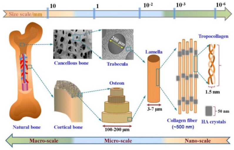
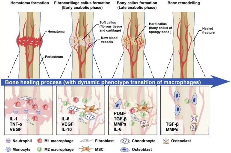
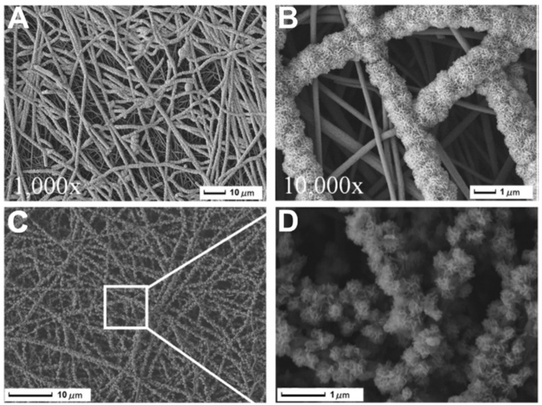
Bone tissue engineering integrates biomaterials, cells, and bioactive agents to propose sophisticated treatment options over conventional choices. Scaffolds have central roles in this scenario, and precisely designed and fabricated structures with the highest similarity to bone tissue have shown promising outcomes. On the other hand, using nanotechnology and nanomaterials as the enabling options confers fascinating properties to the scaffolds, such as precisely tailoring the physicochemical features and better interactions with cells and surrounding tissues. Among different nanomaterials, polymeric nanofibers and carbon nanofibers have attracted significant attention due to their similarity to bone extracellular matrix (ECM) and high surface-to-volume ratio. Moreover, bone ECM is a biocomposite of collagen fibers and hydroxyapatite crystals; accordingly, researchers have tried to mimic this biocomposite using the mineralization of various polymeric and carbon nanofibers and have shown that the mineralized nanofibers are promising structures to augment the bone healing process in the tissue engineering scenario. In this paper, we reviewed the bone structure, bone defects/fracture healing process, and various structures/cells/growth factors applicable to bone tissue engineering applications. Then, we highlighted the mineralized polymeric and carbon nanofibers and their fabrication methods.

Keywords: bone tissue engineering; electrospinning; mineralization; nanofibers.


Conflict of interest statement

The authors declare no conflict of interest.

Figures

Figure 1
Figure 1
The hierarchical multi-scale structure of natural bone ranging from the nanoscale to macroscale. Reproduced with permission from Ref. [38].
Figure 2
Figure 2
A graphical view of the direct bone healing process.
Figure 3
Figure 3
CT pictures of the implanted CNFs/HA nanocomposite that repaired the damaged femur in vivo. After 8 weeks following the accident, diagnostic 3D imaging (CT scan) was conducted of the femur bone defects. The arrow indicates the deficient area that was not healed in the control group (a) and the bone defect that was corrected as a result of the implanted CNFs/HA nanocomposite stimulating the growth of normal tissue (b). Reproduced with permission from Ref. [91].
Figure 4
Figure 4
SEM photos show the development of HAp particles on T-CNF mats treated with NaOH aq. solution at concentrations of (A) 8%, (B) 16%, (C) 20%, and (D) the crystals that resemble flowers. One day for cultivation. Reproduced with permission from Ref. [97].
Figure 5
Figure 5
(A): Digital image of an ARS-stained sample that has been treated with the standard nucleation procedure (SNP) after being dipped in EtOH; bottom: SEM image of the same sample. (B): Digital image of an ARS-stained sample that has been treated with the SNP after being activated by ultrasonic waves; top: SEM image of the same sample. Reproduced with permission from Ref. [98].
Figure 6
Figure 6
The conventional porous bioglass scaffold fabrication.
Figure 7
Figure 7
SEM photos of the distribution of well-dispersed β-TCP nanoparticles that firmly adhered to CNFs at various magnifications (A) 80 µm, (B) 2 µm, (C) 500 nm, (D) 250 nm. (Whereas the CNFs had an average diameter of about 300 nm, the average size of the β-TCP particles ranged from 30 to 60 nm). Reproduced with permission from Ref. [110].
Figure 8
Figure 8
Morphology of PDLCs on the fabricated nanofibers. (A) CLSM image of cells on pristine CNFs, (B) CLSM image of cells on β-TCP-decorated CNFs, (C) SEM image of cells on pristine CNFs after 1 day of cell seeding, (D) SEM image of cells on pristine β-TCP-decorated CNFs after 1 day of cell seeding, (E) SEM image of cells on pristine CNFs after 7 days of cell seeding, and (F) SEM image of cells on pristine β-TCP-decorated CNFs after 7 days of cell seeding. Reproduced with permission from Ref. [112].

Similar articles

Cited by

References

    1. Koons G.L., Diba M., Mikos A.G. Materials design for bone-tissue engineering. Nat. Rev. Mater. 2020;5:584–603. doi: 10.1038/s41578-020-0204-2. - DOI
    1. Qu H., Fu H., Han Z., Sun Y. Biomaterials for bone tissue engineering scaffolds: A review. RSC Adv. 2019;9:26252–26262. doi: 10.1039/C9RA05214C. - DOI - PMC - PubMed
    1. Samadian H., Khastar H., Ehterami A., Salehi M. Bioengineered 3D nanocomposite based on gold nanoparticles and gelatin nanofibers for bone regeneration: In vitro and in vivo study. Sci. Rep. 2021;11:13877. doi: 10.1038/s41598-021-93367-6. - DOI - PMC - PubMed
    1. Nekounam H., Gholizadeh S., Allahyari Z., Samadian H., Nazeri N., Shokrgozar M.A., Faridi-Majidi R. Electroconductive Scaffolds for Tissue Regeneration: Current opportunities, pitfalls, and potential solutions. Mater. Res. Bull. 2021;134:111083. doi: 10.1016/j.materresbull.2020.111083. - DOI
    1. Nazarnezhada S., Abbaszadeh-Goudarzi G., Samadian H., Khaksari M., Ghatar J.M., Khastar H., Rezaei N., Mousavi S.R., Shirian S., Salehi M. Alginate hydrogel containing hydrogen sulfide as the functional wound dressing material: In vitro and in vivo study. Int. J. Biol. Macromol. 2020;164:3323–3331. doi: 10.1016/j.ijbiomac.2020.08.233. - DOI - PubMed

LinkOut - more resources