. 2023 Mar 30;13(7):1236.
doi: 10.3390/nano13071236.

A Novel Drastic Peptide Genetically Adapted to Biomimetic Scaffolds "Delivers" Osteogenic Signals to Human Mesenchymal Stem Cells

Affiliations

A Novel Drastic Peptide Genetically Adapted to Biomimetic Scaffolds "Delivers" Osteogenic Signals to Human Mesenchymal Stem Cells

Aglaia Mantsou et al. Nanomaterials (Basel). .

Abstract

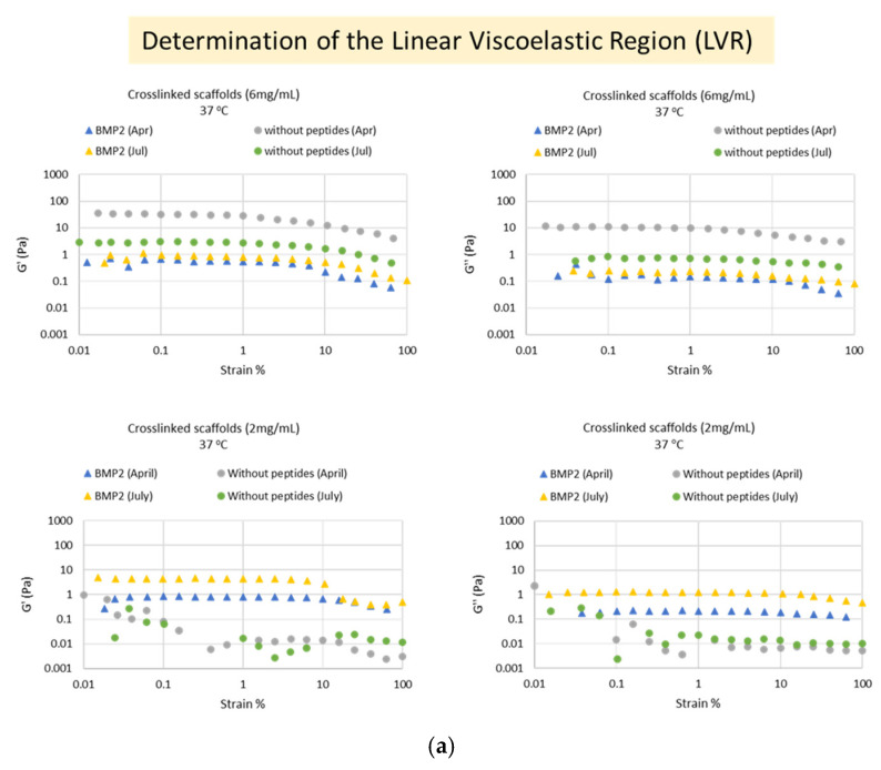
This work describes the design, preparation, and deep investigation of "intelligent nanobiomaterials" that fulfill the safety rules and aim to serve as "signal deliverers" for osteogenesis, harboring a specific peptide that promotes and enhances osteogenesis at the end of their hydrogel fibers. The de novo synthesized protein fibers, besides their mechanical properties owed to their protein constituents from elastin, silk fibroin and mussel-foot adhesive protein-1 as well as to cell-attachment peptides from extracellular matrix glycoproteins, incorporate the Bone Morphogenetic Protein-2 (BMP2) peptide (AISMLYLDEN) that, according to our studies, serves as "signal deliverer" for osteogenesis. The osteogenetic capacity of the biomaterial has been evidenced by investigating the osteogenic marker genes ALP, RUNX2, Osteocalcin, COL1A1, BMPR1A, and BMPR2, which were increased drastically in cells cultured on scaffold-BMP2 for 21 days, even in the absence of osteogenesis medium. In addition, the induction of phosphorylation of intracellular Smad-1/5 and Erk-1/2 proteins clearly supported the osteogenetic capacity of the biomaterial.

Keywords: BMP-2 peptide; biomaterial; bone regeneration; elastin; mussel-foot protein; osteogenesis; scaffold; silk fibroin; tissue engineering.


Conflict of interest statement

The authors declare no conflict of interest.

Figures

Scheme 1
Scheme 1
Depiction of the general structure of the synthesized genes.
Figure 1
Figure 1
Step-by-step synthesis of recombinamer genes. (ac) Electrophoresis in 1% w/v agarose gels of the recombinant plasmids containing building block sequences after digestion with NdeI and XhoI (a,b) or BseRI (c). In (c), lane 1 corresponds to digested pET29c-BMP2 peptide, and lanes 2–5 represent pET29c-heparin-binding peptide. (df) Step-by-step assembly of ELP5 and Silk2-Mussel15. Numbers in brackets state the length of the inserts. (d) Lane (1): pET29c-ELP1 insert (140 bp), lane (2): pET29c-ELP2 insert (215 bp), lane (3): pET29c-ELP4 insert (365 bp), and lane (4): pET29c-ELP5 insert (440 bp). (e) Lane (1): pET29c-Silk1 (164 bp), lane (2): pET29c-Silk2 (260 bp), lane (3): pET29c-Mussel3 (260 bp), lane (4): pET29c-Mussel9 (320 bp), lane (5): pET29c-Mussel15 (500 bp), and lane (6): pET29c–Silk2Mussel15 (710 bp). (f) pET29c–[(ELP10-Silk2-Mussel15)2-Mussel-6xHis] and final plasmids: pET29c–[laminin peptide-(ELP10-Silk2-Mussel15)2-Mussel-6xHis], pET29c–[heparin-binding peptide-(ELP10-Silk2-Mussel15)2-Mussel-6xHis], and pET29c–[BMP-2 peptide-(ELP10-Silk2-Mussel15)2-Mussel-6xHis] digested with NdeI/XhoI. (g) Step-by-step assembly of final plasmid pET29c–[ELP5-RGD-ELP5-Silk2-Mussel15)2-Mussel-6xHis]. Μ1: 50 bp DNA ladder and Μ2: 1 kb DNA ladder.
Figure 1
Figure 1
Step-by-step synthesis of recombinamer genes. (ac) Electrophoresis in 1% w/v agarose gels of the recombinant plasmids containing building block sequences after digestion with NdeI and XhoI (a,b) or BseRI (c). In (c), lane 1 corresponds to digested pET29c-BMP2 peptide, and lanes 2–5 represent pET29c-heparin-binding peptide. (df) Step-by-step assembly of ELP5 and Silk2-Mussel15. Numbers in brackets state the length of the inserts. (d) Lane (1): pET29c-ELP1 insert (140 bp), lane (2): pET29c-ELP2 insert (215 bp), lane (3): pET29c-ELP4 insert (365 bp), and lane (4): pET29c-ELP5 insert (440 bp). (e) Lane (1): pET29c-Silk1 (164 bp), lane (2): pET29c-Silk2 (260 bp), lane (3): pET29c-Mussel3 (260 bp), lane (4): pET29c-Mussel9 (320 bp), lane (5): pET29c-Mussel15 (500 bp), and lane (6): pET29c–Silk2Mussel15 (710 bp). (f) pET29c–[(ELP10-Silk2-Mussel15)2-Mussel-6xHis] and final plasmids: pET29c–[laminin peptide-(ELP10-Silk2-Mussel15)2-Mussel-6xHis], pET29c–[heparin-binding peptide-(ELP10-Silk2-Mussel15)2-Mussel-6xHis], and pET29c–[BMP-2 peptide-(ELP10-Silk2-Mussel15)2-Mussel-6xHis] digested with NdeI/XhoI. (g) Step-by-step assembly of final plasmid pET29c–[ELP5-RGD-ELP5-Silk2-Mussel15)2-Mussel-6xHis]. Μ1: 50 bp DNA ladder and Μ2: 1 kb DNA ladder.
Figure 2
Figure 2
Electrophoresis in 10% w/v SDS-polyacrylamide gels of the fractions after overexpression (20 μL of cell extract per lane) and purification (10 μg of protein per lane) of the polypeptides from BL21 E. coli cells. (a) ELP5-RGD-ELP5-Silk2-Mussel15)2-Mussel-6xHis, (b) Heparin-binding peptide-(ELP10-Silk2-Mussel15)2-Mussel-6xHis, (c) Laminin peptide-(ELP10-Silk2-Mussel15)2-Mussel-6xHis, and (d) BMP-2 peptide -(ELP10-Silk2-Mussel15)2-Mussel-6xHis. 0 h: before induction of overexpression, 5 h: 5 h after induction of overexpression with 1 mM IPTG, and EL1-EL10: elutions with 250 mM imidazole. M: protein marker.
Figure 3
Figure 3
(a) Schematic representation of the three-dimensional scaffolds that were produced after the crosslinking reaction. (b) Imaging of the micromorphology of the porous surface of the scaffold without peptides by scanning electron microscopy (SEM) at 1000× g (left) and 2200× g (right) magnification. Each photograph has a scale bar of 10 μm.
Figure 3
Figure 3
(a) Schematic representation of the three-dimensional scaffolds that were produced after the crosslinking reaction. (b) Imaging of the micromorphology of the porous surface of the scaffold without peptides by scanning electron microscopy (SEM) at 1000× g (left) and 2200× g (right) magnification. Each photograph has a scale bar of 10 μm.
Figure 4
Figure 4
Determination of the Linear Viscoelastic Region of the crosslinked biomaterials. (a) Plots of elastic modulus (G′) or viscous modulus (G″) as a function of applied strain (%) in the crosslinked biomaterials (6 mg/mL in DMEM) at 37 °C. “BMP2”: scaffold-BMP2, “without peptides”: scaffold without peptides, “Apr”: April 2022 measurements, “Jul”: July 2022 measurements. (b) Plot of G′ and G″ as a function of applied strain (%) in the crosslinked biomaterials (6 mg/mL in DMEM) at 37 °C. The G′ to G″ ratio indicates the formation of elastic networks. (c) Schematic representation of the behavior of the biomaterials at 37 °C in DMEM. The “crosslinked” biomaterials have a small LVR region, indicating that they rather behave as semi-rigid (a), rather than fully flexible (b), polymers. Created with BioRender.com. All data correspond to different aliquots of the same samples in order to avoid preshearing effects or different sample preparations in order to show reproducibility.
Figure 4
Figure 4
Determination of the Linear Viscoelastic Region of the crosslinked biomaterials. (a) Plots of elastic modulus (G′) or viscous modulus (G″) as a function of applied strain (%) in the crosslinked biomaterials (6 mg/mL in DMEM) at 37 °C. “BMP2”: scaffold-BMP2, “without peptides”: scaffold without peptides, “Apr”: April 2022 measurements, “Jul”: July 2022 measurements. (b) Plot of G′ and G″ as a function of applied strain (%) in the crosslinked biomaterials (6 mg/mL in DMEM) at 37 °C. The G′ to G″ ratio indicates the formation of elastic networks. (c) Schematic representation of the behavior of the biomaterials at 37 °C in DMEM. The “crosslinked” biomaterials have a small LVR region, indicating that they rather behave as semi-rigid (a), rather than fully flexible (b), polymers. Created with BioRender.com. All data correspond to different aliquots of the same samples in order to avoid preshearing effects or different sample preparations in order to show reproducibility.
Figure 5
Figure 5
The viscoelastic properties of the crosslinked and uncrosslinked biomaterials, as measured within the LVR (at 3% strain). (a): Plots of elastic modulus (G′) and viscous modulus (G″) of the biomaterials, as a function of time (s) at 37 °C. “BMP2”: scaffold-BMP2, “without peptides”: scaffold without peptides, “Apr”: April 2022 measurements, “Jul”: July 2022 measurements. All data correspond to different aliquots of the same samples in order to avoid preshearing effects or different sample preparations in order to show reproducibility. (b): Schematic representation of the possible form of the scaffolds in DMEM at 37 °C, as islands of similar 3D networks instead of a continuous network. Created with BioRender.com.
Figure 6
Figure 6
The effect of temperature on the viscoelastic properties of scaffold-BMP2, in the crosslinked and uncrosslinked state, at a concentration of 6 mg/mL in DMEM. (a) Temperature sweeps on the crosslinked scaffold-BMP2 at the range 10–40 °C and time sweeps on the uncrosslinked scaffold-BMP2 at 25 °C and 37 °C. The viscoelastic properties of crosslinked scaffolds showed a tendency to decrease with increasing temperature. In the uncrosslinked scaffolds, a shift in the G′/G″ ratio was observed between 25 °C and 37 °C, which indicated the possible formation of loose networks. All data correspond to different aliquots of the same samples in order to avoid preshearing effects or different sample preparations in order to show reproducibility. (b) Schematic representation of the potential transition observed in the “uncrosslinked” biomaterials between 25 °C and 37 °C (loose network formation). The schematic representations were created with BioRender.com.
Figure 7
Figure 7
Determination of the shear-thinning properties of the crosslinked biomaterials at 37 °C (flowstep plots). The viscosity of the crosslinked scaffold-BMP2 decreases with increasing shear rate, indicating that the material is strongly shear thinning. The schematics show the proposed gradual acquisition of a common orientation of the fibers of the material with the increasing shear rate. Schematics were created with BioRender.com.
Figure 8
Figure 8
Evaluation of the viability of human dental pulp stem/stromal cells (hDPSCs) on different concentrations of scaffold-BMP-2 or scaffold without peptides by MTT assay. The assay was performed at 3 time points (3, 7, and 14 days of culture). The optical density was measured at 570 nm with a reference filter at 630 nm. “Control”: hDPSCs in full α-MEM (α-MEM supplemented with 15% v/v fetal bovine serum, 100 units/mL penicillin, 100 μg/mL streptomycin, and 100 mM L-ascorbic acid). The data are presented as mean± SD values of % cell viability. Asterisks (*), (**), and (***) indicate statistically significant differences (p ≤ 0.05, p ≤ 0.01, and p ≤ 0.001, respectively) compared to control cells.
Figure 9
Figure 9
(a) Relative quantification of the mRNA levels of osteogenic markers ALP, Osteocalcin, RUNX2, COL1A1, BMPR1A, and BMPR2 after 21 days of differentiation of hDPSCs on the scaffolds and without scaffold. The normalization of Ct values was performed against two housekeeping genes, GAPDH and RPLPO. (b) Western blotting against phospho-Erk1/2, Erk1/2, phospho-Smad-1/5, and Smad-1 in protein extracts after 21 days of differentiation of hDPSCs. The bar charts depict phospho-Erk/Erk and phospho-Smad-1/5/Smad-1 ratios after quantification of band intensities in the blots, using the ImageJ 1.53t software. The data are presented as the mean ± SD values (n = 3). Asterisks (*), (**), and (***) indicate statistically significant differences (p ≤ 0.05, p ≤ 0.01, and p ≤ 0.001, respectively) compared to control cells.
Figure 10
Figure 10
(a) Detection of calcium deposits in the extracellular matrix by Alizarin Red staining after 21 days of culture of hDPSCs on scaffolds and without scaffolds. The photographs were taken at 10× g magnification with a Nikon DS-Fi3 microscope camera, and 20 μm-scale bars have been included. (b) Quantification of Alizarin Red staining at 21 days. Optical absorption was measured at 550 nm with a microplate reader (Biotek Plate Reader).
Figure 11
Figure 11
SEM microphotographs of control hDPSCs, hDPSCs cultured on scaffold without peptides and hDPSCs cultured on scaffold-BMP2 for 21 days. Photographs were taken at 200× g and 1000× g magnification.
Scheme 2
Scheme 2
Schematic representation of the signal transduction induced by the scaffold-BMP2 in human dental pulp stem/stromal cells. Upon binding of the drastic BMP-2 peptide to the BMR1A/BMPR2 receptor complexes, the activated BMPR1A receptor (a serine–threonine kinase) phosphorylates and activates intracellular Smads-1/5/8 and Erk-1/2. Activated Smad-1/5/8 forms a complex with co-Smad (Smad-4), which translocates to the nucleus and acts as transcriptional activator of osteogenesis-inducing transcription factors. In the same way, the Erk-1/2 upon its phosphorylation after its receptor activation translocates similarly to the nucleus acting as a transcriptional activator of osteogenetic factors. In particular, both signaling pathways activate transcriptional factors such as RUNX2, which in turn enhance the production of enzymes and structural proteins involved in the formation of bone extracellular matrix (alkaline phosphatase -ALP-, collagen type I -Col1A-, osteocalcin, BMPR1A, and BMPR2). Schematic representation created with BioRender.com.

References

    1. Janmohammadi M., Nazemi Z., Salehi A.O.M., Seyfoori A., John J.V., Nourbakhsh M.S., Akbari M. Cellulose-based composite scaffolds for bone tissue engineering and localized drug delivery. Bioact. Mater. 2023;20:137–163. doi: 10.1016/j.bioactmat.2022.05.018. - DOI - PMC - PubMed
    1. Saravanan S., Leena R.S., Selvamurugan N. Chitosan based biocomposite scaffolds for bone tissue engineering. Int. J. Biol. Macromol. 2016;93:1354–1365. doi: 10.1016/j.ijbiomac.2016.01.112. - DOI - PubMed
    1. Tao F., Cheng Y., Shi X., Zheng H., Du Y., Xiang W., Deng H. Applications of chitin and chitosan nanofibers in bone regenerative engineering. Carbohydr. Polym. 2020;230:115658. doi: 10.1016/j.carbpol.2019.115658. - DOI - PubMed
    1. Hannink G., Arts J.C. Bioresorbability, porosity and mechanical strength of bone substitutes: What is optimal for bone regeneration? Injury. 2011;42:S22–S25. doi: 10.1016/j.injury.2011.06.008. - DOI - PubMed
    1. Wubneh A., Tsekoura E.K., Ayranci C., Uludağ H. Current state of fabrication technologies and materials for bone tissue engineering. Acta Biomater. 2018;80:1–30. doi: 10.1016/j.actbio.2018.09.031. - DOI - PubMed