Review
. 2023 Apr 1;15(7):1732.
doi: 10.3390/nu15071732.

Renal-Protective Roles of Lipoic Acid in Kidney Disease

Affiliations
Review

Renal-Protective Roles of Lipoic Acid in Kidney Disease

Sulin F Kamt et al. Nutrients. .

Abstract

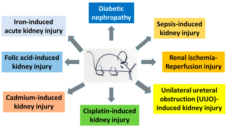
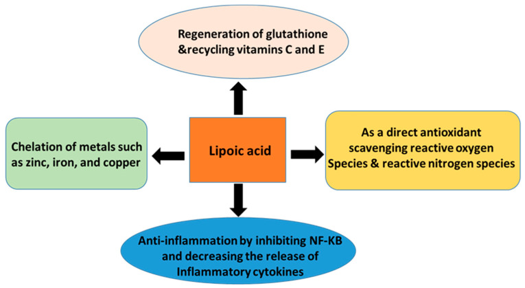
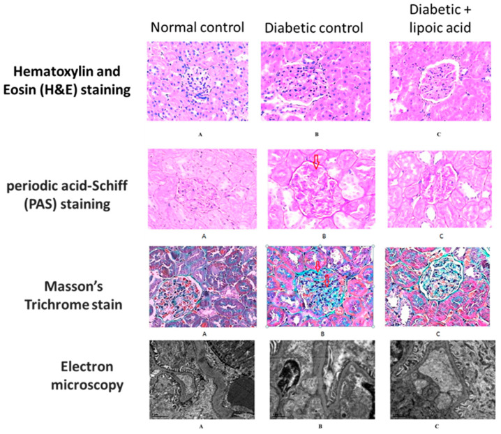
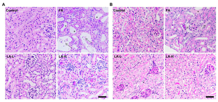
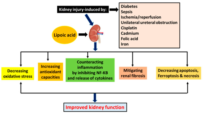
The kidney is a crucial organ that eliminates metabolic waste and reabsorbs nutritious elements. It also participates in the regulation of blood pressure, maintenance of electrolyte balance and blood pH homeostasis, as well as erythropoiesis and vitamin D maturation. Due to such a heavy workload, the kidney is an energy-demanding organ and is constantly exposed to endogenous and exogenous insults, leading to the development of either acute kidney injury (AKI) or chronic kidney disease (CKD). Nevertheless, there are no therapeutic managements to treat AKI or CKD effectively. Therefore, novel therapeutic approaches for fighting kidney injury are urgently needed. This review article discusses the role of α-lipoic acid (ALA) in preventing and treating kidney diseases. We focus on various animal models of kidney injury by which the underlying renoprotective mechanisms of ALA have been unraveled. The animal models covered include diabetic nephropathy, sepsis-induced kidney injury, renal ischemic injury, unilateral ureteral obstruction, and kidney injuries induced by folic acid and metals such as cisplatin, cadmium, and iron. We highlight the common mechanisms of ALA's renal protective actions that include decreasing oxidative damage, increasing antioxidant capacities, counteracting inflammation, mitigating renal fibrosis, and attenuating nephron cell death. It is by these mechanisms that ALA achieves its biological function of alleviating kidney injury and improving kidney function. Nevertheless, we also point out that more comprehensive, preclinical, and clinical studies will be needed to make ALA a better therapeutic agent for targeting kidney disorders.

Keywords: acute kidney injury; chronic kidney disease; diabetic kidney disease; diabetic nephropathy; lipoic acid; nephroprotection.


Conflict of interest statement

The authors declare no conflict of interest.

Figures

Figure 1
Figure 1
Animal models of kidney injury discussed in this article. These models include both acute kidney injury and chronic kidney disease.
Figure 2
Figure 2
Lipoic acid is a cofactor of mitochondrial 2-ketoacid dehydrogenase complex, including pyruvate dehydrogenase complex, α-ketoglutarate dehydrogenase complex, and branched-chain amino acid dehydrogenase complex. The E1 subunit is 2-ketoacid decarboxylase using TPP as a cofactor; the E2 subunit is a dihydrolipoamide acyltransferase using lipoic acid as a cofactor; the E3 subunit is a dihydrolipoamide dehydrogenase, which uses NAD+ as an electron acceptor for the oxidation of the lipoyl group linked to the E2 subunit. E3 catalyzes the formation of the oxidized form of lipoic acid and generates NADH in the meantime [32,33,34].
Figure 3
Figure 3
Lipoic acid is involved in thiol-disulfide exchanges that modulate cell’s redox and energy status.
Figure 4
Figure 4
Biological actions of lipoic acid and its potential mechanisms, which include regeneration of glutathione, vitamins C and E, scavenging ROS, chelating metal ions, and anti-inflammatory power.
Figure 5
Figure 5
Protective effects of lipoic acid on diabetic kidney disease. Histological staining of the kidney tissues derived from control, diabetic, and diabetic + lipoic acid treatment. This figure was reproduced from reference [55]. For all the microscopic images, the amplification magnitude is ×800. (A) normal control group; (B) diabetic control group; (C) diabetic + ALA group. Glomerular hypertrophy, mesangial region expansion, proliferation of mesangial cells, and inflammatory cell filtration could also be observed in H&E staining. Disruption of glomerular basement membrane and mesangial region expansion could also be observed in PAS staining. Additionally, collagen fiber staining shows an increased intensity in the diabetic control group but exhibits a significant decrease in ALA-treated animals. For PAS staining, the red arrow indicates thickening and deformation in the glomerular basement membrane in the diabetic kidney. For Masson’s stained sections, the red arrows indicate the increased intensity of collagen fiber stain in the diabetic kidney.
Figure 6
Figure 6
Protective effects of lipoic acid on folic acid-induced kidney injury. Histological staining of the kidney tissues derived from control, folic acid (FA), and folic acid + lipoic acid (LA). This figure was reproduced from reference [105]. (A) Representative images of hematoxylin & eosin (H&E) staining reflecting the histological changes in folic acid-induced acute kidney injury and low- and high-ALA-treated animal groups. (B) Renal damage assessed by Periodic acid/Schiff staining in folic acid-induced acute kidney injury and low- and high-ALA-treated animal groups.
Figure 7
Figure 7
Mechanisms underlying lipoic acid’s role in various animal models of kidney injury discussed in the text. Lipoic acid alleviates kidney injury and improves kidney function by exerting various biological actions, as depicted in this figure.

Similar articles

Cited by

References

    1. Rennke H.G., Denker B.M. Renal Pathology: The Essentials. 5th ed. Wolters Kluwer; New York, NY, USA: 2020.
    1. Koeppen B.M., Stanton B.A. Renal Physiology. 5th ed. Elsevier; Philadelphia, PA, USA: 2013.
    1. Dipiro J.T., Talbet R.L., Yee G.C., Matzke G.R., Wells B.G., Posey L.M. Pharmacotherapy: A Pathophysiological Approach. 9th ed. McGraw-Hill Education; New York, NY, USA: 2014.
    1. Lieberman M., Marks A.D. Marks’ Basic Medical Biochemistry: A Clinical Approach. 4th ed. Wolters Kluwer/Lippincott Williams & Wilkins; Philadelphia, PA, USA: 2013.
    1. Ammirati A.L. Chronic kidney disease. Rev. Assoc. Med. Bras. (1992) 2020;66((Suppl. S1)):s03–s09. doi: 10.1590/1806-9282.66.s1.3. - DOI - PubMed

MeSH terms