. 2023 Apr 4;15(7):1766.
doi: 10.3390/nu15071766.

Potential Effect of Glutamine in the Improvement of Intestinal Stem Cell Proliferation and the Alleviation of Burn-Induced Intestinal Injury via Activating YAP: A Preliminary Study

Affiliations

Potential Effect of Glutamine in the Improvement of Intestinal Stem Cell Proliferation and the Alleviation of Burn-Induced Intestinal Injury via Activating YAP: A Preliminary Study

Xia Chen et al. Nutrients. .

Abstract

Burn injury is a common form of traumatic injury that leads to high mortality worldwide. A severe burn injury usually induces gut barrier dysfunction, partially resulting from the impairment in the proliferation and self-renewal of intestinal stem cells (ISCs) post burns. As a main energy substance of small intestinal enterocytes, glutamine (Gln) is important for intestinal cell viability and growth, while its roles in ISCs-induced regeneration after burns are still unclear. To demonstrate the potential effects of Gln in improving ISCs proliferation and alleviating burn-induced intestinal injury, in this study, we verified that Gln significantly alleviated small intestine injury in burned mice model. It showed that Gln could significantly decrease the ferroptosis of crypt cells in the ileum, promote the proliferation of ISCs, and repair the crypt. These effects of Gln were also confirmed in the mouse small intestine organoids model. Further research found that Yes-associated protein (YAP) is suppressed after burn injury, and Gln could improve cell proliferation and accelerate the renewal of the damaged intestinal mucosal barrier after burns by activating YAP. YAP is closely associated with the changes in intestinal stem cell proliferation after burn injury and could be served as a potential target for severe burns.

Keywords: burns; crypt; glutamine; intestinal stem cells; proliferation.


Conflict of interest statement

The authors declare no competing interest.

Figures

Figure 1
Figure 1
Effect of burn injury on the pathological structure of small intestinal crypts. The villus length and crypt depth were measured as indicated in the image (×100; n = 6). Yellow arrows indicate goblet cell infiltration in the small intestine. The red bidirectional arrows indicate the thickness of the basal tissue of the small intestinal crypt. (A1A4) Representative images of the small intestine of each group under a light microscope: (A1) Control group; (A2) Burn group; (A3) Gln group; and (A4) Burn+Gln group. (B) Quantitative analysis of the length of the small intestine under different treatments. (C) Quantitative analysis of the number of preservation injury scores of the small intestine of different groups. (D1D4) Representative images of the hematoxylin and eosin (H&E) staining of the small intestine sections in jejunum from control\Gln\Burn and Burn+Gln group at day 3. (E1E4) Representative images of H&E staining of the small intestine sections in ileum from control\Gln\Burn and Burn+Gln group at day 3. (F) The statistical analysis of the length of crypts from images shown in (E1E4). (G) The statistical analysis of the thickness of the lleum basal layer in ileum from images shown at (E1E4). The data are Mean ± SEM with an n = 6. * p < 0.1, ** p < 0.01, **** p < 0.0001 using one-way ANOVA and post hoc Tukey’s test.
Figure 2
Figure 2
Regeneration of the organoids of the mouse small intestine crypts after Gln treatment. (A1A4) are representative images of the organoids of the mouse small intestine crypt in control group, Gln group, Burn group and Burn+Gln group 24 h after burns injury. (B1B4) are representative images of the organoids of the mouse small intestine crypt in control group, Gln group, Burn group, and Burn+Gln group 48 h after burns injury. (C) Comparison of the number of budding crypts in a single organoid of the mouse small intestine crypt 24 h after burns injury. (D) Comparison of the number of budding crypts in a single organoid of mice small intestine crypt post burns 48 h. Data were expressed as mean ± SEM from 6 independent experiments (n = 6). **** p < 0.0001 using 1-way ANOVA and post hoc Tukey’s test.
Figure 3
Figure 3
Effects of Gln treatment on apoptosis and ferroptosis in the small intestine after severe burns. (A) Gln supplementation after severe burns for 3 days. The expressions of Bax and Caspase9 were detected by immunoblotting (n = 6). (B) The relative intensities of Bax on the 3rd day (Day 3) after burn of bands were quantified using ImageJ. (C) The relative intensities of Caspase9 on the 3rd day (Day 3) after burn of bands were quantified using ImageJ. (D) Representative images of immunofluorescence for Glutathione Peroxidase 4 (GPX4) (green) and diamidino-phenyl-indole (DAPI) (blue): Control group; Burn group; Gln group; and Burn+Gln group. Data were expressed as mean ± SEM from 6 independent experiments (n = 6).
Figure 4
Figure 4
Influences of Gln treatment on proliferation and stemness of small intestinal crypts and ISCs of C57 mice after burn injury. (AC) Representative images of immunofluorescence for proliferation cell nuclear antigen (PCNA) (red), olfactomedin 4 (Olfm4) (green), and DAPI (blue) in different groups: (A1A5) Control group. (B1B5) Gln group. (C1C5) Burn group. (D1D5) Burn+Gln group. (E) Comparison of the number of PCNA-positive cells in a single crypt in different groups. (F) Comparison of the number of Olfm4-positive cells in a single crypt in different groups. Data were expressed as mean ± SEM from 6 independent experiments (n = 6).
Figure 5
Figure 5
Gln treatment accelerates the small intestinal stem cell cycle and promotes the dry expression of ISCs C57 mice after burn injury. (AD) Representative images of immunofluorescence for BrdU (green), ATP binding cassette subfamily G member 2 (ABCG2) (red), and DAPI (blue) in different groups: (A1A5) Control group. (B1B5) Gln group. (C1C5) burn group. (D1D5) Burn+Gln group. (E) Comparison of the number of BrdU-positive cells in a single crypt in different groups (n = 6). (F) Comparison of the number of ABCG2-positive cells in a single crypt in different groups. Data were expressed as mean ± SEM from 6 independent experiments (n = 6). * p < 0.1, **** p < 0.0001 using 1-way ANOVA and post hoc Tukey’s test.
Figure 6
Figure 6
Gln could promote proliferation and accelerate cell cycle. (A) Flow cytometry was used to analyze the typical cell cycle of small intestinal stem cells treated in different groups. (B) The proportion of G1 phase of cell cycle in different groups. (C) The proportion of S phase of cell cycle in different groups. Data were expressed as mean ± SEM from 6 independent experiments (n = 6). PE-A represents the curve area of fluorescence intensity. The blue part represents the cell cycle phase of G1 and the yellow part represents the cell cycle phase of S. ** p < 0.01, *** p < 0.001 using 1-way ANOVA and post hoc Tukey’s test.
Figure 7
Figure 7
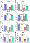
Gln accelerates the small intestine stem cell cycle process and promotes self-renewal. (AC) The expression of the mRNA with stem cell markers in the four different groups according to the quantitative by Real-Time Quantitativeq Polymerase Chain Reaction (RT-qPCR). (A) Olfactomedin 4 (Olfm4) mRNA expression. (B) Sulfur Oxides 9 (SOX9) mRNA expression. (C) Proliferation Cell Nuclear Antigen (PCNA) mRNA expression. (DF) The expression of the mRNA related to cell cycle of mouse small intestine crypts was measured by RT-PCR from the 4 different groups on day 3 after burn: (D) Cyclin-Dependent Kinase12 (CDK1) mRNA expression. (E) Cyclin-Dependent Kinase 2 (CDK2) mRNA expression. (F) Cyclin-Dependent Kinase 4 (CDK4) mRNA expression. (G) Yes-associated Protein (YAP) mRNA expression. (H) Mammalian sterile 20-like kinase 1 (MST1) mRNA expression. Data were expressed as mean ± SEM from 6 independent experiments (n = 6), ns no significance, * p < 0.1, ** p < 0.01 *** p < 0.001, **** p < 0.0001 using 1-way ANOVA and post hoc Tukey’s test.
Figure 8
Figure 8
Gln promotes proliferation by activating the YAP-Hippo signaling pathway. (A) Protein abundances in crypts from C57 mice of Olfm4, PCNA, Cyclin D1, YAP, and p-Yes-associated Protein (p-YAP) were measured by immunoblotting using antibodies in small intestinal stem cell tissue treated with Gln after burn (n = 6). (B) Quantification of Olfm4 protein expression in crypts from C57 mice on Day 3. (C) Representative immunoblot analysis of PCNA in treated Gln. (D) Quantification of Cyclin D1 protein expression in crypts from C57 mice on Day 3. (E) Quantification of YAP expression in crypts from C57 mice on Day 3. (F) Quantification of p-YAP expression in crypts from C57 mice on Day3. Data were expressed as Mean ± SEM from 6 independent experiments (n = 6). * p < 0.1, ** p < 0.01, **** p < 0.0001 using 1-way ANOVA and post hoc Tukey’s test.

Similar articles

Cited by

References

    1. He W., Wang Y., Wang P., Wang F. Intestinal barrier dysfunction in severe burn injury. Burn. Trauma. 2019;7:24. doi: 10.1186/s41038-019-0162-3. - DOI - PMC - PubMed
    1. Hageman J.H., Heinz M.C., Kretzschmar K., van der Vaart J., Clevers H., Snippert H.J.G. Intestinal Regeneration: Regulation by the Microenvironment. Dev. Cell. 2020;54:435–446. doi: 10.1016/j.devcel.2020.07.009. - DOI - PubMed
    1. Takahashi T., Fujishima K., Kengaku M. Modeling Intestinal Stem Cell Function with Organoids. Int. J. Mol. Sci. 2021;22:10912. doi: 10.3390/ijms222010912. - DOI - PMC - PubMed
    1. Jasper H. Intestinal Stem Cell Aging: Origins and Interventions. Annu. Rev. Physiol. 2020;82:203–226. doi: 10.1146/annurev-physiol-021119-034359. - DOI - PubMed
    1. Gehart H., Clevers H. Tales from the crypt: New insights into intestinal stem cells. Nat. Rev. Gastroenterol. Hepatol. 2019;16:19–34. doi: 10.1038/s41575-018-0081-y. - DOI - PubMed