

Pirfenidone Protects from UVB-Induced Photodamage in Hairless Mice
- PMID: 37049691
- PMCID: PMC10096127
- DOI: 10.3390/molecules28072929
Pirfenidone Protects from UVB-Induced Photodamage in Hairless Mice
Abstract
Background: Ultraviolet radiation (UV) is the main environmental factor that causes histological degenerative changes of the skin giving rise to a chronic process called photodamage. Non-melanoma skin cancer induced by UVB radiation is a result of a cascade of molecular events caused by DNA damage in epidermis cells, including persistent inflammation, oxidative stress, and suppression of T cell-mediated immunity. Retinoids such as tretinoin have been widely used in skin to treat photoaging and photodamage, though its secondary adverse effects have been recognized. Pirfenidone (PFD) has emerged as an antifibrogenic, anti-inflammatory and antioxidant agent, and in this work its efficacy was evaluated in a model of UVB-induced photodamage.
Methods: Epidermal, dermal, and inflammatory changes were measured by histomorphometric parameters. In addition, gene, and protein expression of key molecules in these processes were evaluated.
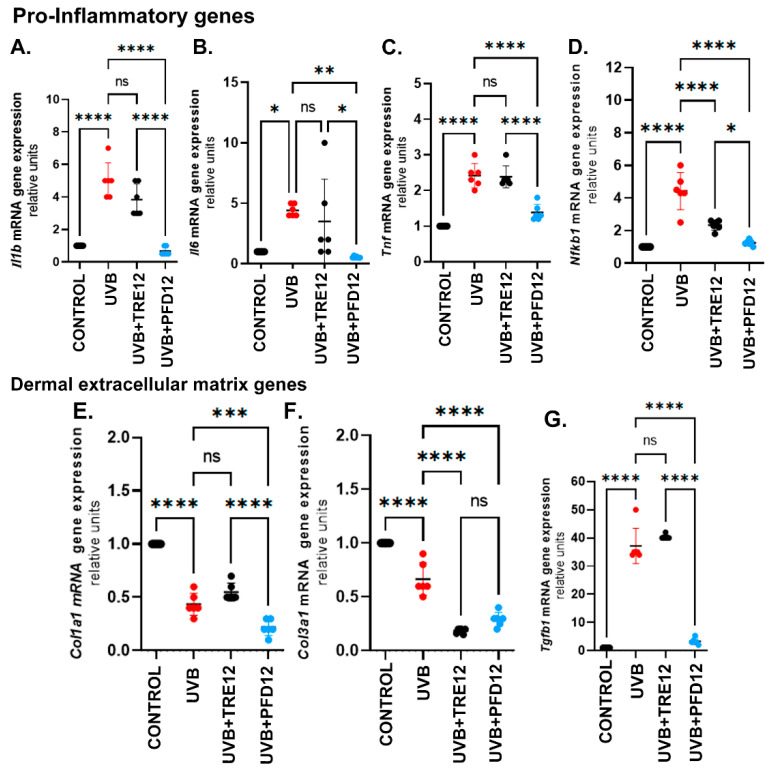
Results: Our results revealed an anti-photodamage effect of topical PFD with absence of inflammatory skin lesions determined by dermoscopy. In addition, PFD reduced elastosis, improved organization, arrangement, and deposition of dermal collagens, downregulated several pro-inflammatory markers such as NF-kB, IL-1, IL-6 and TNFα, and decreased keratinocyte damage.
Conclusion: Topical pirfenidone represents a promising agent for the treatment of cell photodamage in humans. Clinical trials need to be carried out to explore this premise.
Keywords: photodamage; pirfenidone; pro-inflammatory markers; skin; ultraviolet radiation.
Conflict of interest statement
The authors declare no conflict of interest.
Figures


Similar articles
-
Protective effects of Oxya chinensis sinuosa Mishchenko against ultraviolet B-induced photodamage in hairless mice.BMC Complement Altern Med. 2019 Oct 28;19(1):286. doi: 10.1186/s12906-019-2692-4. BMC Complement Altern Med. 2019. PMID: 31660950 Free PMC article.
-
Borago officinalis L. attenuates UVB-induced skin photodamage via regulation of AP-1 and Nrf2/ARE pathway in normal human dermal fibroblasts and promotion of collagen synthesis in hairless mice.Exp Gerontol. 2018 Jul 1;107:178-186. doi: 10.1016/j.exger.2018.02.017. Epub 2018 Feb 27. Exp Gerontol. 2018. PMID: 29499374
-
Coriander leaf extract exerts antioxidant activity and protects against UVB-induced photoaging of skin by regulation of procollagen type I and MMP-1 expression.J Med Food. 2014 Sep;17(9):985-95. doi: 10.1089/jmf.2013.2999. Epub 2014 Jul 14. J Med Food. 2014. PMID: 25019675 Free PMC article.
-
A review of skin ageing and its medical therapy.Br J Dermatol. 1996 Dec;135(6):867-75. doi: 10.1046/j.1365-2133.1996.d01-1088.x. Br J Dermatol. 1996. PMID: 8977705 Review.
-
An organotypic model of skin to study photodamage and photoprotection in vitro.J Am Acad Dermatol. 2008 May;58(5 Suppl 2):S155-9. doi: 10.1016/j.jaad.2007.08.050. J Am Acad Dermatol. 2008. PMID: 18410802 Review.
Cited by
-
Gold@Mesoporous Polydopamine Nanocomposite Hydrogel Loaded with Estrogen for the Treatment of Skin Photoaging.Int J Nanomedicine. 2025 Apr 12;20:4571-4587. doi: 10.2147/IJN.S511388. eCollection 2025. Int J Nanomedicine. 2025. PMID: 40242609 Free PMC article.
-
Assessing changes in ultraviolet radiation-exposed mouse skin using optical coherence tomography.PLoS One. 2025 Aug 11;20(8):e0328647. doi: 10.1371/journal.pone.0328647. eCollection 2025. PLoS One. 2025. PMID: 40788959 Free PMC article.
-
Linoleic Acid Induces Metabolic Reprogramming and Inhibits Oxidative and Inflammatory Effects in Keratinocytes Exposed to UVB Radiation.Int J Mol Sci. 2024 Sep 26;25(19):10385. doi: 10.3390/ijms251910385. Int J Mol Sci. 2024. PMID: 39408715 Free PMC article.
References
-
- Green A., Williams G., Nèale R., Hart V., Leslie D., Parsons P., Marks G.C., Gaffney P., Battistutta D., Frost C., et al. Daily sunscreen application and betacarotene supplementation in prevention of basal-cell and squamous-cell carcinomas of the skin: A randomised controlled trial. Lancet. 1999;354:723–729. doi: 10.1016/S0140-6736(98)12168-2. - DOI - PubMed
MeSH terms
Substances
Grants and funding
LinkOut - more resources
Full Text Sources
Medical
