. 2023 Mar 29;28(7):3057.
doi: 10.3390/molecules28073057.

Cleistocalyx nervosum var. paniala Berry Seed Protects against TNF-α-Stimulated Neuroinflammation by Inducing HO-1 and Suppressing NF-κB Mechanism in BV-2 Microglial Cells

Affiliations

Cleistocalyx nervosum var. paniala Berry Seed Protects against TNF-α-Stimulated Neuroinflammation by Inducing HO-1 and Suppressing NF-κB Mechanism in BV-2 Microglial Cells

Sakawrat Janpaijit et al. Molecules. .

Abstract

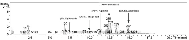
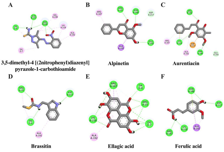
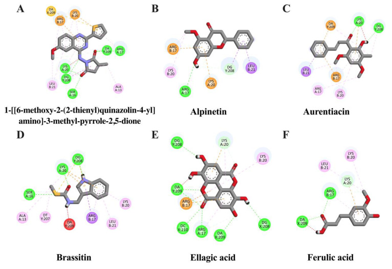
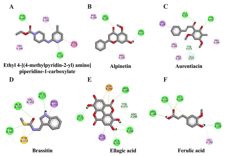
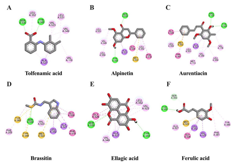
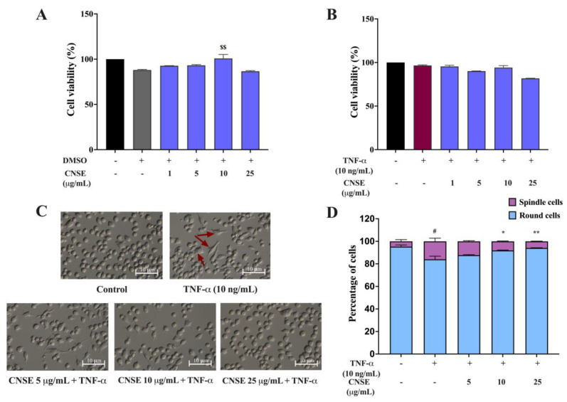
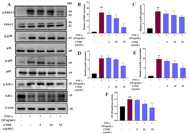
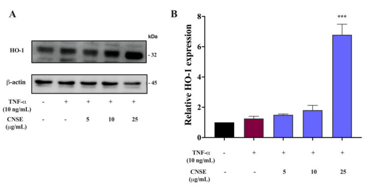
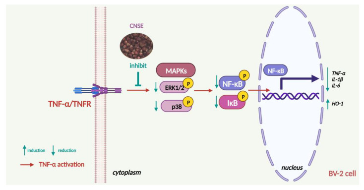
Sustained inflammatory responses have been implicated in various neurodegenerative diseases (NDDs). Cleistocalyx nervosum var. paniala (CN), an indigenous berry, has been reported to exhibit several health-beneficial properties. However, investigation of CN seeds is still limited. The objective of this study was to evaluate the protective effects of ethanolic seed extract (CNSE) and mechanisms in BV-2 mouse microglial cells using an inflammatory stimulus, TNF-α. Using LC-MS, ferulic acid, aurentiacin, brassitin, ellagic acid, and alpinetin were found in CNSE. Firstly, we examined molecular docking to elucidate its bioactive components on inflammation-related mechanisms. The results revealed that alpinetin, aurentiacin, and ellagic acid inhibited the NF-κB activation and iNOS function, while alpinetin and aurentiacin only suppressed the COX-2 function. Our cell-based investigation exhibited that cells pretreated with CNSE (5, 10, and 25 μg/mL) reduced the number of spindle cells, which was highly observed in TNF-α treatment (10 ng/mL). CNSE also obstructed TNF-α, IL-1β, and IL-6 mRNA levels and repressed the TNF-α and IL-6 releases in a culture medium of BV-2 cells. Remarkably, CNSE decreased the phosphorylated forms of ERK, p38MAPK, p65, and IκB-α related to the inhibition of NF-κB binding activity. CNSE obviously induced HO-1 protein expression. Our findings suggest that CNSE offers good potential for preventing inflammatory-related NDDs.

Keywords: Cleistocalyx nervosum var. paniala; MAPKs; NF-κB; TNF-α; microglial cells; neuroinflammation.


Conflict of interest statement

The authors declare no conflict of interest.

Figures

Figure 1
Figure 1
Chromatographic diagram of bioactive ingredients in ethanolic seed extract (CNSE) by liquid chromatography-mass spectrometry (LC-MS) analysis.
Figure 2
Figure 2
Schematic interactions of amino acid residues between reference inhibitor or candidate compounds and NF-κB protein. The original inhibitor of NF-κB protein is (A) 3,5-dimethyl-4-[(2-nitrophenyl)diazenyl]pyrazole-1-carbothioamide and candidate ligands are as follows: (B) alpinetin, (C) aurentiacin, (D) brassitin, (E) ellagic acid, and (F) ferulic acid. The green dashed line represents hydrogen, the pink or purple dashed lines represent hydrophobic bonds, and the yellow dashed line indicates other bonds.
Figure 3
Figure 3
Schematic interactions of amino acid residues between reference inhibitor or candidate compounds and AP-1 protein. The original inhibitor of AP-1 protein is (A) 1-[[6-methoxy-2-(2-thienyl)quinazolin-4-yl]amino]-3-methyl-pyrrole-2,5-dione and candidate ligands are as follows: (B) alpinetin, (C) aurentiacin, (D) brassitin, (E) ellagic acid, and (F) ferulic acid. The green dashed line represents hydrogen, the pink or purple dashed lines represent hydrophobic bonds, and the yellow dashed line indicates other bonds.
Figure 4
Figure 4
Schematic interactions of amino acid residues between reference inhibitor or candidate compounds and iNOS protein. The original inhibitor of iNOS protein is (A) ethyl 4-[(4-methylpyridin-2-yl) amino] piperidine-1-carboxylate and candidate ligands are as follows: (B) alpinetin, (C) aurentiacin, (D) brassitin, (E) ellagic acid, and (F) ferulic acid. The green dashed line represents hydrogen, the pink or purple dashed lines represent hydrophobic bonds, and the yellow dashed line indicates other bonds.
Figure 5
Figure 5
Schematic interactions of amino acid residues between reference inhibitor or candidate compounds and COX-2 protein. The reference inhibitor of COX-2 protein is (A) tolfenamic acid and candidate ligands are as follows: (B) alpinetin, (C) aurentiacin, (D) brassitin, (E) ellagic acid, and (F) ferulic acid. The green dashed line represents hydrogen, the pink or purple dashed lines represent hydrophobic bonds, and the yellow dashed line indicates other bonds.
Figure 6
Figure 6
Effects of CNSE on cell viability and morphological phenotypes in BV-2 cells. Cell viability was determined using an MTT assay. For cytotoxicity assay, (A) BV-2 cells were incubated with various doses of CNSE for 24 h. Black color represents untreated control, grey color represents DMSO control and violet color represents CNSE treatment. (B) CNSE was pretreated for 24 h, followed by TNF-α for another 24 h. Black color represents untreated control, red color represents TNF-α group and violet color represents CNSE treatment. For morphological analysis. (C) Morphological phenotypes were captured using 10× magnification of phase-contrast microscopy after pretreatment of cells with CNSE and TNF-α for 24 h. The red arrows represent the elongated cells. (D) The percentage of round and spindle cells is shown as a bar graph. Data are represented as the mean ± SD from at least three independent experiments. A p-value < 0.05 was considered to show a significant difference between each group ($$ p < 0.01 vs. DMSO control; # p < 0.05 vs. untreated control; ** p < 0.01; * p < 0.05 vs. TNF-𝛼-treated group).
Figure 7
Figure 7
Inhibitory effects of CNSE on the expressions of proinflammatory cytokines’ production in BV-2 cells. The mRNA levels of inflammatory cytokines were measured after treatment of cells with CNSE for 24 h, followed by TNF-α for another 3 h using real-time PCR. RESV was used as a positive control. The relative expression, as shown by fold changes in (A) TNF-𝛼, (B) IL-1β, and (C) IL-6, was normalized with the internal control, β-actin. The release of cytokines, including (D) TNF-𝛼 and (E) IL-6, was also measured using commercial ELISA kits. The concentrations of protein levels (pg/mL) in the cell culture medium after CNSE and TNF-𝛼 treatment for 24 h were calculated by comparing them to the standard curve. Black color represents untreated control, red color represents TNF-α group, violet color represents CNSE treatment and green color represent RESV treatment. Data are represented as the mean ± SD from at least three independent experiments. A p-value < 0.05 was considered to show a significant difference between each group (#### p < 0.0001; ### p < 0.001; ## p < 0.01; # p < 0.05 vs. untreated control; **** p < 0.0001; ** p < 0.01; * p < 0.05 vs. TNF-𝛼-treated group).
Figure 8
Figure 8
Inhibitory effects of CNSE on MAPKs and NF-κB activation. Cells were pretreated with CNSE in response to TNF-α for 5 min, and then (A) the protein levels of MAPKs and NF-κB were evaluated by Western blot analysis. The relative proteins levels of (B) phosphorylated ERK (p-ERK), (C) phosphorylated p38MAPK (p−p38), (D) phosphorylated p65 (p−65), and (E) phosphorylated IκB-α (p-IκB-α) are presented in the histogram graph, and each protein was quantified and normalized with β-actin. NF-κB (p−65) binding activity after treatment cells with the extract, followed by TNF-α, was investigated by performing a dual-luciferase assay. (F) The relative level of p65 binding activity was normalized through the activity of pRL-null and was expressed as fold changes. Black color represents untreated control, red color represents TNF-α group and violet color represents CNSE treatment. Data are represented as the mean ± SD from at least three independent experiments. A p-value < 0.05 was considered to show a significant difference between each group (#### p < 0.0001; ### p < 0.001; ## p < 0.01 vs. untreated control; **** p < 0.0001; *** p < 0.001; ** p < 0.01; * p < 0.05 vs. TNF-𝛼-treated group).
Figure 9
Figure 9
Effects of CNSE on HO-1 induction. BV-2 cells were preincubated with CNSE and then stimulated with TNF-α for 24 h. (A) The protein expression of HO-1 using Western blot analysis. (B) The histogram graph presents the relative expression of HO-1, which was quantified and normalized with β-actin. Black color represents untreated control, red color represents TNF-α group and violet color represents CNSE treatment. Data are represented as the mean ± SD from at least three independent experiments. A p-value < 0.05 was considered to show a significant difference between each group (*** p < 0.001 vs. TNF-𝛼-treated group).
Figure 10
Figure 10
The proposed schematic overview of the effects and molecular mechanism of CNSE on protecting TNF-α-induced neuroinflammation in BV-2 mouse microglial cells (created by BioRender.com).

Similar articles

Cited by

References

    1. Colonna M., Butovsky O. Microglia Function in the Central Nervous System During Health and Neurodegeneration. Annu. Rev. Immunol. 2017;35:441–468. doi: 10.1146/annurev-immunol-051116-052358. - DOI - PMC - PubMed
    1. Kempuraj D., Thangavel R., Natteru P.A., Selvakumar G.P., Saeed D., Zahoor H., Zaheer S., Iyer S.S., Zaheer A. Neuroinflammation Induces Neurodegeneration. J. Neurol. Neurosurg. Spine. 2016;1:1003. - PMC - PubMed
    1. Rauf A., Badoni H., Abu-Izneid T., Olatunde A., Rahman M.M., Painuli S., Semwal P., Wilairatana P., Mubarak M.S. Neuroinflammatory Markers: Key Indicators in the Pathology of Neurodegenerative Diseases. Molecules. 2022;27:3194. doi: 10.3390/molecules27103194. - DOI - PMC - PubMed
    1. Yuste J.E., Tarragon E., Campuzano C.M., Ros-Bernal F. Implications of glial nitric oxide in neurodegenerative diseases. Front. Cell. Neurosci. 2015;9:322. doi: 10.3389/fncel.2015.00322. - DOI - PMC - PubMed
    1. Datta P.K., Gross E.J., Lianos E.A. Interactions between inducible nitric oxide synthase and heme oxygenase-1 in glomerulonephritis. Kidney Int. 2002;61:847–850. doi: 10.1046/j.1523-1755.2002.00231.x. - DOI - PubMed