. 2023 Mar 31;28(7):3128.
doi: 10.3390/molecules28073128.

Anticancer Properties of 3-Dietoxyphosphorylfuroquinoline-4,9-dione

Affiliations

Anticancer Properties of 3-Dietoxyphosphorylfuroquinoline-4,9-dione

Joanna Drogosz-Stachowicz et al. Molecules. .

Abstract

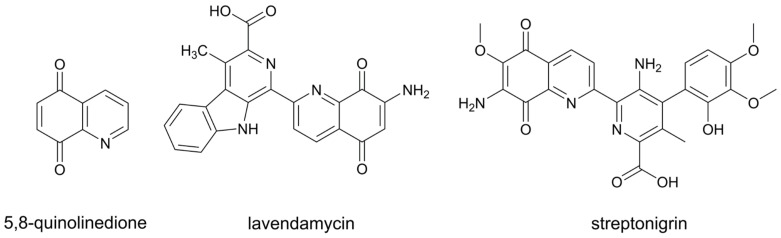
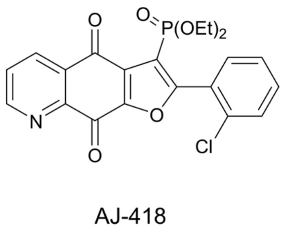
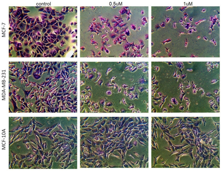
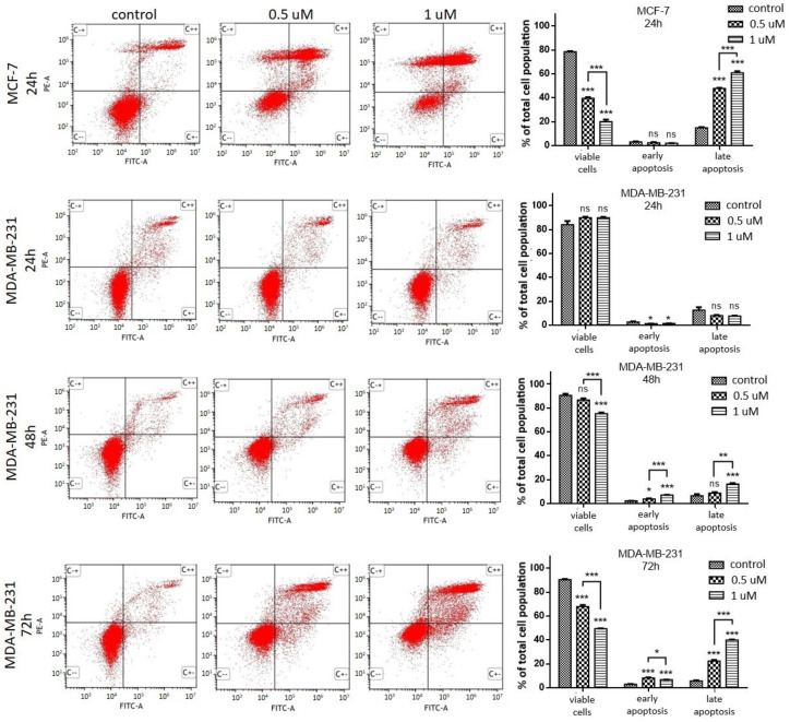
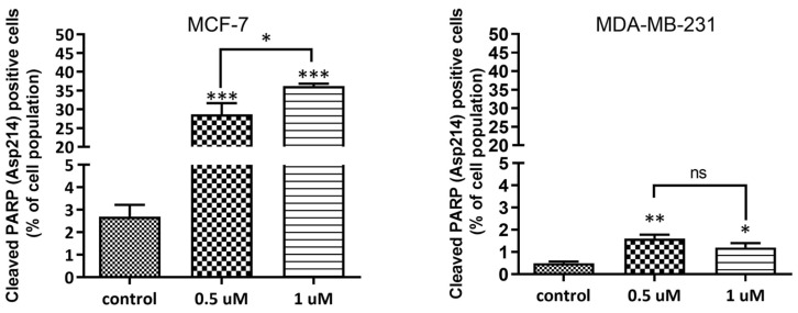
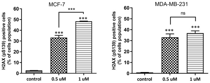
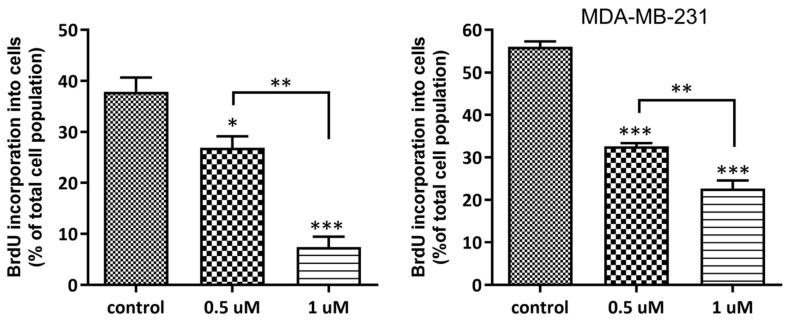
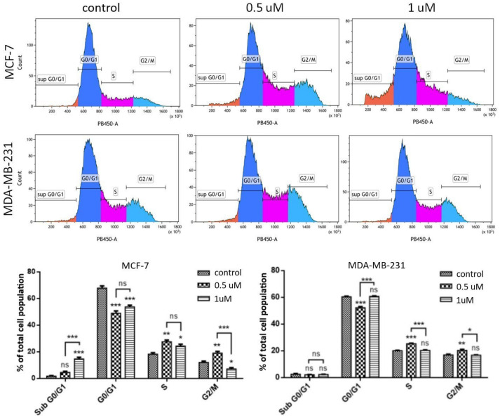
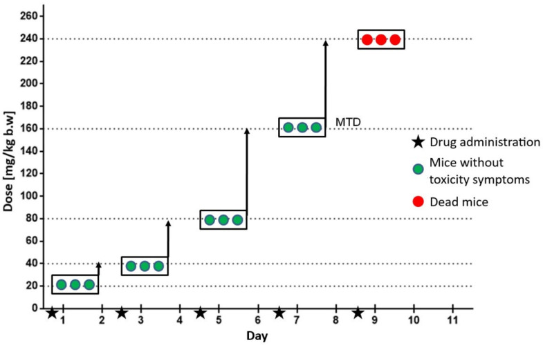
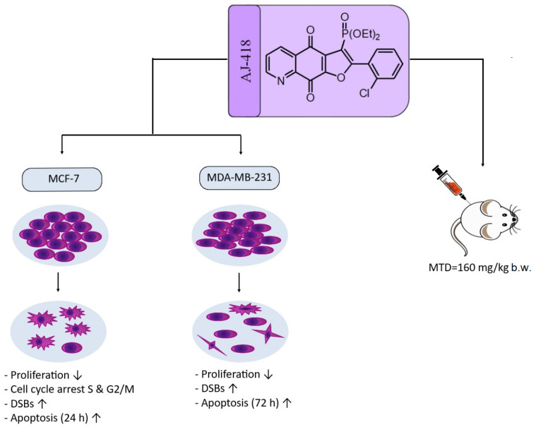
Herein, the antitumor activity of a novel synthetic analog with 5,8-quinolinedione scaffold, diethyl (2-(2-chlorophenyl)-4,9-dioxo-4,9-dihydrofuro [3,2-g]quinolin-3-yl)phosphonate (AJ-418) was investigated on two breast cancer cell lines. This analog was selected from a small library of synthetic quinolinediones on the basis of its strong antiproliferative activity against MCF-7 and MDA-MB-231 cells and 4-5-fold lower cytotoxicity towards healthy MCF-10A cells. The morphology of MCF-7 and MDA-MB-231 cancer cells treated with AJ-418 changed drastically, while non-tumorigenic MCF-10A cells remained unaffected. In MCF-7 cells, after 24 h incubation, the increased number of apoptotic cells coincided with a decrease in proliferation and cell viability. The 24 h treatment of MDA-MB-231 cells with the tested compound reduced their cell viability and proliferation rate; however, a significant pro-apoptotic effect was visible only after longer incubation times (48 h and 72 h). Then, the maximum tolerated dose (MTD) of compound AJ-418 in C3H mice after subcutaneous administration was determined to be 160 mg/kg, showing that this analog was well tolerated and can be further evaluated to assess its potential therapeutic effect in tumor-bearing mice.

Keywords: DNA damage; apoptosis; cytotoxic activity; maximum tolerated dose; quinolinediones.


Conflict of interest statement

The authors declare no conflict of interest.

Figures

Figure 1
Figure 1
Structures of 5,8-quinolinedione and anticancer antibiotics containing this motif.
Figure 2
Figure 2
Structure of diethyl (2-(2-chlorophenyl)-4,9-dioxo-4,9-dihydrofuro[3,2-g]quinolin-3-yl)phosphonate (AJ-418).
Figure 3
Figure 3
Morphological changes in MCF-7, MDA-MB-231 and MCF-10A cells after 24 h incubation with AJ-418 at concentrations of 0.5 µM and 1 µM. After staining with Giemsa dye, the cells were photographed under a light microscope with a built-in camera (magnification 40×).
Figure 4
Figure 4
Induction of apoptosis by AJ-418 in MCF-7 and MDA-MB-231 cells. Cells were exposed to the tested compound at 0.5 µM and 1 µM concentrations, MCF-7 for 24 h and in MDA-MB-231 for 24, 48 and 72 h, then stained with annexin V and PI and analyzed using flow cytometry. Representative cytograms and quantification of apoptotic cell death are shown in the figure; each column represents the mean ± SEM of three independent experiments; * p < 0.05, ** p < 0.01 and *** p < 0.001 were considered statistically significant compared to controls, ns—no statistical significance.
Figure 5
Figure 5
Percentage of the cell population containing cleaved PARP. MCF-7 and MDA-MB-231 cells were exposed to AJ-418 at 0.5 µM and 1 µM for 24 h, then labeled with Anti-Cleaved-PARP antibody (Asp214) and analyzed using flow cytometry. Data represent means ± SEM of three independent experiments. Statistical significance was determined using one-way ANOVA and the Student–Newman–Kleus post-hoc multiple comparison test; *** p < 0.001, ** p < 0.01, * p < 0.05 represent statistically significant differences in comparison to controls, ns—no statistical significance.
Figure 6
Figure 6
Induction of DNA damage by AJ-418 in MCF-7 and MDA-MB-231 cells. Cells were exposed to the tested compound at 0.5 µM and 1 µM concentration for 24 h, stained with anti-H2AX antibody (pS139) and analyzed using flow cytometry. Error bars represent means ± SEM of three independent experiments. *** p < 0.001 were considered statistically significant compared to controls, ns—no statistical significance.
Figure 7
Figure 7
Effect of AJ-418 on the proliferation of MCF-7 and MDA-MB-231 cells. Cells were exposed to AJ-418 at 0.5 µM and 1 µM concentrations for 24 h, stained with BrdU and analyzed using flow cytometry. Error bars represent means of three replicates ± SEM. * p < 0.05, ** p < 0.01 and *** p < 0.001 were considered statistically significant compared to controls.
Figure 8
Figure 8
Effect of AJ-418 on cell cycle distribution in MCF-7 and MDA-MB-231 cells. Cells were exposed to AJ-418 at 0.5 µM and 1 µM concentrations for 24 h, stained with DAPI and analyzed using flow cytometry. Representative histograms and quantification are shown in the figure; error bars represent means ± SEM of three replicates. * p < 0.05, ** p < 0.01 and *** p < 0.001 were considered statistically significant compared to controls, ns—no statistical significance.
Figure 9
Figure 9
Diagram showing the determination of the maximum tolerated dose (MTD). Each box represents a group of 3 mice that were given the tested compound at the specific dose. After each administration, the mice were observed for 48 h.
Figure 10
Figure 10
Schematic representation of the effect of quinolinedione AJ-418 on MCF-7 and MDA-231 cells and evaluation of MTD in C3H mice.

Similar articles

References

    1. Jampilek J. Heterocycles in medicinal chemistry. Molecules. 2019;24:3839. doi: 10.3390/molecules24213839. - DOI - PMC - PubMed
    1. Mancini I., Vigna J., Sighel D., Defant A. Hybrid molecules containing naphthoquinone and quinolinedione scaffolds as antineoplastic agents. Molecules. 2022;27:4948. doi: 10.3390/molecules27154948. - DOI - PMC - PubMed
    1. Kadela-Tomanek M., Bębenek E., Chrobak E., Boryczka S. 5, 8-Quinolinedione scaffold as a promising moiety of bioactive agents. Molecules. 2019;24:4115. doi: 10.3390/molecules24224115. - DOI - PMC - PubMed
    1. Rao K.V., Cullen W.P. Streptonigrin, an antitumor substance. I. Isolation and characterization. Antibiot. Annu. 1959;7:950–953. - PubMed
    1. Balitz D.M., Bush J.A., Bradner W.T., Doyle T.W., O’Herron F.A., Nettleton D.E. Isolation of lavendamycin, a new antibiotic from Streptomyces lavendulae. J. Antibiot. 1982;35:259–265. doi: 10.7164/antibiotics.35.259. - DOI - PubMed

Substances