. 2023 Apr 4;28(7):3228.
doi: 10.3390/molecules28073228.

Therapeutic Potential of Albumin Nanoparticles Encapsulated Visnagin in MDA-MB-468 Triple-Negative Breast Cancer Cells

Affiliations

Therapeutic Potential of Albumin Nanoparticles Encapsulated Visnagin in MDA-MB-468 Triple-Negative Breast Cancer Cells

Abdullah Alsrhani et al. Molecules. .

Abstract

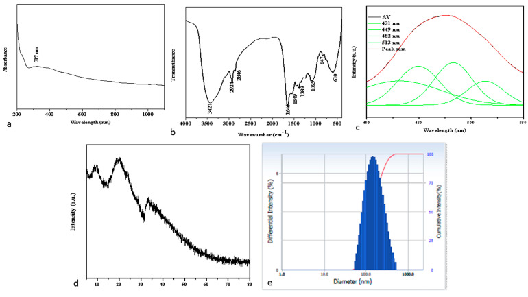
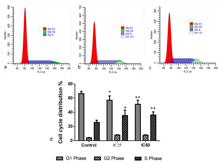
Breast cancer is among the most recurrent malignancies, and its prevalence is rising. With only a few treatment options available, there is an immediate need to search for better alternatives. In this regard, nanotechnology has been applied to develop potential chemotherapeutic techniques, particularly for cancer therapy. Specifically, albumin-based nanoparticles are a developing platform for the administration of diverse chemotherapy drugs owing to their biocompatibility and non-toxicity. Visnagin, a naturally derived furanochromone, treats cancers, epilepsy, angina, coughs, and inflammatory illnesses. In the current study, the synthesis and characterization of albumin visnagin (AV) nanoparticles (NPs) using a variety of techniques such as transmission electron microscopy, UV-visible, Fourier transform infrared, energy dispersive X-ray composition analysis, field emission scanning electron microscopy, photoluminescence, X-Ray diffraction, and dynamic light scattering analyses have been carried out. The MTT test, dual AO/EB, DCFH-DA, Annexin-V-FITC/PI, Propidium iodide staining techniques as well as analysis of apoptotic proteins, antioxidant enzymes, and PI3K/Akt/mTOR signaling analysis was performed to examine the NPs' efficacy to suppress MDA-MB-468 cell lines. The NPs decreased cell viability increased the amount of ROS in the cells, disrupted membrane integrity, decreased the level of antioxidant enzymes, induced cell cycle arrest, and activated the PI3K/Akt/mTOR signaling cascade, ultimately leading to cell death. Thus, AV NPs possesses huge potential to be employed as a strong anticancer therapy alternative.

Keywords: MDA-MB-468 cell line; albumin visnagin nanoparticles; apoptosis; breast cancer; nanotechnology.


Conflict of interest statement

The authors declare no conflict of interest.

Figures

Figure 1
Figure 1
Characterization of albumin visnagin NPs. UV-Vis spectrophotometer (a), FTIR Transmittance vs. wavenumber chart (b), and PL spectrum (c) analysis of synthesized albumin visnagin NPs. (d) X-ray diffraction Pattern and (e) DLS spectrum of albumin visnagin NPs.
Figure 2
Figure 2
Electron microscopic imaging of albumin visnagin NPs lower and higher magnification TEM images (ac) and SEAD patterns (d). Lower and higher magnification FESEM (e,f) and EDAX (g) spectrums.
Figure 3
Figure 3
Albumin visnagin NPs cause cytotoxicity in MDA-MB-468 cells. Cells were treated with various concentrations (2.5–160 µg/mL) of albumin visnagin NPs for 24, 48, and 72 h and the viability was assessed by MTT assay. Data are illustrated as mean ± SD of triplicates. * Signifies p < 0.05 when compared with control.
Figure 4
Figure 4
Effect of albumin visnagin NPs on apoptosis in MDA-MB-468 cells for 24 h. The cells were stained with ethidium bromide and acridine orange (1:1) and assessed using fluorescence microscopy (Labomed, Los Angeles, CA, USA). As shown by the white arrow, the control cells exhibited green fluorescence (indicating viable cells). As shown by the blue arrow, albumin visnagin NPs-treated cells revealed yellow/orange fluorescence, indicating early and late apoptosis, respectively. Necrosis is indicated by the red arrow. Control (untreated cells) (Left), albumin and visnagin NPs-treated cells; IC25 (Middle) and IC50 concentrations (Right). This is a representative image of the experiment performed in triplicate with 20X magnification (scale bar = 100 mm).
Figure 5
Figure 5
Cell cycle analysis was performed using flow cytometry after staining with Propidium Iodide (PI). Albumin and visnagin NPs were administered to MDA-MB-468 cells at IC50 concentrations for 48 h compared with controls. An analysis of cell cycle patterns and apoptosis distribution in untreated or control (a) and albumin and visnagin NP-treated cells, and their concentrations of IC25 (b) and IC50 (c). Percentage of cell cycle distribution (d). (* signifies p < 0.05 when compared with control, ** signifies p < 0.001 when compared with control).
Figure 6
Figure 6
Flow cytometry analysis of MDA-MB-468 cancer cells. The cells were exposed to IC50 concentration of albumin visnagin NPs for 48 h. At least two independent experiments were conducted to produce these figures. Live cells, early apoptotic, and necrotic cells were revealed in the lower left quadrant, the lower right quadrant, and the upper (Annexin-V+/PI+) quadrant, respectively (a). After albumin visnagin NP administration to MDA-MB-468 cells, the proportion of early and late apoptotic cells was determined (b). Two independent experiments were conducted to obtain the mean ± SD. (* signifies p < 0.05 when compared with control, ** signifies p < 0.001 when compared with control).
Figure 7
Figure 7
MDA-MB-468 cells were administered albumin visnagin NPs to measure DNA damage. The cells were left untreated (a), low dosage treated with albumin visnagin NPs (b), and high dosage treated with albumin visnagin NPs (c). Comet assays, mean comet scores and comet classes were used to assess DNA damage in the control and albumin visnagin NPs exposed groups. A mean score was calculated from three independent samples (d) based on the tail size and shape, ranging from 0 (undamaged) to 4 (extremely damaged). Two independent experiments were conducted to obtain the mean ± SD. (* signifies p < 0.05 when compared with control).
Figure 8
Figure 8
A fluorescence microscope image of endogenous ROS accumulation promoted by albumin visnagin NPs stained with DCFH-DA. Control (a), cells exposed to albumin visnagin NPs and IC25 (b) and IC50 (c). Image representing a triplicate experiment at 200X magnification (scale bar = 100 mm).
Figure 9
Figure 9
MDA-MB-468 cells treated with albumin or visnagin NPs for the treatment of CAT, SOD, GSH, and MDA. Each unit of CAT activity is equal to 1 mol H2O2 decomposed every second. It can be calculated that one unit of SOD activity equals the quantity of protein that inhibits 50% of the super oxygen radical oxidation of hydroxylamine to nitrite by superoxide radicals. A mean and standard deviation are presented for each of the values. It is significant to see the differences in bars with different letters, ** p < 0.01, * p < 0.05, between the treated and control groups.
Figure 10
Figure 10
Albumin visnagin NPs inhibit cell proliferation in MDA-MB-468 cells (b) and promote apoptosis. Activities of caspase-3, 8, 9, Bax, Bcl-2, CytC, and P53 in both cells were determined using kits. All the experiments were performed in triplicates. The data are given as mean ± SD of triplicates. One-way ANOVA was utilized to analyze the values. ** p < 0.01 and * p < 0.05 compared with control.
Figure 11
Figure 11
Effect of albumin and visnagin NPs on the PI3K/AKT/mTOR pathway in MDA-MB-468 cells. Values were presented as mean ± SD of the three samples. Data are analyzed by one-way ANOVA and Tukey postdoc assay using SPSS software. *** p < 0.0001 compared with control, ** p < 0.005 compared with control, * p < 0.05 compared with control.

References

    1. Han Y., Fan S., Qin T., Yang J., Sun Y., Lu Y., Mao J., Li L. Role of autophagy in breast cancer and breast cancer stem cells (Review) Int. J. Oncol. 2018;52:1057–1070. doi: 10.3892/ijo.2018.4270. - DOI - PubMed
    1. Akram M., Iqbal M., Daniyal M., Khan A.U. Awareness and current knowledge of breast cancer. Biol. Res. 2017;50:33. doi: 10.1186/s40659-017-0140-9. - DOI - PMC - PubMed
    1. Silvestri M., Cristaudo A., Morrone A., Messina C., Bennardo L., Nisticò S.P., Mariano M., Cameli N. Emerging skin toxicities in patients with breast cancer treated with new cyclin-dependent kinase 4/6 inhibitors: A systematic review. Drug Saf. 2021;44:725–732. doi: 10.1007/s40264-021-01071-1. - DOI - PubMed
    1. Danhier F. To exploit the tumor microenvironment: Since the EPR effect fails in the clinic, what is the future of nanomedicine? J. Control. Release. 2016;244:108–121. doi: 10.1016/j.jconrel.2016.11.015. - DOI - PubMed
    1. Indira K., Mudali U.K., Nishimura T., Rajendran N. A review on TiO2 nanotubes: Influence of anodization parameters, formation mechanism, properties, corrosion behavior, and biomedical applications. J. Bio-Tribo-Corros. 2015;1:28. doi: 10.1007/s40735-015-0024-x. - DOI