. 2023 Mar 28;23(7):3539.
doi: 10.3390/s23073539.

The MotoNet: A 3 Tesla MRI-Conditional EEG Net with Embedded Motion Sensors

Affiliations

The MotoNet: A 3 Tesla MRI-Conditional EEG Net with Embedded Motion Sensors

Joshua Levitt et al. Sensors (Basel). .

Abstract

We introduce a new electroencephalogram (EEG) net, which will allow clinicians to monitor EEG while tracking head motion. Motion during MRI limits patient scans, especially of children with epilepsy. EEG is also severely affected by motion-induced noise, predominantly ballistocardiogram (BCG) noise due to the heartbeat.

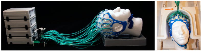
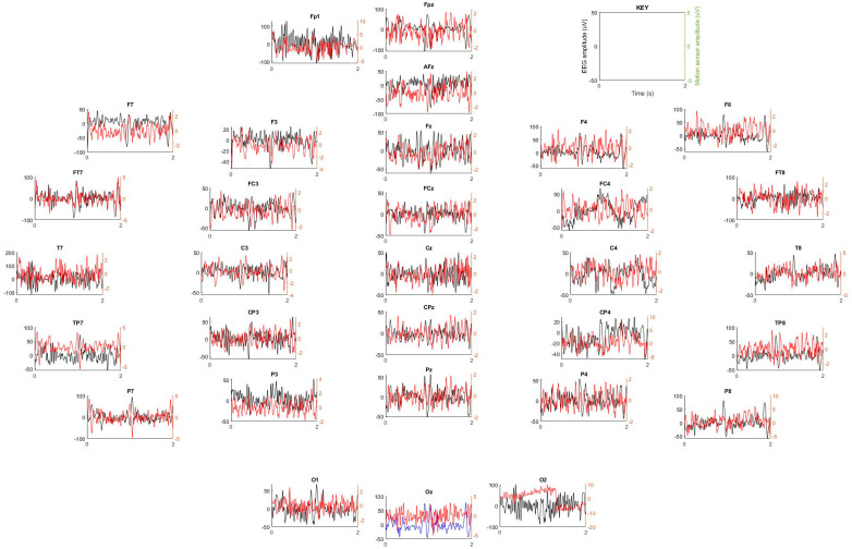
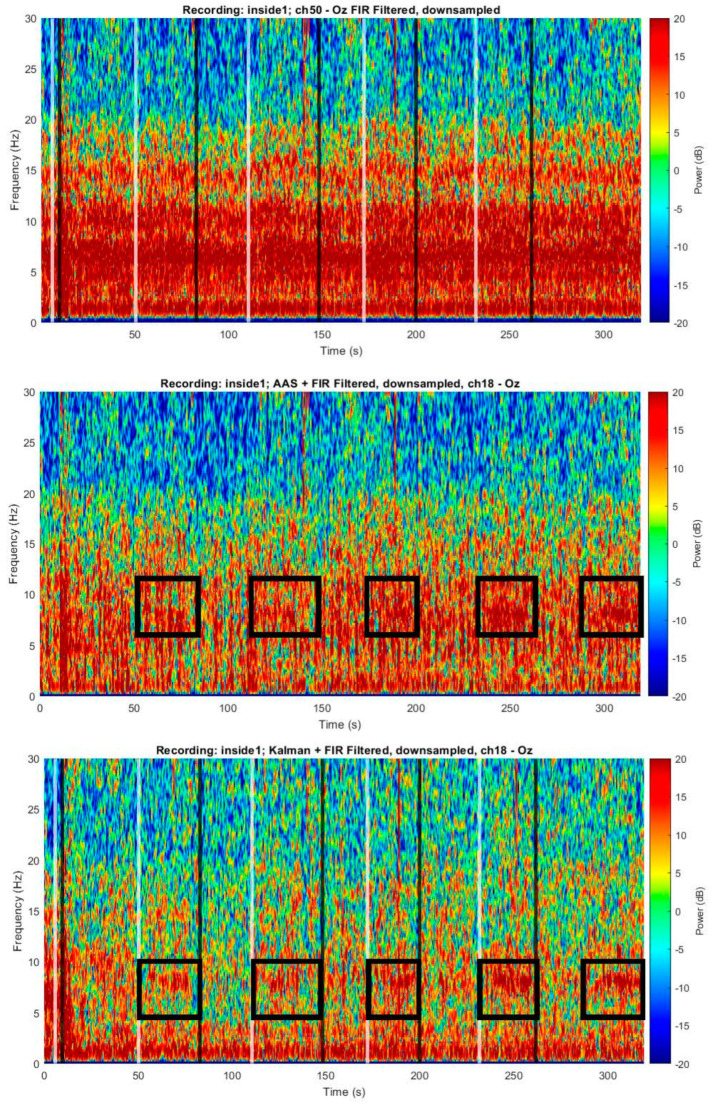
Methods: The MotoNet was built using polymer thick film (PTF) EEG leads and motion sensors on opposite sides in the same flex circuit. EEG/motion measurements were made with a standard commercial EEG acquisition system in a 3 Tesla (T) MRI. A Kalman filtering-based BCG correction tool was used to clean the EEG in healthy volunteers.

Results: MRI safety studies in 3 T confirmed the maximum heating below 1 °C. Using an MRI sequence with spatial localization gradients only, the position of the head was linearly correlated with the average motion sensor output. Kalman filtering was shown to reduce the BCG noise and recover artifact-clean EEG.

Conclusions: The MotoNet is an innovative EEG net design that co-locates 32 EEG electrodes with 32 motion sensors to improve both EEG and MRI signal quality. In combination with custom gradients, the position of the net can, in principle, be determined. In addition, the motion sensors can help reduce BCG noise.

Keywords: EEG/fMRI; Kalman adaptive noise cancellation; ballistocardiogram; position estimation.


Conflict of interest statement

The authors declare no conflict of interest. The funders had no role in the design of the study; in the collection, analyses, or interpretation of data; in the writing of the manuscript, or in the decision to publish the results.

Figures

Figure 1
Figure 1
The 32-channels/motion sensors net. Image of the actual MotoNet (A). Image of the double sided PTF leads with a PTF coil on one side (B) and a traditional EEG electrode (C) on the opposite side.
Figure 2
Figure 2
MotoNet setup. The MotoNet is connected to two commercial EEG amplifiers through a custom-made interface (left). The traces have been designed to slide into the Siemens 64-channel head/neck coil (orange arrow on the right).
Figure 3
Figure 3
Flowchart for the EEG processing and Kalman filtering.
Figure 4
Figure 4
Temperature measurements with head-sized agar phantom. (A) The spatial distribution of 32-channel EEG electrodes where temperature was monitored. The eight thermal probes were positioned in the hot-spots identified by simulations, circled in green. (B) The temperature elevation in each probe during a high-power turbo spin-echo sequence with the maximum SAR allowed in a clinical scan for 30 min using a birdcage body transmit coil; (C) table of the total change in temperature (ΔT) during the scan for each measurement location. The temperature increase was less than 1 °C, well within the safety limits.
Figure 5
Figure 5
Example of motion sensor signal outputs for the x-axis gradient.
Figure 6
Figure 6
Relationship between the MotoNet motion sensors’ average amplitude (Figure S5) and the mean x-axis position, corresponding to 9.552 µV/cm. The circles with a dotted line indicate measured data; the solid line indicates the best-fit line.
Figure 7
Figure 7
Kalman filter input signals. Each EEG signal (e.g., Oz in blue) corrupted by BCG is cleaned by subtracting a weighted sum of all the motion sensor signals (i.e., in red). The algorithm is then repeated for all EEG signals (i.e., in black).
Figure 8
Figure 8
Example of signal cleaning in a single EEG channel. Raw EEG recording inside a 3 T MRI, without scanning (top); EEG after low pass filtering (middle); and cleaned EEG data after Kalman filtering, using the motion sensor signals (bottom).
Figure 9
Figure 9
Example of a single EEG channel with adaptive BCG artifact cancellation. Spectrogram of an EEG recording at 3 T with the MotoNet after FIR filtering (top) and after Kalman filtering (bottom). After cleaning, ~10 Hz alpha rhythms are detected during the eyes-closed periods (black boxes in bottom panel). White vertical lines indicate instructions to close eyes; black vertical lines indicate eye opening.
Figure 10
Figure 10
Power spectra per electrode across cleaning methods: unfiltered/raw, AAS [34], Kalman filter, and clean signals acquired outside the MRI scanner field.

References

    1. Jorge J., Grouiller F., Gruetter R., Van Der Zwaag W., Figueiredo P. Towards high-quality simultaneous EEG-fMRI at 7 T: Detection and reduction of EEG artifacts due to head motion. Neuroimage. 2015;120:143–153. doi: 10.1016/j.neuroimage.2015.07.020. - DOI - PubMed
    1. Schulte-Uentrop L., Goepfert M.S. Anaesthesia or sedation for MRI in children. Curr. Opin. Anaesthesiol. 2010;23:513–517. doi: 10.1097/ACO.0b013e32833bb524. - DOI - PubMed
    1. Mulert C., Lemieux L. In: EEG-fMRI: Physiological Basis, Technique, and Applications. 2nd ed. Mulert C., Lemieux L., editors. Volume IX. Springer; Cham, Switzerland: 2023. p. 789.
    1. Kuperman J.M., Brown T.T., Ahmadi M.E., Erhart M.J., White N.S., Roddey J.C., Shankaranarayanan A., Han E.T., Rettmann D., Dale A.M. Prospective motion correction improves diagnostic utility of pediatric MRI scans. Pediatr. Radiol. 2011;41:1578–1582. doi: 10.1007/s00247-011-2205-1. - DOI - PMC - PubMed
    1. Placidi G. MRI: Essentials for Innovative Technologies. CRC Press; Boca Raton, FL, USA: 2012. 192p

LinkOut - more resources