

Effects of aging and macrophages on mice stem Leydig cell proliferation and differentiation in vitro
- PMID: 37051204
- PMCID: PMC10083278
- DOI: 10.3389/fendo.2023.1139281
Effects of aging and macrophages on mice stem Leydig cell proliferation and differentiation in vitro
Abstract
Background: Testosterone plays a critical role in maintaining reproductive functions and well-beings of the males. Adult testicular Leydig cells (LCs) produce testosterone and are generated from stem Leydig cells (SLCs) during puberty through adulthood. In addition, macrophages are critical in the SLC regulatory niche for normal testicular function. Age-related reduction in serum testosterone contributes to a number of metabolic and quality-of-life changes in males, as well as age-related changes in immunological functions. How aging and testicular macrophages may affect SLC function is still unclear.
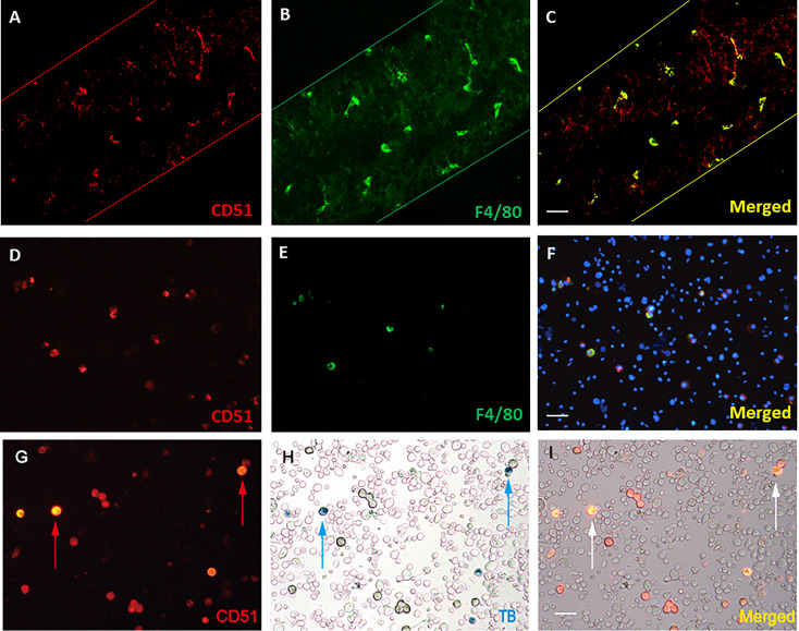
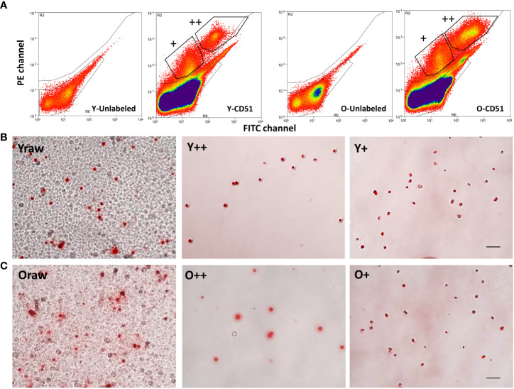
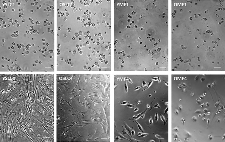
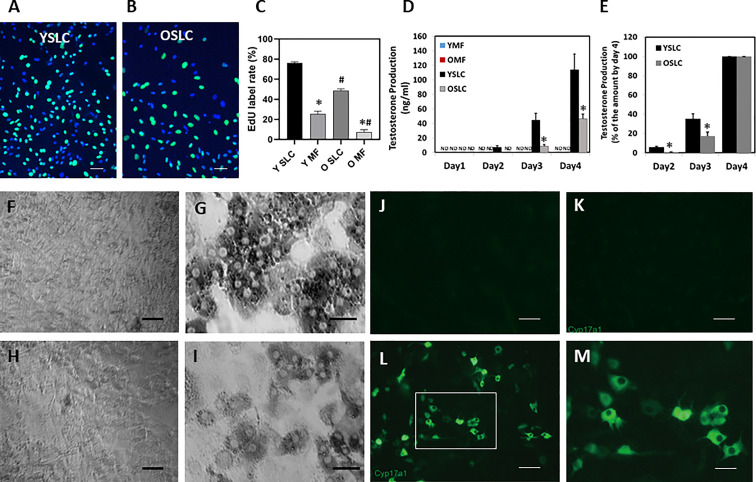
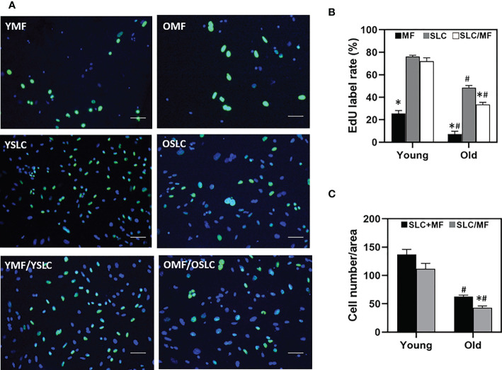
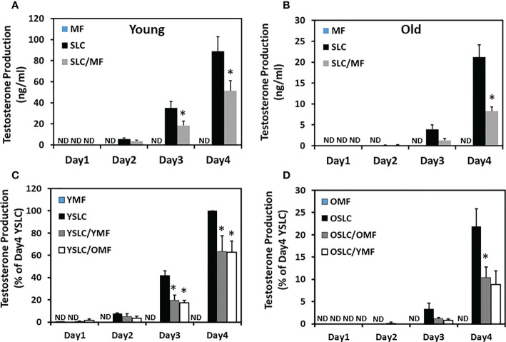
Methods: SLCs and macrophages were purified from adult and aged mice via FACS using CD51 as a marker protein. The sorted cells were first characterized and then co-cultured in vitro to examine how aging and macrophages may affect SLC proliferation and differentiation. To elucidate specific aging effects on both cell types, co-culture of sorted SLCs and macrophages were also carried out across two ages.
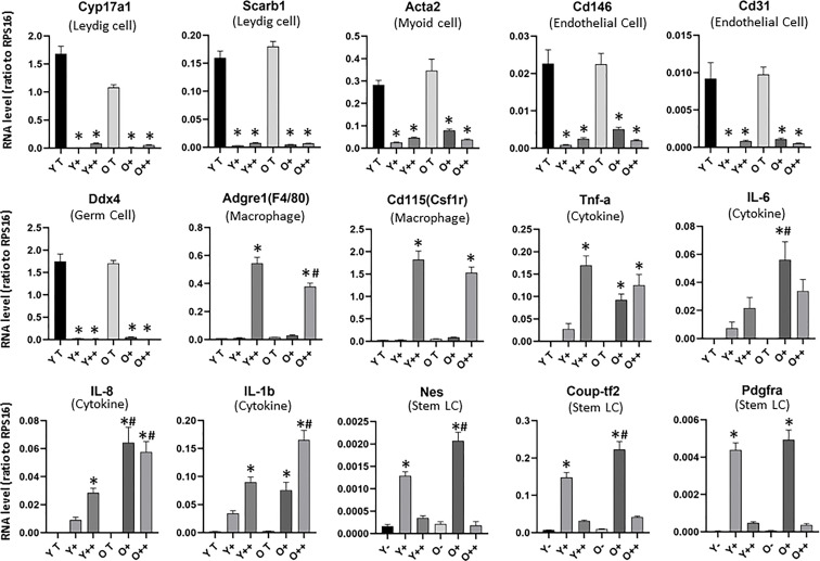
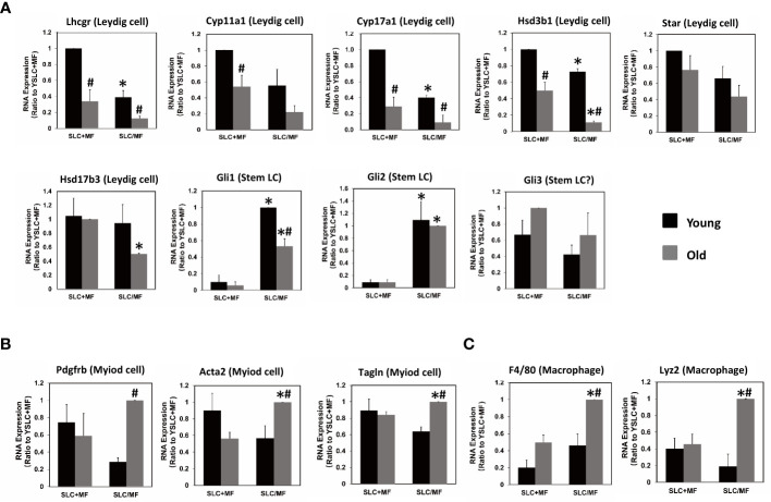
Results: CD51+ (weakly positive) and CD51++ (strongly positive) cells expressed typical SLC and macrophage markers, respectively. However, with aging, both cell types increased expression of multiple cytokine genes, such as IL-1b, IL-6 and IL-8. Moreover, old CD51+ SLCs reduced their proliferation and differentiation, with a more significant reduction in differentiation (2X) than proliferation (30%). Age matched CD51++ macrophages inhibited CD51+ SLC development, with a more significant reduction in old cells (60%) than young (40%). Crossed-age co-culture experiments indicated that the age of CD51+ SLCs plays a more significant role in determining age-related inhibitory effects. In LC lineage formation, CD51+ SLC had both reduced LC lineage markers and increased myoid cell lineage markers, suggesting an age-related lineage shift for SLCs.
Conclusion: The results suggest that aging affected both SLC function and their regulatory niche cell, macrophages.
Keywords: CD51; aging; macrophages; stem Leydig cells; testis; testosterone.
Copyright © 2023 Shao, Wang, Wen, Xie, Huang, Guan, Hao, Duan, Chen and Chen.
Conflict of interest statement
The authors declare that the research was conducted in the absence of any commercial or financial relationships that could be construed as a potential conflict of interest.
Figures
Similar articles
-
Transplantation of CD51+ Stem Leydig Cells: A New Strategy for the Treatment of Testosterone Deficiency.Stem Cells. 2017 May;35(5):1222-1232. doi: 10.1002/stem.2569. Epub 2017 Mar 5. Stem Cells. 2017. PMID: 28090714
-
Characterization and differentiation of CD51+ Stem Leydig cells in adult mouse testes.Mol Cell Endocrinol. 2019 Aug 1;493:110449. doi: 10.1016/j.mce.2019.110449. Epub 2019 May 15. Mol Cell Endocrinol. 2019. PMID: 31102608
-
Characterization of Nestin-positive stem Leydig cells as a potential source for the treatment of testicular Leydig cell dysfunction.Cell Res. 2014 Dec;24(12):1466-85. doi: 10.1038/cr.2014.149. Epub 2014 Nov 21. Cell Res. 2014. PMID: 25418539 Free PMC article.
-
Insights into the Regulation on Proliferation and Differentiation of Stem Leydig Cells.Stem Cell Rev Rep. 2021 Oct;17(5):1521-1533. doi: 10.1007/s12015-021-10133-x. Epub 2021 Feb 17. Stem Cell Rev Rep. 2021. PMID: 33598893 Review.
-
Leydig cell stem cells: Identification, proliferation and differentiation.Mol Cell Endocrinol. 2017 Apr 15;445:65-73. doi: 10.1016/j.mce.2016.10.010. Epub 2016 Oct 12. Mol Cell Endocrinol. 2017. PMID: 27743991 Free PMC article. Review.
Cited by
-
Stem Leydig cells support macrophage immunological homeostasis through mitochondrial transfer in mice.Nat Commun. 2024 Mar 8;15(1):2120. doi: 10.1038/s41467-024-46190-2. Nat Commun. 2024. PMID: 38459012 Free PMC article.
-
Age-related testosterone decline: mechanisms and intervention strategies.Reprod Biol Endocrinol. 2024 Nov 14;22(1):144. doi: 10.1186/s12958-024-01316-5. Reprod Biol Endocrinol. 2024. PMID: 39543598 Free PMC article. Review.
-
Genome-wide landscape of miRNA-mRNA-lncRNA-circRNA ceRNA network in Nanos2 deficient mice.PLoS One. 2025 Jun 27;20(6):e0325260. doi: 10.1371/journal.pone.0325260. eCollection 2025. PLoS One. 2025. PMID: 40577378 Free PMC article.
-
Characterization of ovarian progenitor cells for their potential to generate steroidogenic theca cells in vitro.Reproduction. 2024 May 31;168(1):e230407. doi: 10.1530/REP-23-0407. Print 2024 Jul 1. Reproduction. 2024. PMID: 38718815 Free PMC article.
References
Publication types
MeSH terms
Substances
LinkOut - more resources
Full Text Sources