Toward high-throughput engineering techniques for improving CAR intracellular signaling domains
- PMID: 37051270
- PMCID: PMC10083361
- DOI: 10.3389/fbioe.2023.1101122
Toward high-throughput engineering techniques for improving CAR intracellular signaling domains
Abstract
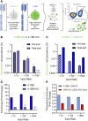
Chimeric antigen receptors (CAR) are generated by linking extracellular antigen recognition domains with one or more intracellular signaling domains derived from the T-cell receptor complex or various co-stimulatory receptors. The choice and relative positioning of signaling domains help to determine chimeric antigen receptors T-cell activity and fate in vivo. While prior studies have focused on optimizing signaling power through combinatorial investigation of native intracellular signaling domains in modular fashion, few have investigated the prospect of sequence engineering within domains. Here, we sought to develop a novel in situ screening method that could permit deployment of directed evolution approaches to identify intracellular domain variants that drive selective induction of transcription factors. To accomplish this goal, we evaluated a screening approach based on the activation of a human NF-κB and NFAT reporter T-cell line for the isolation of mutations that directly impact T cell activation in vitro. As a proof-of-concept, a model library of chimeric antigen receptors signaling domain variants was constructed and used to demonstrate the ability to discern amongst chimeric antigen receptors containing different co-stimulatory domains. A rare, higher-signaling variant with frequency as low as 1 in 1000 could be identified in a high throughput setting. Collectively, this work highlights both prospects and limitations of novel mammalian display methods for chimeric antigen receptors signaling domain discovery and points to potential strategies for future chimeric antigen receptors development.
Keywords: 41BB; OX40; chimeric antigen receptor; directed evolution; mammalian display.
Copyright © 2023 Butler, Hartman, Huang and Ackerman.
Conflict of interest statement
The authors declare that the research was conducted in the absence of any commercial or financial relationships that could be construed as a potential conflict of interest.
Figures


Similar articles
-
Chimeric Antigen Receptor Library Screening Using a Novel NF-κB/NFAT Reporter Cell Platform.Mol Ther. 2019 Feb 6;27(2):287-299. doi: 10.1016/j.ymthe.2018.11.015. Epub 2018 Nov 20. Mol Ther. 2019. PMID: 30573301 Free PMC article.
-
Co-Stimulatory Receptor Signaling in CAR-T Cells.Biomolecules. 2022 Sep 15;12(9):1303. doi: 10.3390/biom12091303. Biomolecules. 2022. PMID: 36139142 Free PMC article. Review.
-
Chimeric antigen receptors containing the OX40 signalling domain enhance the persistence of T cells even under repeated stimulation with multiple myeloma target cells.J Hematol Oncol. 2022 Apr 1;15(1):39. doi: 10.1186/s13045-022-01244-0. J Hematol Oncol. 2022. PMID: 35365211 Free PMC article.
-
4-1BB and optimized CD28 co-stimulation enhances function of human mono-specific and bi-specific third-generation CAR T cells.J Immunother Cancer. 2021 Oct;9(10):e003354. doi: 10.1136/jitc-2021-003354. J Immunother Cancer. 2021. PMID: 34706886 Free PMC article.
-
CARs: Beyond T Cells and T Cell-Derived Signaling Domains.Int J Mol Sci. 2020 May 15;21(10):3525. doi: 10.3390/ijms21103525. Int J Mol Sci. 2020. PMID: 32429316 Free PMC article. Review.
Cited by
-
Bringing cell therapy to tumors: considerations for optimal CAR binder design.Antib Ther. 2023 Sep 12;6(4):225-239. doi: 10.1093/abt/tbad019. eCollection 2023 Oct. Antib Ther. 2023. PMID: 37846297 Free PMC article. Review.
-
Expanding the CAR toolbox with high throughput screening strategies for CAR domain exploration: a comprehensive review.J Immunother Cancer. 2025 Apr 9;13(4):e010658. doi: 10.1136/jitc-2024-010658. J Immunother Cancer. 2025. PMID: 40210240 Free PMC article. Review.
-
Challenges and future perspectives for high-throughput chimeric antigen receptor T cell discovery.Curr Opin Biotechnol. 2024 Dec;90:103216. doi: 10.1016/j.copbio.2024.103216. Epub 2024 Oct 21. Curr Opin Biotechnol. 2024. PMID: 39437676 Review.
-
Spatiotemporal imaging of immune dynamics: rethinking drug efficacy evaluation in cancer immunotherapy.Front Immunol. 2025 May 16;16:1609606. doi: 10.3389/fimmu.2025.1609606. eCollection 2025. Front Immunol. 2025. PMID: 40453091 Free PMC article. No abstract available.
References
Grants and funding
LinkOut - more resources
Full Text Sources
Other Literature Sources
Research Materials

