

CDC25A inhibition suppresses cell proliferation and induces G1/S‑phase cell cycle arrest in nasopharyngeal carcinoma
- PMID: 37052240
- PMCID: PMC10119622
- DOI: 10.3892/mmr.2023.12996
CDC25A inhibition suppresses cell proliferation and induces G1/S‑phase cell cycle arrest in nasopharyngeal carcinoma
Abstract
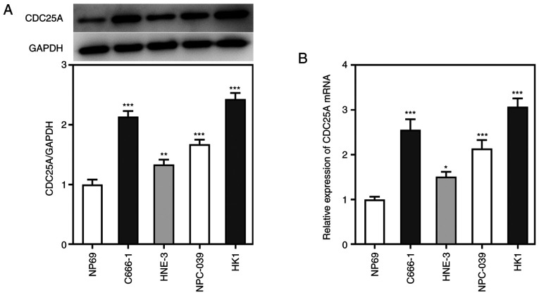
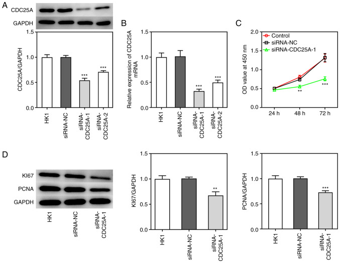
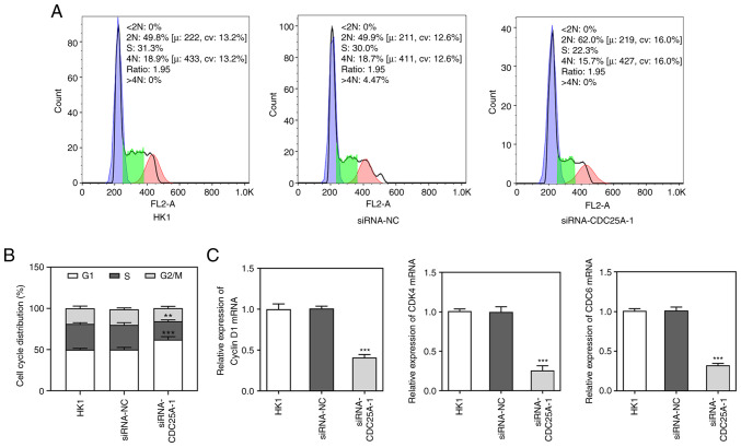
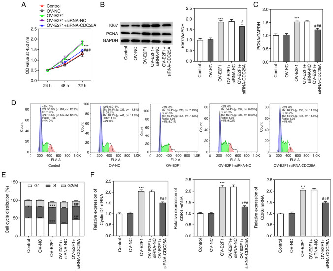
Nasopharyngeal carcinoma (NPC) is a primary malignancy that originates from the nasopharyngeal region. It has been demonstrated that a decrease in the expression level of cell division cycle gene 25A (CDC25A) suppresses cell viability and induces apoptosis in a variety of different types of cancer. However, at present, the role of CDC25A in NPC has yet to be fully elucidated. Therefore, the aim of the present study was to investigate the role of CDC25A in NPC progression and to explore the potential underlying mechanism. Reverse transcription‑quantitative PCR was performed to detect the relative mRNA levels of CDC25A and E2F transcription factor 1 (E2F1). Western blot analysis was subsequently used to determine the expression levels of CDC25A, Ki67, proliferating cell nuclear antigen (PCNA) and E2F1. CCK8 assay was employed to measure cell viability and flow cytometric analysis was employed to analyze the cell cycle. The binding sites between the CDC25A promoter and E2F1 were predicted using bioinformatics tools. Finally, luciferase reporter gene and chromatin immunoprecipitation assays were performed to verify the interaction between CDC25A and E2F1. The results obtained suggested that CDC25A is highly expressed in NPC cell lines and CDC25A silencing was found to inhibit cell proliferation, reduce the protein expression levels of Ki67 and PCNA and induce G1 arrest of NPC cells. Furthermore, E2F1 could bind CDC25A and positively regulate its expression at the transcriptional level. In addition, CDC25A silencing abolished the effects of E2F1 overexpression on cell proliferation and the cell cycle in NPC. Taken together, the findings of the present study showed that CDC25A silencing attenuated cell proliferation and induced cell cycle arrest in NPC and CDC25A was regulated by E2F1. Hence, CDC25A may be a promising therapeutic target for treatment of NPC.
Keywords: E2F transcription factor 1; cell cycle; cell division cycle gene 25A; nasopharyngeal carcinoma; proliferation.
Conflict of interest statement
The authors declare that they have no competing interests.
Figures

Similar articles
-
MicroRNA let-7c Inhibits Cell Proliferation and Induces Cell Cycle Arrest by Targeting CDC25A in Human Hepatocellular Carcinoma.PLoS One. 2015 Apr 24;10(4):e0124266. doi: 10.1371/journal.pone.0124266. eCollection 2015. PLoS One. 2015. Retraction in: PLoS One. 2023 Jan 6;18(1):e0280394. doi: 10.1371/journal.pone.0280394. PMID: 25909324 Free PMC article. Retracted.
-
MicroRNA-99a-5p suppresses breast cancer progression and cell-cycle pathway through downregulating CDC25A.J Cell Physiol. 2019 Apr;234(4):3526-3537. doi: 10.1002/jcp.26906. Epub 2018 Nov 15. J Cell Physiol. 2019. PMID: 30443946
-
PCNA-associated factor KIAA0101 transcriptionally induced by ELK1 controls cell proliferation and apoptosis in nasopharyngeal carcinoma: an integrated bioinformatics and experimental study.Aging (Albany NY). 2020 Apr 9;12(7):5992-6017. doi: 10.18632/aging.102991. Epub 2020 Apr 9. Aging (Albany NY). 2020. PMID: 32275642 Free PMC article.
-
Over-expression of the special AT rich sequence binding protein 1 (SATB1) promotes the progression of nasopharyngeal carcinoma: association with EBV LMP-1 expression.J Transl Med. 2013 Sep 18;11:217. doi: 10.1186/1479-5876-11-217. J Transl Med. 2013. PMID: 24047082 Free PMC article.
-
The role of Cdc25A in the regulation of cell proliferation and apoptosis.Anticancer Agents Med Chem. 2012 Jul;12(6):631-9. doi: 10.2174/187152012800617678. Anticancer Agents Med Chem. 2012. PMID: 22263797 Free PMC article. Review.
Cited by
-
Advances in the mechanism of CDK4/6 inhibitor resistance in HR+/HER2- breast cancer.Ther Adv Med Oncol. 2024 Sep 30;16:17588359241282499. doi: 10.1177/17588359241282499. eCollection 2024. Ther Adv Med Oncol. 2024. PMID: 39371618 Free PMC article. Review.
-
HSPA12A promotes c-Myc lactylation-mediated proliferation of tubular epithelial cells to facilitate renal functional recovery from kidney ischemia/reperfusion injury.Cell Mol Life Sci. 2024 Sep 15;81(1):404. doi: 10.1007/s00018-024-05427-5. Cell Mol Life Sci. 2024. PMID: 39277835 Free PMC article.
References
-
- Zheng ZQ, Li ZX, Zhou GQ, Lin L, Zhang LL, Lv JW, Huang XD, Liu RQ, Chen F, He XJ, et al. Long noncoding RNA FAM225A promotes nasopharyngeal carcinoma tumorigenesis and metastasis by acting as ceRNA to sponge miR-590-3p/miR-1275 and upregulate ITGB3. Cancer Res. 2019;79:4612–4626. doi: 10.1158/0008-5472.CAN-19-0799. - DOI - PubMed
MeSH terms
Substances
LinkOut - more resources
Full Text Sources
Miscellaneous
