

Casticin suppresses RANKL‑induced osteoclastogenesis and prevents ovariectomy‑induced bone loss by regulating the AKT/ERK and NF‑κB signaling pathways
- PMID: 37052260
- PMCID: PMC10198048
- DOI: 10.3892/ijmm.2023.5246
Casticin suppresses RANKL‑induced osteoclastogenesis and prevents ovariectomy‑induced bone loss by regulating the AKT/ERK and NF‑κB signaling pathways
Abstract
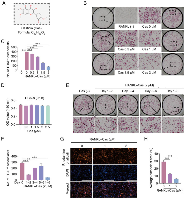
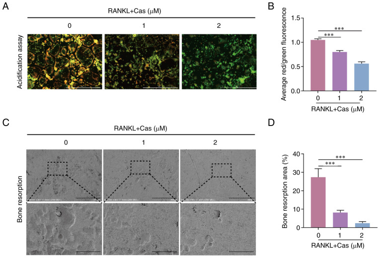
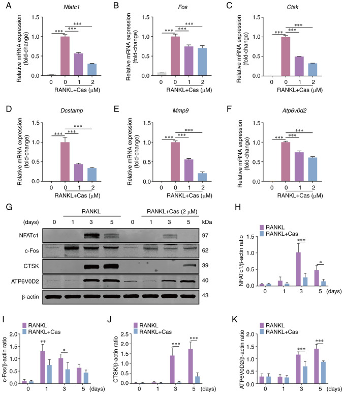
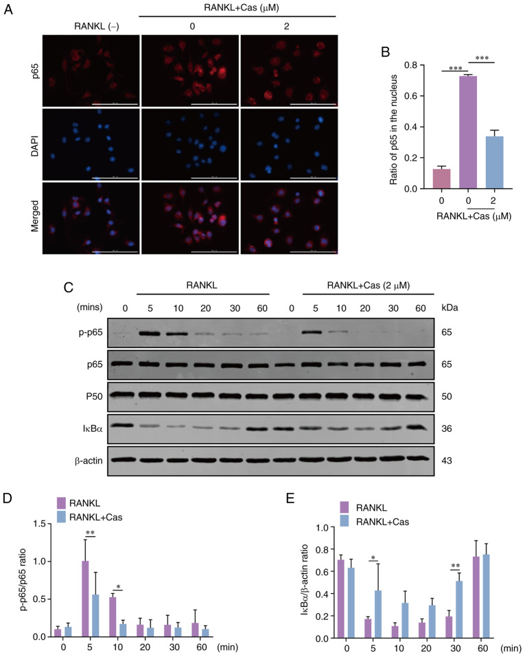
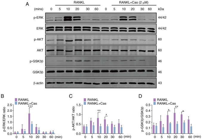
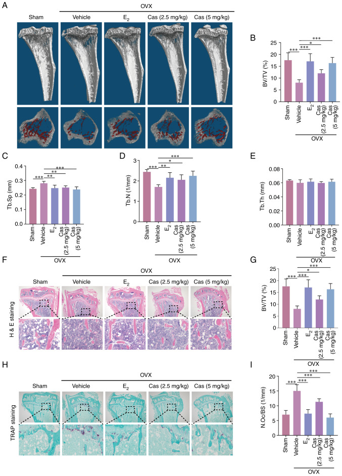
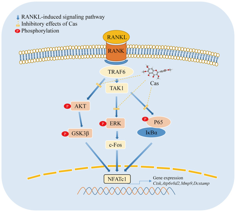
Postmenopausal osteoporosis is a systemic metabolic disease that chronically endangers public health and is typically characterized by low bone mineral density and marked bone fragility. The excessive bone resorption activity of osteoclasts is a major factor in the pathogenesis of osteoporosis; therefore, strategies aimed at inhibiting osteoclast activity may prevent bone decline and attenuate the process of osteoporosis. Casticin (Cas), a natural compound, has anti‑inflammatory and antitumor properties. However, the role of Cas in bone metabolism remains largely unclear. The present study found that the receptor activator of nuclear factor‑κΒ (NF‑κB) ligand‑induced osteoclast activation and differentiation were inhibited by Cas. Tartrate‑resistant acid phosphatase staining revealed that Cas inhibited osteoclast differentiation, and bone resorption pit assays demonstrated that Cas affected the function of osteoclasts. Cas significantly reduced the expression of osteoclast‑specific genes and related proteins, such as nuclear factor of activated T cells, cytoplasmic 1 and c‑Fos at the mRNA and protein level in a concentration‑dependent manner. Cas inhibited osteoclast formation by blocking the AKT/ERK and NF‑κB signaling pathways, according to the intracellular signaling analysis. The microcomputed tomography and tissue staining of tibiae from ovariectomized mice revealed that Cas prevented the bone loss induced by estrogen deficiency and reduced osteoclast activity in vivo. Collectively, these findings indicated that Cas may be used to prevent osteoporosis.
Keywords: AKT; ERK; NF‑κB; casticin; osteoclast; osteoporosis.
Conflict of interest statement
The authors declare that they have no competing interests.
Figures
Similar articles
-
Glaucocalyxin A suppresses osteoclastogenesis induced by RANKL and osteoporosis induced by ovariectomy by inhibiting the NF-κB and Akt pathways.J Ethnopharmacol. 2021 Aug 10;276:114176. doi: 10.1016/j.jep.2021.114176. Epub 2021 Apr 30. J Ethnopharmacol. 2021. PMID: 33933570
-
Anethole inhibits RANKL-induced osteoclastogenesis by downregulating ERK/AKT signaling and prevents ovariectomy-induced bone loss in vivo.Int Immunopharmacol. 2021 Nov;100:108113. doi: 10.1016/j.intimp.2021.108113. Epub 2021 Sep 13. Int Immunopharmacol. 2021. PMID: 34530203
-
Shikonin mitigates ovariectomy-induced bone loss and RANKL-induced osteoclastogenesis via TRAF6-mediated signaling pathways.Biomed Pharmacother. 2020 Jun;126:110067. doi: 10.1016/j.biopha.2020.110067. Epub 2020 Apr 6. Biomed Pharmacother. 2020. PMID: 32272431
-
The role of lactoferrin in bone remodeling: evaluation of its potential in targeted delivery and treatment of metabolic bone diseases and orthopedic conditions.Front Endocrinol (Lausanne). 2023 Aug 23;14:1218148. doi: 10.3389/fendo.2023.1218148. eCollection 2023. Front Endocrinol (Lausanne). 2023. PMID: 37680888 Free PMC article. Review.
-
TNF receptor-associated factors: promising targets of natural products for the treatment of osteoporosis.Front Physiol. 2025 May 27;16:1527814. doi: 10.3389/fphys.2025.1527814. eCollection 2025. Front Physiol. 2025. PMID: 40496246 Free PMC article. Review.
Cited by
-
Hypoxia-Inducible Factor and Oxidative Stress in Tendon Degeneration: A Molecular Perspective.Antioxidants (Basel). 2024 Jan 10;13(1):86. doi: 10.3390/antiox13010086. Antioxidants (Basel). 2024. PMID: 38247510 Free PMC article. Review.
-
Anti-osteoporosis activity of casticin in ovariectomized rats.Toxicol Res (Camb). 2024 Apr 26;13(2):tfae064. doi: 10.1093/toxres/tfae064. eCollection 2024 Apr. Toxicol Res (Camb). 2024. PMID: 38680951 Free PMC article.
-
Denatonium inhibits RANKL-induced osteoclast differentiation and rescues the osteoporotic phenotype by blocking p65 signaling pathway.Mol Med. 2024 Dec 19;30(1):248. doi: 10.1186/s10020-024-01031-2. Mol Med. 2024. PMID: 39701944 Free PMC article.
References
MeSH terms
Substances
LinkOut - more resources
Full Text Sources
Medical
Miscellaneous
