. 2023 May;49(5):109.
doi: 10.3892/or.2023.8546. Epub 2023 Apr 13.

MicroRNA‑130a‑3p inhibition suppresses cervical cancer cell progression

Affiliations

MicroRNA‑130a‑3p inhibition suppresses cervical cancer cell progression

Rhafaela Lima Causin et al. Oncol Rep. 2023 May.

Abstract

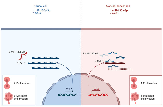
MicroRNAs (miRNAs or miRs) play essential roles in the initiation and progression of human tumors, including cervical cancer. However, the mechanisms underlying their actions in cervical cancer remain unclear. The present study aimed to evaluate the functional role of miR‑130a‑3p in cervical cancer. Cervical cancer cells were transfected with a miRNA inhibitor (anti‑miR‑130a‑3p) and a negative control. Adhesion‑independent cell proliferation, migration and invasion were evaluated. The findings presented herein demonstrated that miR‑130a‑3p was overexpressed in HeLa, SiHa, CaSki, C‑4I and HCB‑514 cervical cancer cells. The inhibition of miR‑130a‑3p significantly reduced the proliferation, migration and invasion of cervical cancer cells. The canonical delta‑like Notch1 ligand (DLL1) was identified as a possible direct target of miR‑103a‑3p. The DLL1 gene was further found to be significantly downregulated in cervical cancer tissues. On the whole, the present study demonstrates that miR‑130a‑3p contributes to the proliferation, migration and invasion of cervical cancer cells. Therefore, miR‑130a‑3p may be used as a biomarker to determine cervical cancer progression.

Keywords: biomarker; cervical cancer; microRNA‑130a‑3p; tumor progression.


Conflict of interest statement

The authors declare that they have not competing interests.

Figures

Figure 1.
Figure 1.
miR-130a-3p is highly expressed in cervical cancer cells. The HeLa, SiHa, CaSki, C-4I and HCB-514 cell lines had a significantly higher expression of miR-130a-3p than the non-tumorigenic epithelial cell line, HaCaT. Bars represent the standard deviation. *P<0.05, **P<0.01 and ***P<0.001, vs. HaCaT cells. ns, not significant; miR, microRNA.
Figure 2.
Figure 2.
Anti-miR-130a-3p knocks down the expression of miR-130a-3p in cervical cancer cells. The expression of miR-130a-3p in CaSki, C-4I and HCB-514 cells following transfection was reduced, indicating that anti-miR-130a-3p suppressed the expression of miR-130a-3p. The bars represent the standard deviation. ****P<0.0001. miR, microRNA.
Figure 3.
Figure 3.
Anti-miR-130a-3p inhibited the proliferation of cervical cancer cells. Colony formation assay was used to evaluate the proliferation of CaSki and C-4I cells, which exhibited a reduced clone number in the anti-miR-130a-3p group. (A) Images of the colony formation assay. (B) Bar plots of the data from panel A. Bars represent the standard deviation. *P<0.05 and ****P<0.0001. miR, microRNA.
Figure 4.
Figure 4.
Anti-miR-130a-3p inhibits the migration of cervical cancer cells. (A) Images of Transwell assay in CaSki, C-4I and HCB-514 cell lines, which exhibited a decreased migration after miR-130a-3p was knocked down. (B) Bar plots of the data from panel A. Bars represent the standard deviation. **P<0.01 and ****P<0.0001. miR, microRNA.
Figure 5.
Figure 5.
Anti-miR-130a-3p inhibits the invasion of cervical cancer cells. (A) Images of Transwell assay in the CaSki, C-4I and HCB-514 cell lines, which exhibited a decreased invasion after miR-130a-3p was knocked down. (B) Bar plots of the data from panel A. Bars represent the standard deviation. ****P<0.0001. miR, microRNA.
Figure 6.
Figure 6.
Gene expression profile in HCB-514 cells using nCounter® technologies. Heatmap was used for gene expression profiling of the HCB-514 primary cell line vs. HaCaT control cells. Cell lines were arranged in columns, while gene expression levels were arranged in rows, and both were hierarchically clustered using the Euclidean distance with the average linkage of nodes. Red shades indicate an increased relative expression, while blue shades indicate a reduced expression. The bars above indicate the groups used for the analysis. Purple indicates samples from the HaCaT control cells (n=3), while green refers to the HCB-514 cells (n=3) (FC≥2; P<0.01).
Figure 7.
Figure 7.
In silico analysis of DLL1 and WNT10A, and their association with the progression of patients with CESC and with cervical cancer cell lines. (A) Putative miR-130a-3p binding site in the 3′untranslated regions of DLL1 and WNT10A, as predicted using TargetScan. (B,C) Expression boxplots of the genes using the GEPIA2 database (P<0.05; CESC tissues, n=306; normal tissues, n=13) (B) DLL1 expression; (C) WNT10A expression. (D) Association between DLL1 expression, and pathological stage in patients with CESC using the GEPIA2 database (P<0.05). (E) Association between WNT10A expression, and pathological stage in patients with CESC using the GEPIA2 database (P<0.05). (F) Data from the GENT2 database regarding DLL1 expression in a cervical cancer cell line panel in comparison with HaCaT cells. ns, not significant. *P<0.05, **P<0.01, ***P<0.001 and ****P<0.0001. (G) Data from the GENT2 database regarding WNT10A expression in a cervical cancer cell line panel in comparison with HaCaT cells. **P<0.01, ***P<0.001. (H,I) Spearman's pair-wise correlation analysis of miRNA-mRNA target pairs in cervical cancer cell lines paired in triplicate (n=5). (H) Correlation analysis revealed that increased miR-130a-p expression levels were associated with DLL1 downregulation in cervical cancer cell lines. In the correlation plots, the solid line represents the least square estimate, and P-values represent global significance P<0.05; (I) correlation analysis revealed that increased miR-130a-p expression levels were associated with WNT10A downregulation in cervical cancer cell lines. In the correlation plots, the solid line represents the least square estimate, and P-values represent global significance P<0.05. (J) Enrichment pathways using the DLL1 (right panel, red) and WNT10A (left panel, blue) genes in CESC. Significantly enriched pathways terms are shown in -log10 with Benjamini-Hochberg false discovery rate-corrected P-values. The letter in parentheses after each pathway gene set name corresponds to the source of the pathway annotations. B, BioCarta; K Kyoto Encyclopedia of Genes and Genomes; R, Reactome; P, Panther; CESC, cervical squamous cell carcinoma and endocervical adenocarcinoma; miR, microRNA; GEPIA, Gene Expression Profiling Interactive Analysis; DLL1, δ-like Notch1 ligand.
Figure 8.
Figure 8.
GSEA. GSEA was performed in the HCB-514 and HaCaT cells. The GSEA algorithm calculates an enrichment score reflecting the degree of overrepresentation at the top or bottom of the ranked list of the genes included in a gene set in a ranked list of all genes present in the nCounter PanCancer Pathways Panel. The analysis demonstrates that the signaling pathways (A) gap junction, (B) calcium signaling pathway, (C) long-term potentiation and (D) apoptosis were enriched in HCB-514 cells. GSEA, gene set enrichment analysis
Figure 9.
Figure 9.
Graphical summary of the possible mechanism of the regulation of miR-130a-3p in cervical cancer cells. The high expression of miR-130a-3p in cervical cancer cells may promote the proliferation, migration and invasion of cervical cancer cells. By contrast, the inhibition of miR-130a-3p expression exerts opposite effects. miR, microRNA; DLL1, delta-like notch 1 ligand.

References

    1. Sung H, Ferlay J, Siegel RL, Laversanne M, Soerjomataram I, Jemal A, Bray F. Global cancer statistics 2020: GLOBOCAN estimates of incidence and mortality worldwide for 36 cancers in 185 countries. CA Cancer J Clin. 2021;71:209–249. doi: 10.3322/caac.21660. - DOI - PubMed
    1. Espenel S, Garcia MA, Trone JC, Guillaume E, Harris A, Rehailia-Blanchard A, He MY, Ouni S, Vallard A, Rancoule C, et al. From IB2 to IIIB locally advanced cervical cancers: report of a ten-year experience. Radiat Oncol. 2018;13:16. doi: 10.1186/s13014-018-0999-9. - DOI - PMC - PubMed
    1. Robin TP, Amini A, Schefter TE, Behbakht K, Fisher CM. Disparities in standard of care treatment and associated survival decrement in patients with locally advanced cervical cancer. Gynecol Oncol. 2016;143:319–325. doi: 10.1016/j.ygyno.2016.09.009. - DOI - PubMed
    1. Koh WJ, Abu-Rustum NR, Bean S, Bradley K, Campos SM, Cho KR, Chon HS, Chu C, Clark R, Cohn D, et al. Cervical cancer, Version 3.2019, NCCN clinical practice guidelines in oncology. J Natl Compr Canc Netw. 2019;17:64–84. doi: 10.6004/jnccn.2019.0001. - DOI - PubMed
    1. He Y, Han SB, Liu Y, Zhang JJ, Wu YM. Role of APOA1 in the resistance to platinum-based chemotherapy in squamous cervical cancer. BMC Cancer. 2022;22:411. doi: 10.1186/s12885-022-09528-x. - DOI - PMC - PubMed

LinkOut - more resources