Review
. 2023 Apr 13;480(7):471-488.
doi: 10.1042/BCJ20220073.

The biology and type I/III hybrid nature of type I-D CRISPR-Cas systems

Affiliations
Review

The biology and type I/III hybrid nature of type I-D CRISPR-Cas systems

Tess M McBride et al. Biochem J. .

Abstract

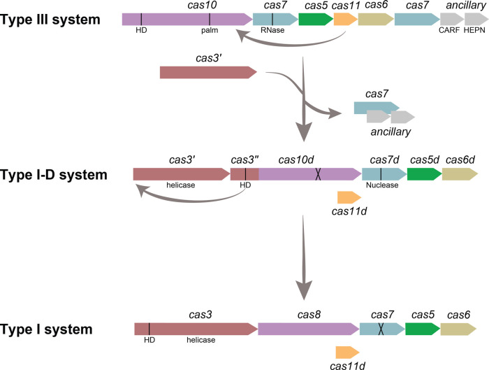
Prokaryotes have adaptive defence mechanisms that protect them from mobile genetic elements and viral infection. One defence mechanism is called CRISPR-Cas (clustered regularly interspaced short palindromic repeats and CRISPR-associated proteins). There are six different types of CRISPR-Cas systems and multiple subtypes that vary in composition and mode of action. Type I and III CRISPR-Cas systems utilise multi-protein complexes, which differ in structure, nucleic acid binding and cleaving preference. The type I-D system is a chimera of type I and III systems. Recently, there has been a burst of research on the type I-D CRISPR-Cas system. Here, we review the mechanism, evolution and biotechnological applications of the type I-D CRISPR-Cas system.

Keywords: CRISPR; RNA-binding proteins; bacteriophages; protein structure.


Conflict of interest statement

All authors are inventors on a patent application based on this research titled ‘Type I-D CRISPR–Cas Systems and Uses Thereof,’ which has been assigned Australian Patent Application No. PCT/NZ2023/050034, and the filing date of 17 March 2023.

Figures

Figure 1.
Figure 1.. CRISPR–Cas stages of defence.
Adaptation occurs when the adaptation complex (blue) captures DNA, which it then incorporates into the CRISPR array as a spacer (grey line). The expression stage occurs when the cas genes (yellow) are expressed, and the CRISPR array is transcribed into pre-crRNA, which is subsequently processed by a Cas protein into crRNA. Interference occurs when the Cas proteins and crRNA assemble into the interference complex and then binds to complementary DNA and degrades it. Stages of defence based on type I CRISPR–Cas systems.
Figure 2.
Figure 2.. Classification Type I and type III CRISPR–Cas systems.
(A) Comparison of type I and III CRISPR–Cas systems, with their ‘signature’ genes in bold. Genes are organised by role; adaptation (yellow), processing (olive green), interference (purple), and ancillary (grey). Genes absent from some subtypes are indicated with white diagonal lines. Diagram adapted from Makarova et al. [16]. (B) Schematic of type I and type III interference complexes with proteins annotated.
Figure 3.
Figure 3.. Schematic of representative type I-D cas genes and CRISPR arrays.
CRISPR repeats are indicated by green diamonds, and spacers are indicated by green boxes. CRISPR arrays represent the position in locus, and the length is not correlated to the actual size of the CRISPR array.
Figure 4.
Figure 4.. Predicted model of naïve type I-D adaptation.
(A) The adaptation Cas proteins form asymmetrical complexes Cas12:Cas22 and Cas12:Cas41, which capture a prespacer. The PAM end is removed by Cas4, and opposite 3′ overhang is trimmed by a host nuclease (1). Half-site integration occurs via nucleophilic attack of the prespacer into the leader-repeat junction (2). Full-site integration occurs via nucleophilic attack of the second strand of the prespacer into the repeat-spacer junction (3). DNA repair completes integration to form a repeat-spacer-repeat at the leader proximal end of the array (4). Adapted from the model proposed by Kieper et al. [70]. (B) Schematic of the Synechocystis leader sequence and associated motifs.
Figure 5.
Figure 5.. Predicted model of type I-D expression and processing.
The cas (purple and brown) and regulator (pink) genes are transcribed and translated while the CRISPR array is transcribed, forming the pre-crRNA. Sll7009 may regulate type I-D interference via any of the promoters. Cas6 associates with the stem–loops of the pre-crRNA and cleaves at the base of the stem, forming type I-like crRNA (green background), as observed by McBride et al. [19]. Shorter type I-D crRNA also occurs (Scholz et al. [83]), resembling type III-like mature crRNA (yellow background).
Figure 6.
Figure 6.. Model of type I-D interference for dsDNA and single-stranded nucleic acids (ssNA).
(A) Structure of Synechocystis type I-D Cascade (Protein Data Bank ID, 7SBA). Subunits displayed: Cas10d (green); Cas5d (pink); Cas11d (orange); Cas7d (grey and blue); crRNA (purple); target strand (TS; red); non-target strand (NTS; cyan). Structure (B) and schematic (C) of the PAM binding pocket, including the glycine loop and lysine finger from Cas10d (top) and the glutamine wedge from Cas5d positioned where dsDNA bifurcation occurs (bottom). R-loop formation is likely stabilised by a positive patch within Cas10d and Cas11d. (C) adapted from Hayes et al. [94]. (D) The predicted mechanism of type I-D interference of dsDNA. Type I-D Cascade samples dsDNA looking for a GTN PAM (1), the PAM is identified with the PAM binding pocket in Cas10d (2), and an R-loop is formed (3), Cas3′ is recruited to Cascade (4), bidirectional degradation of both strands occurs (5). (E) The predicted mechanism of type I-D interference of single-stranded nucleic acids (ssNA). Type I-D Cascade binds the ssNA (1), followed by the cleavage of the ssNA by Cas7d (2).
Figure 7.
Figure 7.. Predicted model of the type I-D system as an evolutionary intermediate between type III and type I systems.
Type I-D likely evolved from a type III-like ancestor system through the acquisition of a helicase domain, the loss of the circular permutation in the cas10d HD, cas11 becoming alternatively expressed from the large subunit, and the loss of one cas7 and the auxiliary genes. Type I systems may have evolved from type I-D via the transfer of the HD domain, cas3″, onto the helicase domain, cas3′, to form the typical cas3 nuclease-helicase. In some type I systems, duplication of cas11 occurs, forming a separate gene. A black line indicates key domains, and inactive domains are indicated by a cross.

References

    1. Price, J.E. and Chapman, M.R. (2018) Phaged and confused by biofilm matrix. Nat. Microbiol. 3, 2–3 10.1038/s41564-017-0078-2 - DOI - PubMed
    1. Hampton, H.G., Watson, B.N. and Fineran, P.C. (2020) The arms race between bacteria and their phage foes. Nature 577, 327–336 10.1038/s41586-019-1894-8 - DOI - PubMed
    1. Barrangou, R., Fremaux, C., Deveau, H., Richards, M., Boyaval, P., Moineau, S.et al. (2007) CRISPR provides acquired resistance against viruses in prokaryotes. Science 315, 1709–1712 10.1126/science.1138140 - DOI - PubMed
    1. Makarova, K.S., Wolf, Y.I., Alkhnbashi, O.S., Costa, F., Shah, S.A., Saunders, S.J.et al. (2015) An updated evolutionary classification of CRISPR-Cas systems. Nat. Rev. Microbiol. 13, 722–736 10.1038/nrmicro3569 - DOI - PMC - PubMed
    1. Safari, F., Sharifi, M., Farajnia, S., Akbari, B., Karimi Baba Ahmadi, M., Negahdaripour, M.et al. (2020) The interaction of phages and bacteria: the co-evolutionary arms race. Crit. Rev. Biotechnol. 40, 119–137 10.1080/07388551.2019.1674774 - DOI - PubMed

Publication types

Substances