. 2023 May 31;193(2):146-165.
doi: 10.1093/toxsci/kfad033.

Pulmonary immune response regulation, genotoxicity, and metabolic reprogramming by menthol- and tobacco-flavored e-cigarette exposures in mice

Affiliations

Pulmonary immune response regulation, genotoxicity, and metabolic reprogramming by menthol- and tobacco-flavored e-cigarette exposures in mice

Thivanka Muthumalage et al. Toxicol Sci. .

Abstract

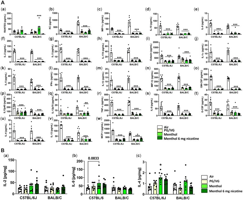
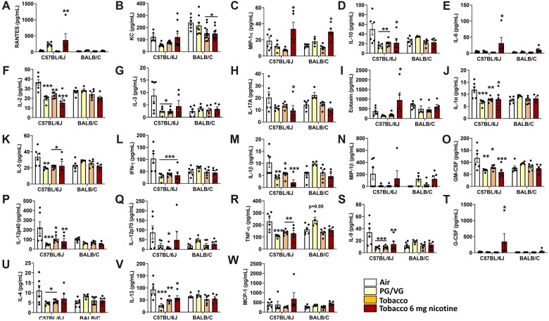
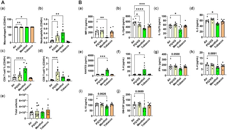
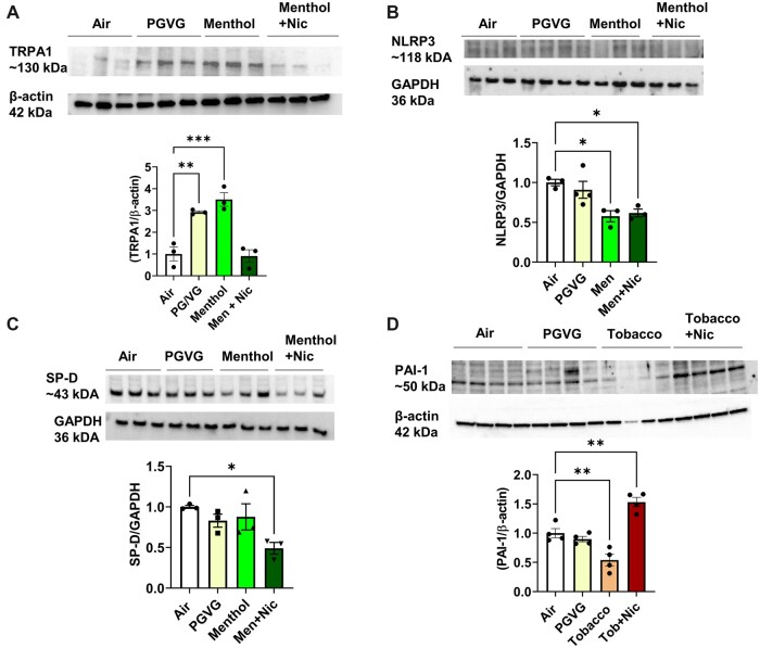
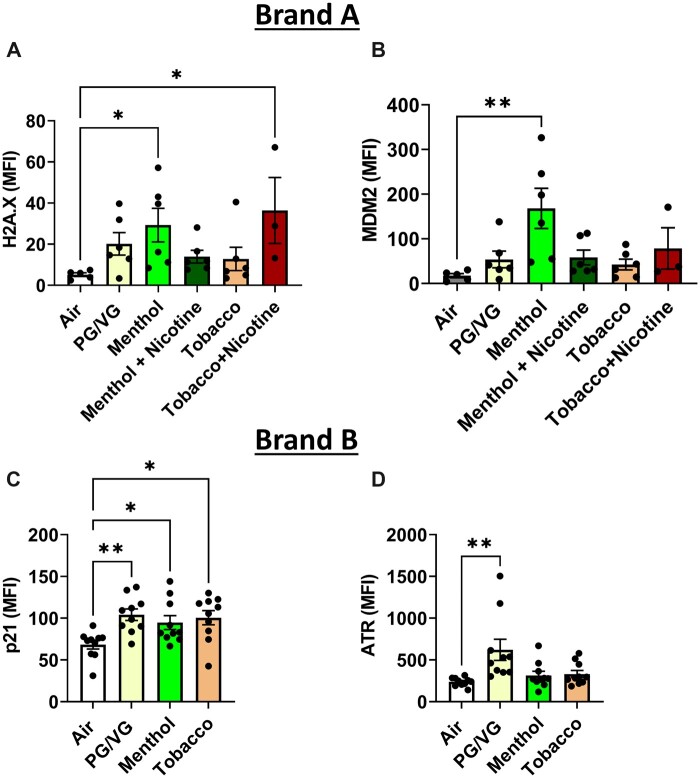
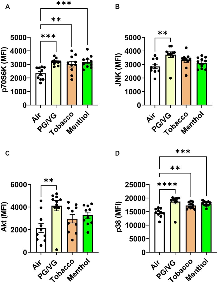
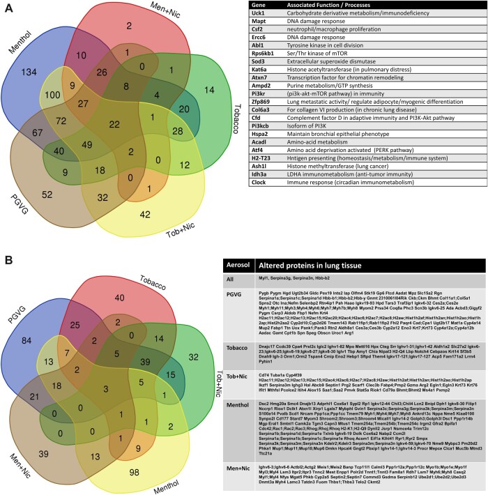
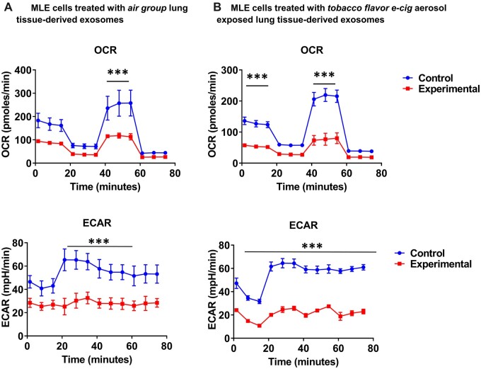
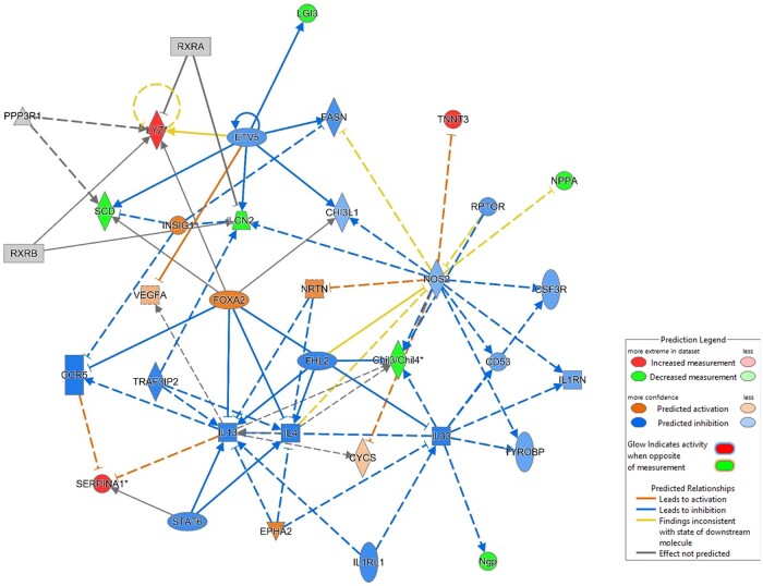
Menthol and tobacco flavors are available for almost all tobacco products, including electronic cigarettes (e-cigs). These flavors are a mixture of chemicals with overlapping constituents. There are no comparative toxicity studies of these flavors produced by different manufacturers. We hypothesized that acute exposure to menthol and tobacco-flavored e-cig aerosols induces inflammatory, genotoxicity, and metabolic responses in mouse lungs. We compared two brands, A and B, of e-cig flavors (PG/VG, menthol, and tobacco) with and without nicotine for their inflammatory response, genotoxic markers, and altered genes and proteins in the context of metabolism by exposing mouse strains, C57BL/6J (Th1-mediated) and BALB/cJ (Th2-mediated). Brand A nicotine-free menthol exposure caused increased neutrophils and differential T-lymphocyte influx in bronchoalveolar lavage fluid and induced significant immunosuppression, while brand A tobacco with nicotine elicited an allergic inflammatory response with increased Eotaxin, IL-6, and RANTES levels. Brand B elicited a similar inflammatory response in menthol flavor exposure. Upon e-cig exposure, genotoxicity markers significantly increased in lung tissue. These inflammatory and genotoxicity responses were associated with altered NLRP3 inflammasome and TRPA1 induction by menthol flavor. Nicotine decreased surfactant protein D and increased PAI-1 by menthol and tobacco flavors, respectively. Integration of inflammatory and metabolic pathway gene expression analysis showed immunometabolic regulation in T cells via PI3K/Akt/p70S6k-mTOR axis associated with suppressed immunity/allergic immune response. Overall, this study showed the comparative toxicity of flavored e-cig aerosols, unraveling potential signaling pathways of nicotine and flavor-mediated pulmonary toxicological responses, and emphasized the need for standardized toxicity testing for appropriate premarket authorization of e-cigarette products.

Keywords: ENDS; e-cigarettes; flavors; hypersensitivity; immunometabolism; menthol; nicotine; tobacco.


Figures

Figure 1.
Figure 1.
A, Venn diagram depicting the number of detected chemicals tobacco and menthol flavors. E-liquids, tobacco and menthol, from two brands (A and B) were analyzed by GC-MS for their chemical composition. Tobacco and menthol flavors have distinctly 65 and 58 flavor constituents with 33 being common constituents. B, Venn diagram depicting the number of detected volatile organic compounds in tobacco and menthol flavored aerosols. Aerosols, tobacco and menthol, from two brands (A and B), were analyzed by GC-MS for their chemical composition. Tobacco and menthol flavors had two and seven constituents exclusively, with 82 being common overlapping constituents. C, Serum cotinine levels of mice exposed to menthol and tobacco aerosols with and without nicotine. Cotinine levels in serum quantified in C57BL/6J and BALB/cJ male and female mice exposed to air, PG/VG, menthol 0 and 6 mg nicotine, and tobacco 0 and 6 mg nicotine of brand A. Serum from menthol and tobacco groups with nicotine were collected at 2 and 24 h post 3-day exposure (N = 3–8 per group, ****p < .0001, t-test).
Figure 2.
Figure 2.
Differential cell counts in bronchoalveolar lavage fluid by exposure to brand A, PG/VG, menthol, and tobacco flavors with and without nicotine in C57BL/6J and BALB/cJ mice. C57BL/6J and BALB/cJ male and female mice were exposed to PG/VG, menthol, menthol 6 mg nicotine, tobacco, and tobacco 6 mg nicotine 2 h/day for three consecutive days in SCIREQ whole-body exposure chamber. Mice were euthanized ∼24 h postexposure and the BALF was collected. A and B, Total cell counts were performed by staining with AO/PI dye. C and D, Macrophages (F4/80+), E and F, neutrophils (Ly6B.2), G and H, Th lymphocytes (CD4+), and I and J, Tc lymphocytes (CD8+) were assessed as percentages of CD45+ parent cell population by flow cytometry. Data are shown as mean ± SEM. *p < .05, **p < .01, and ***p < .001 versus respective air group per strain (2-way ANOVA) (N = 4–8 per group).
Figure 3.
Figure 3.
A, Exposure to brand A, PG/VG, menthol, and menthol with nicotine flavors in C57BL/6J and BALB/cJ mice elicited a regulatory inflammatory cytokine response in bronchoalveolar lavage. C57BL/6J and BALB/cJ, male and female, mice were exposed to PG/VG, menthol, and menthol 6 mg nicotine, 2 h/day for three consecutive days in SCIREQ whole-body exposure chamber. Mice were euthanized ∼24 h postexposure and bronchoalveolar lavage was collected by instilling 0.6 ml 3× and pooled. Cytokine levels were measured by Luminex. a, RANTES; b, KC; c, MIP-1α; d, IL-10; i, IL-6; f, IL2; g, IL-3; h, IL-17A; i, Eotaxin; j, IL-1α; k, IL5; l, IFNγ; m, IL-1β; n, MIP-1β; o, GM-CSF; p, IL12p40; q, IL12p70; r, TNFα; s, IL9; t, G-CSF; u, IL-4; v, IL-13; w, MCP-1. Data are shown as mean ± SEM. *p < .05, **p < .01, ***p < .001 versus respective air group per strain (2-way ANOVA) (N = 7–8 per group). B, Exposure to brand A, PG/VG, menthol, and tobacco flavors with and without nicotine in C57BL/6J and BALB/cJ mice elicited a cytokine response in mouse lung homogenate. C57BL/6J and BALB/cJ, male and female, mice were exposed to PG/VG, menthol, menthol 6 mg nicotine, tobacco, and tobacco 6 mg nicotine 2 h/day for three consecutive days in SCIREQ whole-body exposure chamber. Mice were euthanized ∼24 h postexposure and lung tissues were collected. Homogenized lungs in RIPA buffer were used to determine inflammatory mediators by Luminex and normalized by total protein (BCA assay). a, IL-2; b, IL-9; c, IL-4. Datat are shown as mean ± SEM. *p < .05 versus respective air group per strain (2-way ANOVA) (N = 7–8 per group).
Figure 4.
Figure 4.
Exposure to brand A, PG/VG, tobacco, and tobacco with nicotine flavors in C57BL/6J and BALB/cJ mice elicited a regulatory inflammatory cytokine response in bronchoalveolar lavage. C57BL/6J and BALB/cJ, male and female mice, were exposed to PG/VG, tobacco, and tobacco 6 mg nicotine 2 h/day for three consecutive days in SCIREQ whole-body exposure chamber. Mice were euthanized ∼24 h postexposure and bronchoalveolar lavage was collected by instilling 0.6 ml 3× and pooled. Cytokine levels were measured by Luminex. A, RANTES; B, KC; C, MIP-1α; D, IL-10; E, IL-6; F, IL2; G, IL-3; H, IL-17A; I, Eotaxin; J, IL-1α; K, IL5; L, IFNγ; M, IL-1β; N, MIP-1β; O, GM-CSF; P, IL12p40; Q, IL12p70; R, TNFα; S, IL9; T, G-CSF; U, IL-4; V, IL-13; W, MCP-1. Data are presented as mean ± SEM. *p < .05, **p < .01, and ***p < .001 versus respective air group per strain (2-way ANOVA) (N = 7–8 per group).
Figure 5.
Figure 5.
A, Differential cell counts in bronchoalveolar lavage by exposure to brand B, PG/VG, menthol, and tobacco flavors in C57BL/6J. C57BL/6J male and female mice were exposed to PG/VG, menthol, menthol 6 mg nicotine, tobacco, and tobacco 6 mg nicotine 2 h/day for three consecutive days in SCIREQ whole-body exposure chamber. Mice were euthanized ∼24 h postexposure and the BALF was collected. a, Macrophages (F4/80+); b, neutrophils (Ly6B.2); c, Th lymphocytes (CD4+); d, Tc lymphocytes (CD8+), were assessed as percentages of CD45+ parent cell population by flow cytometry based on the (e) total cell counts determined by staining with AO/PI dye. Data are shown as mean ± SEM. *p < .05, **p < .01, ***p < .001 versus respective air group per strain (2-way ANOVA) (N = 8 per group). B, Exposure to brand B, PG/VG, menthol, and tobacco flavors in C57BL/6J mice elicited a differential inflammatory cytokine response in bronchoalveolar lavage. C57BL/6J male and female mice were exposed to PG/VG, menthol, menthol 6 mg nicotine, tobacco, and tobacco 6 mg nicotine 2 h/day for three consecutive days in SCIREQ whole-body exposure chamber. Mice were euthanized ∼24 h postexposure and bronchoalveolar lavage was collected by instilling 0.6 ml 3× and pooled. Cytokine levels were measured by Luminex. a, MIP-1β; b, IL-12p40; c, IL-12p70; d, IL-3; e, RANTES; f, IL-6; g, IFNγ; h, IL-4; i, IL-10; j, GM-CSF. Data are shown as mean ± SEM. *p < .05, **p < .01, ***p < .001, and ****p < .0001 versus respective air group per strain (2-way ANOVA) (N = 8 per group).
Figure 6.
Figure 6.
ENDS flavors induced acute lung injury-associated biomarkers. C57BL/6J male and female mice were exposed to PG/VG, menthol, menthol 6 mg nicotine, tobacco, and tobacco 6 mg nicotine 2 h/day for three consecutive days in SCIREQ whole-body exposure chamber. Mice were euthanized ∼24 h postexposure, and lung tissues were homogenized for immunoblotting by SDS-PAGE. A, TRPA-1; B, NLRP3; C, SP-D; D, PAI-1 protein abundance were measured and normalized to GAPDH and β-actin loading controls. Data shown for respective bands and the densitometry values were plotted as mean ± SEM. *p < .05, **p < .01, and ***p < .001 versus respective air group per strain (2-way ANOVA) (N = 3–4 per group).
Figure 7.
Figure 7.
Genotoxicity by exposure to PG/VG, menthol, and tobacco flavors in C57BL/6J mouse lung tissue. C57BL/6J male and female mice were exposed to PG/VG, menthol, menthol 6 mg nicotine, tobacco, and tobacco 6 mg nicotine 2 h/day for three consecutive days in SCIREQ whole-body exposure chamber. Mice were euthanized ∼24 h postexposure and mouse lung tissues were homogenized and measured genotoxicity markers, A, H2AX; B, MDM2; C, p21; and D, ATR by measuring net median florescence intensity (MFI) normalized to total protein. Data are shown as mean ± SEM. *p < .05, **p < .01, and ***p < .001 versus respective air group per strain (1-way ANOVA) (N = 3–10 per group).
Figure 8.
Figure 8.
Exposure to PG/VG, menthol, and tobacco flavors in C57BL/6J mouse lung tissue induced PI3K-Akt-mTOR pathway signaling. C57BL/6J male and female mice were exposed to PG/VG, menthol, menthol 6 mg nicotine, tobacco, and tobacco 6 mg nicotine 2 h/day for three consecutive days in SCIREQ whole-body exposure chamber. Mice were euthanized ∼24 h postexposure and mouse lung tissues were homogenized and measured cell signaling pathway markers, A, P70S6K; B, JNK; C, Akt; and D, p38 by measuring net median florescence intensity (MFI) normalized to total protein. Data shown as mean ± SEM. *p < .05, **p < .01, and ***p < .001 versus respective air group per strain (1-way ANOVA) (N = 6–10 per group).
Figure 9.
Figure 9.
A, Inflammatory and Metabolic gene expression alterations by acute exposure to PG/VG, menthol, menthol with nicotine, tobacco, tobacco with nicotine aerosols. C57BL/6J male and female mice were exposed to three days (2 h/day) air, PG/VG, menthol, menthol + nicotine, tobacco, and tobacco + nicotine aerosols. RNA isolated from lungs were hybridized with Nanostring codesets and the inflammatory and metabolic gene alterations were determined. Genes that ±1.5-fold change altered significantly compared to the Air group were compared. p < .05, 1-way ANOVA, N = 6 per group. B, Protein alterations by acute exposure to PG/VG, menthol, menthol with nicotine, tobacco, tobacco with nicotine aerosols. C57BL/6J male and female mice were exposed to three days (2h/day) air, PG/VG, menthol, menthol + nicotine, tobacco, and tobacco flavor + nicotine aerosols. Protein changes were determined by proteomics analysis and presented in a Venn diagram. Proteins significantly altered by 1.5-fold are listed (p < .05 vs air group), N = 3 per group.
Figure 10.
Figure 10.
Mitochondrial respiration is affected by extracellular vesicles derived from the lungs of mice exposed to brand B tobacco flavor. C57BL/6J male and female mice were exposed air and tobacco-flavored e-cig aerosols 2 h/day for three consecutive days in SCIREQ whole-body exposure chamber. Mice were euthanized ∼24 h postexposure and exosomes were isolated from the lung tissue. MLE15 cells were grown in Seahorse plates and treated with isolated exosomes. Twenty-four hours later, Sea horse mitostress assay was run and oxygen consumption rate (OCR) extracellular acidification rate (ECAR) parameters were acquired, and normalized by total cells. A, MLE cells treated with air group extracellular vesicles (EVs) and B, MLE cells treated with tobacco flavor exposed EVs. t-test, ***p < .001 versus control, N = 3.
Figure 11.
Figure 11.
Potential pathways of immunometabolic and allergic inflammatory response via PI3K-Akt-mTOR. C57BL/6J male and female representative canonical pathway of menthol e-cig flavor exposure-induced lung response by ingenuity pathway analysis of proteomics data demonstrating inhibition of inflammatory pathways and upregulating immunoregulatory pathways.

Similar articles

Cited by

References

    1. Alam R., York J., Boyars M., Stafford S., Grant J., A., Lee J., Forsythe P., Sim T., Ida N. (1996). Increased MCP-1, RANTES, and MIP-1alpha in bronchoalveolar lavage fluid of allergic asthmatic patients. Am. J. Respir. Crit. Care Med. 153, 1398–1404. - PubMed
    1. Alexander L. E. C., Bellinghausen A. L., Eakin M. N. (2020). What are the mechanisms underlying vaping-induced lung injury? J. Clin. Invest. 130, 2754–2756. - PMC - PubMed
    1. Ali F. R. M., Seaman E. L., Diaz M. C., Ajose J., King B. A. (2022). Trends in unit sales of cooling flavoured e-cigarettes, USA, 2017-2021. Tob. Control. 1052–1056. doi:10.1136/tc-2022-057395. - DOI - PubMed
    1. Birrell M. A., Belvisi M. G., Grace M., Sadofsky L., Faruqi S., Hele D. J., Maher S., A., Freund-Michel V., Morice A. H. (2009). TRPA1 agonists evoke coughing in guinea pig and human volunteers. Am. J. Respir. Crit. Care Med. 180, 1042–1047. - PMC - PubMed
    1. Bitzer Z., T., Goel R., Reilly S. M., Foulds J., Muscat J., Elias R. J., Richie J. P. Jr (2018). Effects of solvent and temperature on free radical formation in electronic cigarette aerosols. Chem. Res. Toxicol. 31, 4–12. - PMC - PubMed

Publication types