Utilizing 3D printing to assist pre-procedure planning of transjugular intrahepatic portosystemic shunt (TIPS) procedures: a pilot study
- PMID: 37052816
- PMCID: PMC10099647
- DOI: 10.1186/s41205-023-00176-w
Utilizing 3D printing to assist pre-procedure planning of transjugular intrahepatic portosystemic shunt (TIPS) procedures: a pilot study
Abstract
Background: 3D (three-dimensional) printing has been adopted by the medical community in several ways, procedure planning being one example. This application of technology has been adopted by several subspecialties including interventional radiology, however the planning of transjugular intrahepatic portosystemic shunt (TIPS) placement has not yet been described. The impact of a 3D printed model on procedural measures such as procedure time, radiation exposure, intravascular contrast dosage, fluoroscopy time, and provider confidence has also not been reported.
Methods: This pilot study utilized a quasi-experimental design including patients who underwent TIPS. For the control group, retrospective data was collected on patients who received a TIPS prior to Oct 1, 2020. For the experimental group, patient-specific 3D printed models were integrated in the care of patients that received TIPS between Oct 1, 2020 and April 15, 2021. Data was collected on patient demographics and procedural measures. The interventionalists were surveyed on their confidence level and model usage following each procedure in the experimental group.
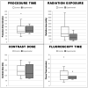
Results: 3D printed models were created for six TIPS. Procedure time (p = 0.93), fluoroscopy time (p = 0.26), and intravascular contrast dosage (p = 0.75) did not have significant difference between groups. Mean radiation exposure was 808.8 mGy in the group with a model compared to 1731.7 mGy without, however this was also not statistically significant (p = 0.09). Out of 11 survey responses from interventionists, 10 reported "increased" or "significantly increased" confidence after reviewing the 3D printed model and all responded that the models were a valuable tool for trainees.
Conclusions: 3D printed models of patient anatomy can consistently be made using consumer-level, desktop 3D printing technology. This study was not adequately powered to measure the impact that including 3D printed models in the planning of TIPS procedures may have on procedural measures. The majority of interventionists reported that patient-specific models were valuable tools for teaching trainees and that confidence levels increased as a result of model inclusion in procedure planning.
Keywords: 3D printing; Anatomic segmentation; Pilot study.; Procedure planning; TIPS.
© 2023. The Author(s).
Conflict of interest statement
The authors declare that they have no competing interests.
Figures


Similar articles
-
Transjugular intrahepatic portosystemic shunt placement: portal vein puncture guided by 3D/2D image registration of contrast-enhanced multi-detector computed tomography and fluoroscopy.Abdom Radiol (NY). 2020 Nov;45(11):3934-3943. doi: 10.1007/s00261-020-02589-1. Abdom Radiol (NY). 2020. PMID: 32451673 Free PMC article.
-
Image fusion-guided portal vein puncture during transjugular intrahepatic portosystemic shunt placement.Diagn Interv Imaging. 2016 Nov;97(11):1095-1102. doi: 10.1016/j.diii.2016.06.015. Epub 2016 Aug 5. Diagn Interv Imaging. 2016. PMID: 27503116
-
Intravascular US-Guided Portal Vein Access: Improved Procedural Metrics during TIPS Creation.J Vasc Interv Radiol. 2016 Aug;27(8):1140-7. doi: 10.1016/j.jvir.2015.12.002. Epub 2016 Feb 4. J Vasc Interv Radiol. 2016. PMID: 26852944
-
Standardizing evaluation of patient-specific 3D printed models in surgical planning: development of a cross-disciplinary survey tool for physician and trainee feedback.BMC Med Educ. 2022 Aug 12;22(1):614. doi: 10.1186/s12909-022-03581-7. BMC Med Educ. 2022. PMID: 35953840 Free PMC article. Review.
-
Complications of transjugular intrahepatic portosystemic shunt (TIPS) in the era of the stent graft - What the interventionists need to know?Eur J Radiol. 2021 Nov;144:109986. doi: 10.1016/j.ejrad.2021.109986. Epub 2021 Sep 29. Eur J Radiol. 2021. PMID: 34619618 Review.
Cited by
-
Investigation of bending angle algorithm and path planning for puncture needles in transjugular intrahepatic portosystemic shunt.Biomed Eng Online. 2025 May 27;24(1):66. doi: 10.1186/s12938-025-01397-2. Biomed Eng Online. 2025. PMID: 40426132 Free PMC article.
References
-
- Youssef R, Spradling K, Yoon R et al. “Applications of three-dimensional printing technology in urological practice.” BJU Int(2015; 116):697–702. - PubMed
-
- Malik H, Darwood A, Shaunak S et al. “Three-dimensional printing in surgery: a review of current surgical applications.” Journal of Surgical Research (2015):119; 512–522. - PubMed
-
- Chang D, Tummala S, Sotero D et al. “Three-Dimensional Printing for Procedure Rehersal/Simulation/Planning in Interventional Radiology.” Techniques in Vascular and Inteventional Radiology (2018). - PubMed
Grants and funding
LinkOut - more resources
Full Text Sources
Miscellaneous
