. 2023 Jun 26;35(7):2615-2634.
doi: 10.1093/plcell/koad108.

Blue light promotes ascorbate synthesis by deactivating the PAS/LOV photoreceptor that inhibits GDP-L-galactose phosphorylase

Affiliations

Blue light promotes ascorbate synthesis by deactivating the PAS/LOV photoreceptor that inhibits GDP-L-galactose phosphorylase

Céline Bournonville et al. Plant Cell. .

Abstract

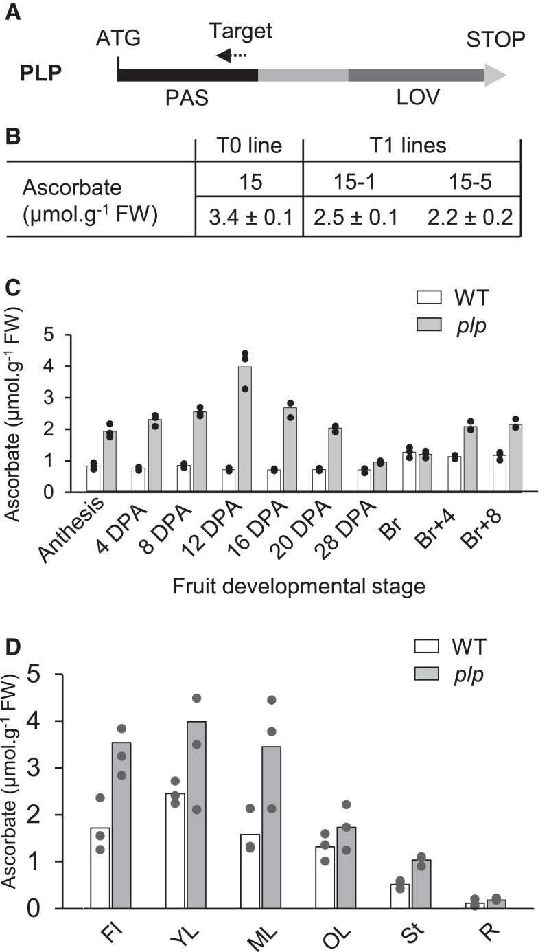
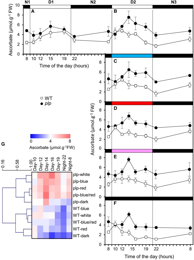
Ascorbate (vitamin C) is an essential antioxidant in fresh fruits and vegetables. To gain insight into the regulation of ascorbate metabolism in plants, we studied mutant tomato plants (Solanum lycopersicum) that produce ascorbate-enriched fruits. The causal mutation, identified by a mapping-by-sequencing strategy, corresponded to a knock-out recessive mutation in a class of photoreceptor named PAS/LOV protein (PLP), which acts as a negative regulator of ascorbate biosynthesis. This trait was confirmed by CRISPR/Cas9 gene editing and further found in all plant organs, including fruit that accumulated 2 to 3 times more ascorbate than in the WT. The functional characterization revealed that PLP interacted with the 2 isoforms of GDP-L-galactose phosphorylase (GGP), known as the controlling step of the L-galactose pathway of ascorbate synthesis. The interaction with GGP occurred in the cytoplasm and the nucleus, but was abolished when PLP was truncated. These results were confirmed by a synthetic approach using an animal cell system, which additionally demonstrated that blue light modulated the PLP-GGP interaction. Assays performed in vitro with heterologously expressed GGP and PLP showed that PLP is a noncompetitive inhibitor of GGP that is inactivated after blue light exposure. This discovery provides a greater understanding of the light-dependent regulation of ascorbate metabolism in plants.


Conflict of interest statement

Conflict of interest statement. None declared.

Figures

Figure 1.
Figure 1.
Identification of the mutation responsible for the ascorbate-enriched fruit phenotype. A) Identification of the chromosome associated with the ascorbate-enriched phenotype. Pattern of the mutation allelic frequencies obtained in the mutant and WT-like bulks are represented along tomato chromosomes. The plot represents allelic frequencies (y axis) against genome positions (x axis). A sliding window of 5 SNPs was used. The x axis displays the 12 tomato chromosomes; the arrow indicates the peak of allelic frequency (AF) of the chromosome 5 region carrying the putative causal mutations, since it displayed an AF > 0.95 in the ascorbate bulk and an AF < 0.4 in the WT-like bulk. B) Fine mapping of the causal mutation using the BC1F2 population. Recombinant analysis of 44 BC1F2 individuals displaying the ascorbate-enriched phenotype allowed us to locate the causal mutation at position 1,610,253 nucleotides. Marker positions are indicated by triangles. The number of recombinants is shown below the position of the markers. C) A single nucleotide transversion, G to A at position 1,610,253 in the Solyc05g007020 of the last codon of the fourth exon sequence, led to a change from glutamine to STOP codon. The sequence corresponding to the PAS and LOV domains is stained in the first exon and the third to the seventh exons, respectively.
Figure 2.
Figure 2.
Validation of PAS/LOV as the candidate gene involved in regulating ascorbate content in developing fruit and several tomato plant organs. A) Schematic representation of PLP showing its PAS and LOV domains. The dashed arrow in the PAS domain indicates the position of the target sequence for the CRISPR/Cas9 construct. B) Ascorbate in red ripe fruit (means ± SD, n = 4) of WT, T0 line 15, and progeny T1 lines 15-1 and 15-4. C) Ascorbate content in fruit of WT and plp mutant plants during development, from anthesis to ripeness. D) Ascorbate content in flowers, leaves at 3 stage of development, stem and roots of the 15-5 line, and control 1-mo-old plants. Data are the means (histograms) and 3 biological replicates representing 3 individual plants per organ and 3 organs per plant, except for anthesis (100 organs) and at 4 DPA (20 organs). PLP, PAS/LOV; DPA, days postanthesis; Br, breaker stage; FI, flowers; YL, young leaf; ML, mature leaf; OL, old leaf; St, stem; R, roots.
Figure 3.
Figure 3.
Changes in ascorbate and GDP-L-galactose phosphorylase and PAS/LOV mRNA during a day and night cycle in plp mutant and WT plants. The plp mutant (T2 line 15-5) and WT plants were cultured in the greenhouse for 1 mo. The night before the beginning of the experiment, all plants were moved outside and maintained under natural light conditions during 32 h. This experiment was carried out twice, on May 19 and July 10, 2018; they both lead remarkably to the same results. Here is presented the data obtained on July 10. A) Ambient temperature (diamonds) and light intensity (bars). B) Ascorbate content. C)PLP mRNA abundance. D)GGP1 mRNA abundance. Data are expressed as means ± SD of a total 3 mature leaves from 3 individual plants from the 15-5 plp line and WT control.
Figure 4.
Figure 4.
Subcellular localization of PAS/LOV and GDP-L-galactose phosphorylase and their interaction. A) Diagram of the full-length PLP and the truncated PLP (trPLP). B) Localization of free GFP and 35S-GFP-fused proteins transiently expressed in N. benthamiana leaves 4 d after agroinfiltration, co-transformed with nuclear NLS-mcherry as nuclear marker. Scale bar = 50 µm. C) Analysis of PLP-GGP1/2 interactions and its light dependency in a heterologous mammalian split transcription factor system. Fifty thousand HEK-293 T cells were seeded in 24-well plates and transfected after 24 h with the plasmids pMZ1214, pMZ1215, pMZ1216, pMZ1217, pMZ1218, pMZ1219, pMZ1240, pMZ1241, pSAM, pRSET, and pKM006. Twenty-four hours posttransfection, the medium was exchanged by fresh medium and the cells were illuminated at 455 nm light (10 μmol m−2s−1) or kept in the dark for 24 h prior to SEAP quantification. Data are represented as means (histograms) and four biological replicates (dots). HEK-293 T, human embryonic kidney cells; SEAP, secreted alkaline phosphatase.
Figure 5.
Figure 5.
Effect of light on ascorbate evolution in WT and plp mutant leaves during a day–night cycle. A) In leaves of plants grown in a greenhouse then transferred to a growth chamber under a white light (with visible wavelengths from 420 nm to 760 nm) intensity of 260 to 270 µmol.m−2.s−1 for 24 h. B) Following cycle, still under white light. C) Following cycle under blue light (with a peak wavelength of 440 nm). D) Following cycle under red light (with peak wavelengths of 660 nm and 730 nm). E) Following cycle under blue (50%) and red (40%) light. F) Following cycle in the dark. G) Heat map representing a clustering analysis performed with mean values in MEV4.9. Columns correspond to time, and lines correspond to clustered content of ascorbate based on Pearson's correlation coefficient. All data shown in A to F) are expressed as means ± SD (n = 4).
Figure 6.
Figure 6.
In vitro inhibition of GDP-L-galactose phosphorylase by PAS/LOV and effect of blue light on PAS/LOV. A) Relation between the PLP/GGP ratio and the inhibition of GGP. B) Hanes–Woolf plot GDP-L-galactose phosphorylase inhibition by PLP. GDP-glucose at concentrations of 12, 30, 60, 120, and 300 µM was used as substrate. C) Effect of blue light exposure duration on GGP inhibition by PLP. The light was applied before mixing the 2 proteins. All data shown are expressed as means (dashed lines) and technical duplicates (dots).
Figure 7.
Figure 7.
Schematic model describing the activation of ascorbate synthesis by blue light. Newly synthesized PAS/LOV protein binds GDP-L-galactose phosphorylase unless deactivated by blue light. Its deactivated form is stable for several hours, while its active form irreversibly inhibits its target, possibly leading to its degradation. PLP, PAS/LOV; GGP, GDP-L-galactose phosphorylase.

Comment in

References

    1. Ahmad M, Cashmore AR. HY4 gene of A-Thaliana encodes a protein with characteristics of a blue-light photoreceptor. Nature 1993:366(6451):162–166. 10.1038/366162a0 - DOI - PubMed
    1. Alimohammfadi M, de Silva K, Ballu C, Ali N, Khodakovskaya MV. Reduction of inositol (1,4,5)–trisphosphate affects the overall phosphoinositol pathway and leads to modifications in light signalling and secondary metabolism in tomato plants. J Exp Bot. 2012:63(2):825–835. 10.1093/jxb/err306 - DOI - PMC - PubMed
    1. Bartoli CG, Tambussi EA, Diego F, Foyer CH. Control of ascorbic acid synthesis and accumulation and glutathione by the incident light red/far red ratio in Phaseolus vulgaris leaves. FEBS Lett. 2009:583(1):118–122. 10.1016/j.febslet.2008.11.034 - DOI - PubMed
    1. Belouah I, Bénard C, Denton A, Blein-Nicolas M, Balliau T, Teyssier E, Gallusci P, Bouchez O, Usadel B, Zivy M, et al. . Transcriptomic and proteomic data in developing tomato fruit. Data Brief 2020:28:105015. 10.1016/j.dib.2019.105015 - DOI - PMC - PubMed
    1. Bergmeyer HU. Methods of enzymatic analysis. Weinheim: VCH. 1987

Publication types