. 2023 May 1;324(5):E461-E475.
doi: 10.1152/ajpendo.00059.2023. Epub 2023 Apr 13.

Central androgen action reverses hypothalamic astrogliosis and atherogenic risk factors induced by orchiectomy and high-fat diet feeding in male mice

Affiliations

Central androgen action reverses hypothalamic astrogliosis and atherogenic risk factors induced by orchiectomy and high-fat diet feeding in male mice

Mauricio D Dorfman et al. Am J Physiol Endocrinol Metab. .

Abstract

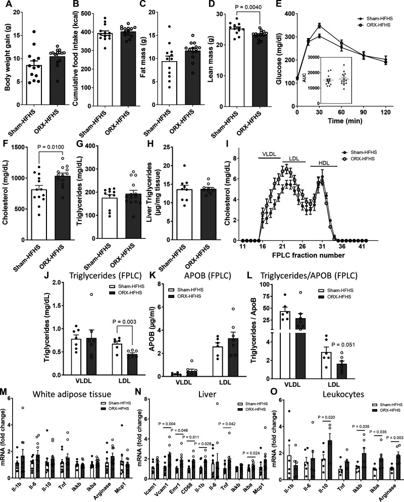
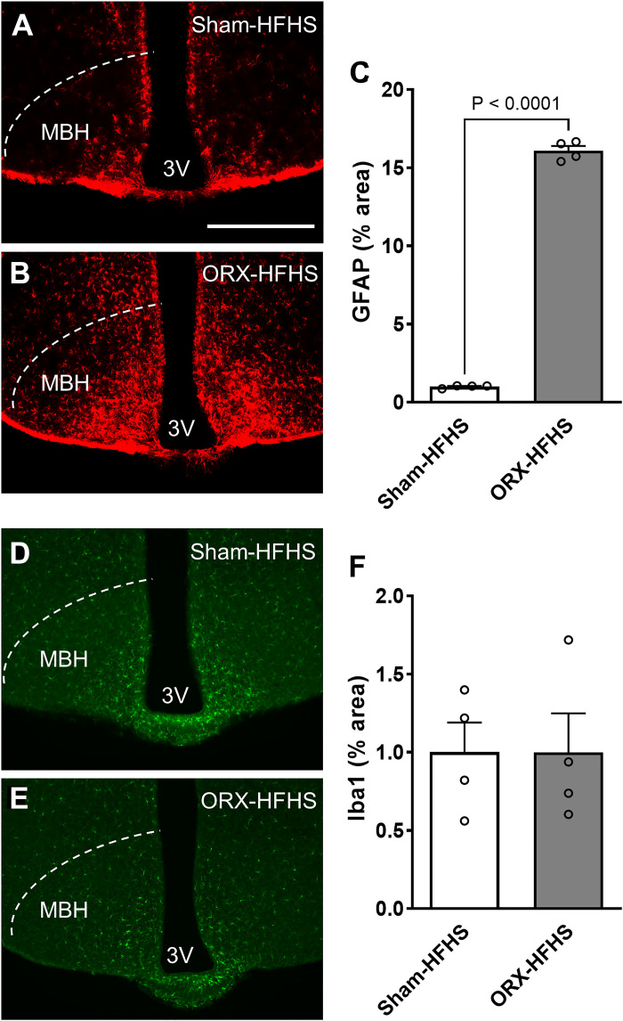
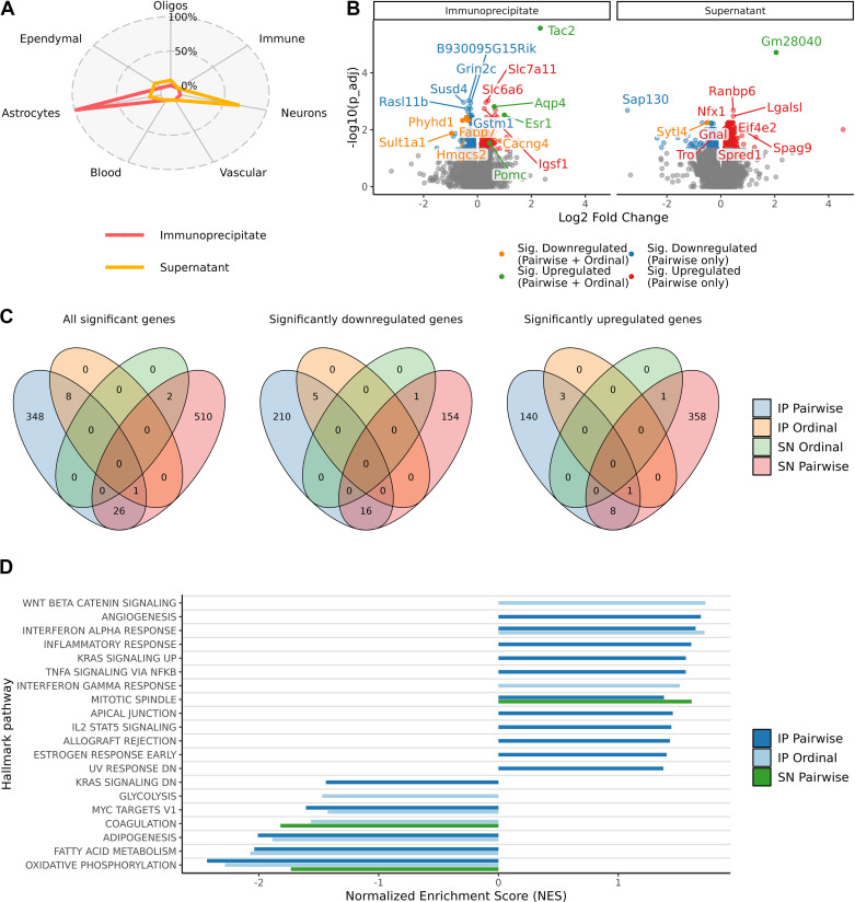
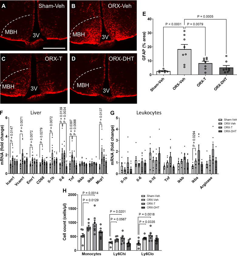
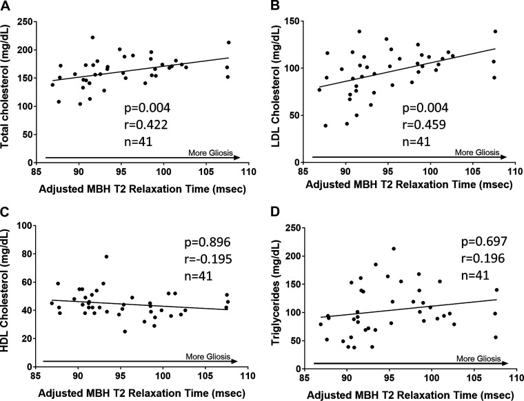
Hypogonadism in males confers elevated cardiovascular disease (CVD) risk by unknown mechanisms. Recent radiological evidence suggests that low testosterone (T) is associated with mediobasal hypothalamic (MBH) gliosis, a central nervous system (CNS) cellular response linked to metabolic dysfunction. To address mechanisms linking CNS androgen action to CVD risk, we generated a hypogonadal, hyperlipidemic mouse model with orchiectomy (ORX) combined with hepatic PCSK9 overexpression. After 4 wk of high-fat, high-sucrose diet (HFHS) consumption, despite equal body weights and glucose tolerance, androgen-deficient ORX mice had a more atherogenic lipid profile and increased liver and leukocyte inflammatory signaling compared with sham-operated control mice. Along with these early CVD risk indicators, ORX markedly amplified HFHS-induced astrogliosis in the MBH. Transcriptomic analysis further revealed that ORX and high-fat diet feeding induced upregulation of inflammatory pathways and downregulation of metabolic pathways in hypothalamic astrocytes. To interrogate the role of sex steroid signaling in the CNS in cardiometabolic risk and MBH inflammation, central infusion of T and dihydrotestosterone (DHT) was performed on ORX mice. Central DHT prevented MBH astrogliosis and reduced the liver inflammatory signaling and monocytosis induced by HFHS and ORX; T had a partial protective effect. Finally, a cross-sectional study in 41 adult men demonstrated a positive correlation between radiological evidence of MBH gliosis and plasma lipids. These findings demonstrate that T deficiency in combination with a Western-style diet promotes hypothalamic gliosis concomitant with increased atherogenic risk factors and provide supportive evidence for regulation of lipid metabolism and cardiometabolic risk determinants by the CNS action of sex steroids.NEW & NOTEWORTHY This study provides evidence that hypothalamic gliosis is a key early event through which androgen deficiency in combination with a Western-style diet might lead to cardiometabolic dysregulation in males. Furthermore, this work provides the first evidence in humans of a positive association between hypothalamic gliosis and LDL-cholesterol, advancing our knowledge of CNS influences on CVD risk progression.

Keywords: astrocyte; cardiovascular risk; hypogonadism; hypothalamic gliosis; testosterone.


Conflict of interest statement

T. Monfeuga, A. Chandran, and T. H. Meek are employees of Novo Nordisk Ltd. None of the other authors has any conflicts of interest, financial or otherwise, to disclose.

Figures

Figure 1.
Figure 1.
Orchiectomized (ORX) mice exposed to high-fat, high-sucrose diet (HFHS) for 4 wk have increased markers of cardiovascular disease (CVD) risk despite no metabolic changes. Metabolic and inflammatory parameters measured in sham-operated (Sham) and ORX mice injected with AAV- D377Y-mPCSK9 and fed 4 wk of HFHS. A–D: body weight gain (A), cumulative food intake (B), fat mass (C), and lean mass (D) (n = 13 or 14/group). E: intraperitoneal glucose tolerance test (2 g/kg). Inset, area under the curve (AUC). F–H: plasma cholesterol (F), plasma triglycerides (TG) (G), and liver TG content (H) (n = 9 or 10/group). I: lipoprotein distribution of cholesterol measured by fast-phase liquid chromatography (FPLC) (pooled plasma of 2 or 3 mice/n, n = 4 and n = 6 for Sham-HFHS and ORX-HFHS, respectively). Fractions 15–20 contain VLDL, fractions 21–27 contain LDL, and fractions 28–35 contain HDL. J–L: TG (J) and apolipoprotein B (APOB) (K) measured in the VLDL peak fraction (17) and the LDL peak fraction (22) and TG measured in VLDL and LDL fractions normalized to APOB (L), providing an estimate of TG molecules/lipoprotein particle. M–O: mRNA levels of inflammatory markers in white adipose tissue (M), liver (N), and circulating leukocytes (O). In A–L, data are presented as means ± SE. In M–O, data are presented as fold change relative to Sham-HFHS. HDL, high-density lipoprotein; LDL, low-density lipoprotein; VLDL, very low-density lipoprotein.
Figure 2.
Figure 2.
Orchiectomized (ORX) mice exposed to high-fat, high-sucrose diet (HFHS) have increased astrocytosis but not microgliosis in the mediobasal hypothalamus. A and B: representative images showing glial fibrillary acidic protein (GFAP) immunoreactivity in the mediobasal hypothalamus (MBH) of sham-operated (Sham)-HFHS (A) and ORX-HFHS (B) mice. 3V, third ventricle. Scale bar, 500 µm. C: quantification of GFAP-positive area percentage in the MBH from 6 sections per animal. D and E: representative images showing Iba1 immunoreactivity in the MBH of Sham-HFHS (D) and ORX-HFHS (E) mice. F: quantification of Iba1-positive area percentage in the MBH from 6 sections per animal. Data are presented as means ± SE of 4 mice per group.
Figure 3.
Figure 3.
Androgen deficiency in combination with high-fat diet (HFD) feeding promotes a proinflammatory and hypometabolic transcriptomic profile in hypothalamic astrocytes. A: cell type proportion estimations with the deconvolution software MuSiC for each cell fraction [immunoprecipitate (IP) and supernatant (SN)] performed in hypothalamic samples from 3 groups of Aldh1l1eGFP-L10a mice [sham operated (Sham)-regular chow (Chow), Sham-HFD, and orchiectomized (ORX)-HFD]. B: volcano plot representing the significantly altered genes (blue = down; red = up) in the pairwise comparison (Sham-HFD vs. ORX-HFD) and with the additional ordinal regression analysis (Sham-Chow > Sham-HFD > ORX-HFD = orange or Sham-Chow < Sham-HFD < ORX-HFD = green) in both the IP (left) and SN (right) fractions. Genes with the most significant changes are labeled. C: Venn diagrams indicating overlap of genes with altered expression levels in the different analyses performed. Left: all genes. Center: downregulated genes. Right: upregulated genes. D: gene set enrichment analysis showing over- and underrepresentation of expression changes in hallmark pathways (all genes included). The top 10 over- and underenriched in each analysis (adjusted P value ≤ 0.05) are presented (no pathways were significantly enriched for the SN Ordinal test). Data obtained from individual hypothalamic lysates (Sham-Chow n = 7, Sham-HFD n = 6, ORX-HFD n = 7).
Figure 4.
Figure 4.
Chronic central infusion of testosterone (T) and dihydrotestosterone (DHT) reduces hypothalamic astrogliosis and peripheral risk markers in orchiectomized (ORX) mice exposed to high-fat, high-sucrose diet (HFHS). A–D: representative images showing glial fibrillary acidic protein (GFAP) immunoreactivity in the mediobasal hypothalamus (MBH) of sham operated (Sham)-vehicle (Veh) (A), ORX-Veh (B), ORX-T (C), and ORX-DHT (D). 3V, third ventricle. Scale bar, 500 µm. All mice were injected with AAV-D377Y-mPCSK9 and fed with HFHS and received intracerebroventricular infusion for 28 days. E: quantification of GFAP-positive area percentage in the MBH from 6 sections per animal. F and G: mRNA levels of inflammatory markers in liver (F) and circulating leukocytes (G). H: quantification of total monocyte and monocyte subset cell numbers determined by flow cytometry. In E and H, data are presented as means ± SE of 8 mice per group. In F and G, data are presented as fold change relative to Sham-Veh.
Figure 5.
Figure 5.
Radiological evidence that hypothalamic gliosis is associated with total cholesterol and LDL-cholesterol in men. Scatterplot and regression line showing correlations between mean bilateral mediobasal hypothalamus (MBH) T2 relaxation time and plasma concentrations of total cholesterol (A), LDL-cholesterol (B), HDL-cholesterol (C), and triglycerides (D). Correlation P values were determined through generalized estimating equations. Data are adjusted for T2 relaxation times in control regions in the putamen and amygdala. HDL, high-density lipoprotein; LDL, low-density lipoprotein.

References

    1. Shahani S, Braga-Basaria M, Basaria S. Androgen deprivation therapy in prostate cancer and metabolic risk for atherosclerosis. J Clin Endocrinol Metab 93: 2042–2049, 2008. doi:10.1210/jc.2007-2595. - DOI - PubMed
    1. Corona G, Rastrelli G, Monami M, Guay A, Buvat J, Sforza A, Forti G, Mannucci E, Maggi M. Hypogonadism as a risk factor for cardiovascular mortality in men: a meta-analytic study. Eur J Endocrinol 165: 687–701, 2011. doi:10.1530/EJE-11-0447. - DOI - PubMed
    1. Hak AE, Witteman JC, de Jong FH, Geerlings MI, Hofman A, Pols HA. Low levels of endogenous androgens increase the risk of atherosclerosis in elderly men: the Rotterdam Study. J Clin Endocrinol Metab 87: 3632–3639, 2002. doi:10.1210/jcem.87.8.8762. - DOI - PubMed
    1. Akishita M, Hashimoto M, Ohike Y, Ogawa S, Iijima K, Eto M, Ouchi Y. Low testosterone level as a predictor of cardiovascular events in Japanese men with coronary risk factors. Atherosclerosis 210: 232–236, 2010. doi:10.1016/j.atherosclerosis.2009.10.037. - DOI - PubMed
    1. Mulligan T, Frick MF, Zuraw QC, Stemhagen A, McWhirter C. Prevalence of hypogonadism in males aged at least 45 years: the HIM study. Int J Clin Pract 60: 762–769, 2006. doi:10.1111/j.1742-1241.2006.00992.x. - DOI - PMC - PubMed

Publication types