

Condyloma acuminata: An evaluation of the immune response at cellular and molecular levels
- PMID: 37053156
- PMCID: PMC10101375
- DOI: 10.1371/journal.pone.0284296
Condyloma acuminata: An evaluation of the immune response at cellular and molecular levels
Abstract
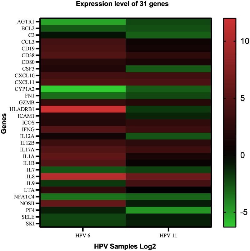
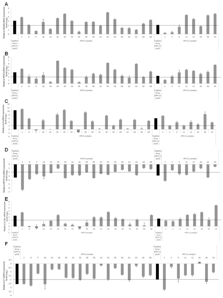
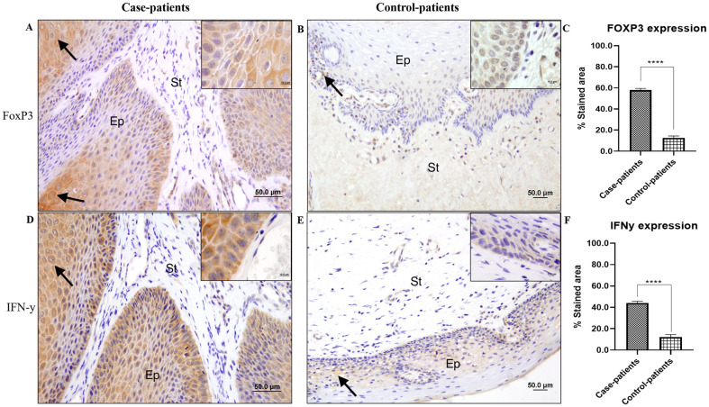
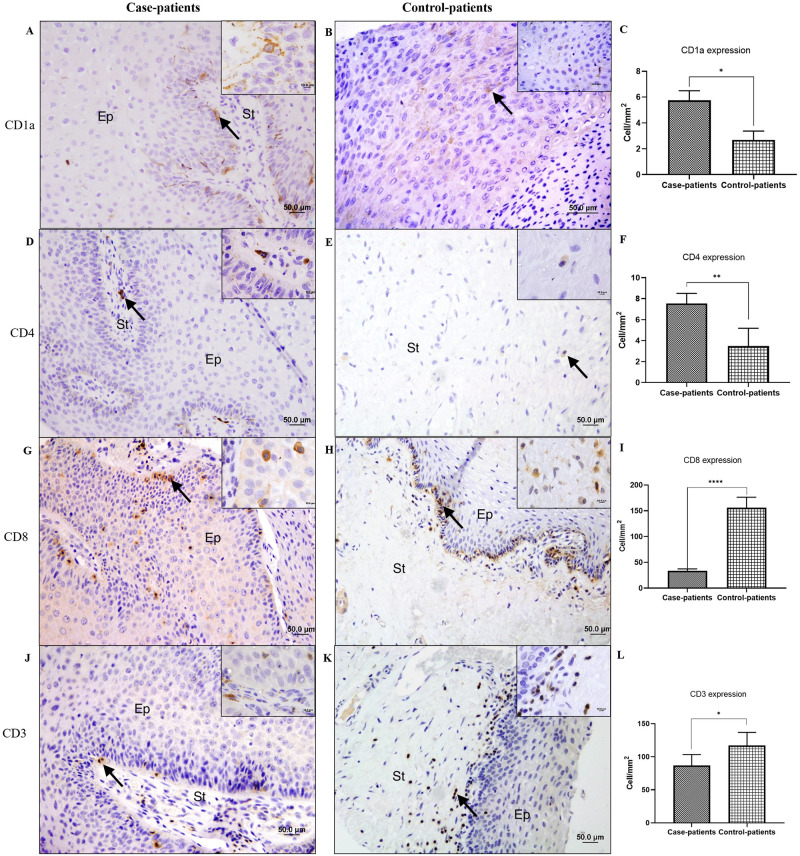
Condyloma acuminata (CA) is a benign proliferative disease mainly affecting in non-keratinized epithelia. Most cases of CA are caused by low-risk human papillomavirus (HPV), mainly HPV 6 and 11. The aim of the current study was to highlight the candidate genes and pathways associated with immune alterations in individuals who did not spontaneously eliminate the virus and, thus, develop genital warts. Paraffin-embedded condyloma samples (n = 56) were analyzed by immunohistochemistry using antibodies against CD1a, FOXP3, CD3, CD4, CD8, and IFN-γ. The immunomarkers were chosen based on the evaluation of the innate and adaptive immune pathways using qPCR analysis of 92 immune-related genes, applying a TaqMan Array Immune Response assay in HPV 6 or HPV 11 positive samples (n = 27). Gene expression analysis revealed 31 differentially expressed genes in CA lesions. Gene expression validation revealed upregulation of GZMB, IFNG, IL12B, and IL8 and downregulation of NFATC4 and IL7 in CA samples. Immunohistochemical analysis showed increased FOXP3, IFN-γ, CD1a, and CD4 expression in CA than in the control tissue samples. In contrast, CD3 and CD8 expression was decreased in CA lesion samples. Increased levels of pro-inflammatory cytokines in HPV-positive patients compared with HPV-negative patients seem to reflect the elevated immunogenicity of HPV-positive CA lesions. Host defense against HPV begins during the early stages of the innate immune response and is followed by activation of T lymphocytes, which are mainly represented by CD4+ and regulatory T cells. The low CD8+ T cell count in CA may contribute to this recurrent behavior. Additional studies are needed to elucidate the mechanism of host defense against HPV infection in CA.
Copyright: © 2023 Stuqui et al. This is an open access article distributed under the terms of the Creative Commons Attribution License, which permits unrestricted use, distribution, and reproduction in any medium, provided the original author and source are credited.
Conflict of interest statement
The authors have declared that no competing interests exist.
Figures
References
-
- Brianti P, De Flammineis E, Mercuri SR. Review of HPV-related diseases and cancers. New Microbiol. 2017;40(2):80–5. Epub 2017/04/04. . - PubMed
Publication types
MeSH terms
Substances
LinkOut - more resources
Full Text Sources
Medical
Research Materials
Miscellaneous
