

L-type calcium channel blocker increases VEGF concentrations in retinal cells and human serum
- PMID: 37053203
- PMCID: PMC10101440
- DOI: 10.1371/journal.pone.0284364
L-type calcium channel blocker increases VEGF concentrations in retinal cells and human serum
Abstract
Objective: Vascular endothelial growth factor (VEGF) plays a key role in diabetic retinopathy (DR). Previously, we have reported an association between mutations in a gene coding for the L-type calcium channel subunit, VEGF and DR. L-type calcium channel blockers (LTCCBs) have been widely used as antihypertensive medication (AHM), but their association with VEGF and DR is still unclear. Therefore, we explored the effect of LTCCBs compared to other AHMs on VEGF concentrations in retinal cells and human serum. Furthermore, we evaluated the association between the use of LTCCBs and the risk of severe diabetic eye disease (SDED).
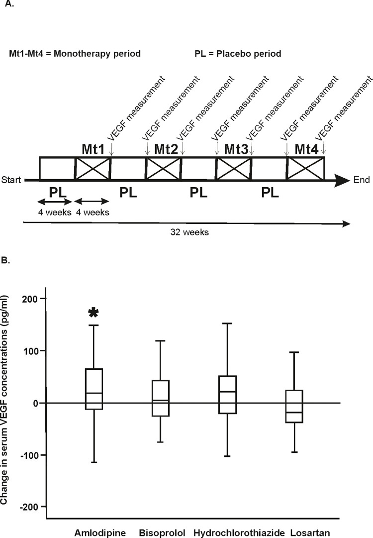
Research design and methods: Müller cells (MIO-M1) were cultured as per recommended protocol and treated with LTCCBs and other AHMs. VEGF secreted from cells were collected at 24 hours intervals. In an interventional study, 39 individuals received LTCCBs or other AHM for four weeks with a four-week wash-out placebo period between treatments. VEGF was measured during the medication and placebo periods. Finally, we evaluated the risk of SDED associated with LTCCB usage in 192 individuals from the FinnDiane Study in an observational setting.
Results: In the cell cultures, the medium VEGF concentration increased time-dependently after amlodipine (P<0.01) treatment, but not after losartan (P>0.01), or lisinopril (P>0.01). Amlodipine, but no other AHM, increased the serum VEGF concentration (P<0.05) during the interventional clinical study. The usage of LTCCB was not associated with the risk of SDED in the observational study.
Conclusions: LTCCB increases VEGF concentrations in retinal cells and human serum. However, the usage of LTCCBs does not appear to be associated with SDED in adults with type 1 diabetes.
Copyright: © 2023 Kumar et al. This is an open access article distributed under the terms of the Creative Commons Attribution License, which permits unrestricted use, distribution, and reproduction in any medium, provided the original author and source are credited.
Conflict of interest statement
EBP reports receiving lecture honorariums from Eli Lilly, Abbott, Astra Zeneca, Sanofi, Boehringer Ingelheim and is an advisory board member of Sanofi. PHG reports receiving lecture honorariums from Astellas, Astra Zeneca, Boehringer Ingelheim, Eli Lilly, Elo Water, Medscape, MSD, Mundipharma, Novo Nordisk, PeerVoice, Sanofi, Sciarc, and being an advisory board member of Astellas, Astra Zeneca, Bayer, Boehringer Ingelheim, Eli Lilly, Janssen, Medscape, MSD, Mundipharma, Novo Nordisk, and Sanofi. This does not alter our adherence to PLOS ONE policies on sharing data and materials. AK, SM, TH, VH, RL, SiMa, CF, ML and KK declare no conflict of interest.
Figures

References
Publication types
MeSH terms
Substances
LinkOut - more resources
Full Text Sources
Medical
