. 2023 Apr 13;18(4):e0282732.
doi: 10.1371/journal.pone.0282732. eCollection 2023.

Distension evoked mucosal secretion in human and porcine colon in vitro

Affiliations

Distension evoked mucosal secretion in human and porcine colon in vitro

Kristin Elfers et al. PLoS One. .

Abstract

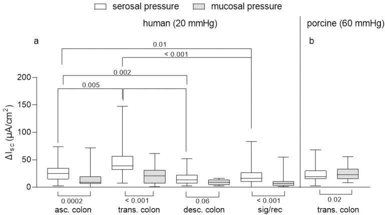
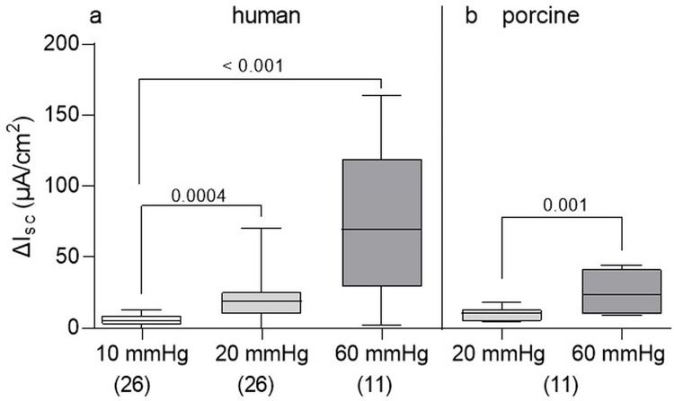
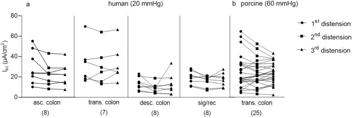
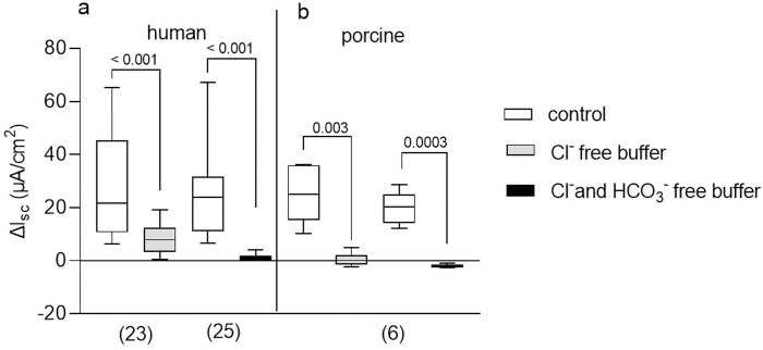
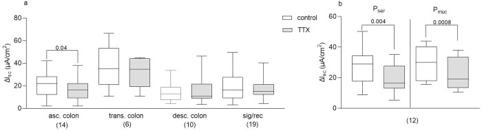
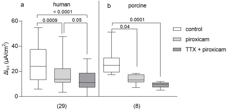
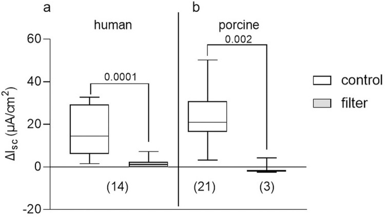
It was suggested that intestinal mucosal secretion is enhanced during muscle relaxation and contraction. Mechanisms of mechanically induced secretion have been studied in rodent species. We used voltage clamp Ussing technique to investigate, in human and porcine colonic tissue, secretion evoked by serosal (Pser) or mucosal (Pmuc) pressure application (2-60 mmHg) to induce distension into the mucosal or serosal compartment, respectively. In both species, Pser or Pmuc caused secretion due to Cl- and, in human colon, also HCO3- fluxes. In the human colon, responses were larger in proximal than distal regions. In porcine colon, Pmuc evoked larger responses than Pser whereas the opposite was the case in human colon. In both species, piroxicam revealed a strong prostaglandin (PG) dependent component. Pser and Pmuc induced secretion was tetrodotoxin (TTX) sensitive in porcine colon. In human colon, a TTX sensitive component was only revealed after piroxicam. However, synaptic blockade by ω-conotoxin GVIA reduced the response to mechanical stimuli. Secretion was induced by tensile rather than compressive forces as preventing distension by a filter inhibited the secretion. In conclusion, in both species, distension induced secretion was predominantly mediated by PGs and a rather small nerve dependent response involving mechanosensitive somata and synapses.


Conflict of interest statement

I have read the journal’s policy and the authors of this manuscript have the following competing interests:The authors declare no competing interests.

Figures

Fig 1
Fig 1. Traces of the secretory response to serosal pressure application (Pser).
Representative short circuit current traces evoked by Pser in (a) human sigmoid (20 mmHg) and (b) porcine transverse (60 mmHg) colonic tissue. Arrows indicate onset of Pser lasting for 60 s.
Fig 2
Fig 2. Secretory responses to serosal (Pser) and mucosal (Pmuc) pressure application.
(a) human tissue: Responses to Pser 20 mmHg were significantly greater than those Pmuc application in all colonic regions investigated despite of the descending colon (bottom brackets; ascending colon = asc. colon, n = 51; transverse colon = trans. colon, n = 45; descending colon = desc. colon; n = 37, sigma–rectum regions = sig/rec, n = 109; Mann-Whitney test, numbers below brackets indicate p values). In the transverse colon the distension-induced secretory response was significantly greater than in all other regions (top brackets, Kruskal-Wallis (p < 0.001) test with Dunn’s multiple comparison test and p values above brackets). (b) porcine tissue: Responses to Pser 60 mmHg were significantly smaller than those to Pmuc in the transverse colon (n = 80); Wilcoxon signed rank test, p value below bracket). Data shown are the medians with the 25th and 75th quartiles as a box plot and the minima and maxima as a whisker plot.
Fig 3
Fig 3. Secretory response to different pressure values.
In human (a) and porcine (b) colonic tissue preparations, secretory responses to Pser significantly increased with increasing stimulus strength. Human: Intestinal regions pooled; Kruskal-Wallis test (p < 0.001) with Dunn’s multiple comparison test and p values above brackets; porcine: Paired t test, p value above bracket. Data shown are the medians with the 25th and 75th quartiles as a box plot and the minima and maxima as a whisker plot. N numbers are given in parenthesis.
Fig 4
Fig 4. Secretory response to three times repeated Pser.
In human (a) and porcine tissue (b), responses to three times repeated Pser 20 mmHg (human) and 60 mmHg (porcine) were not significantly different from each other (human: Repeated measures ANOVA: n.s. with Holm-Sidak’s comparison test: n.s.; porcine: Friedman test: n.s. with Dunn‘s multiple comparison test: n.s.). Ascending colon = asc. colon; transverse colon = trans. colon; descending colon = desc. colon; sigma–rectum regions = sig/rec. Data shown are the individual values for the first, second and third pressure application, respectively. N numbers are given in parenthesis.
Fig 5
Fig 5. Ionic basis of the distension induced secretory response.
Distension induced secretory responses in human (a) and porcine (b) colonic tissue evoked by Pser was significantly reduced by depletion of chloride (Cl-) and Cl- and bicarbonate (HCO3-), respectively (human: Wilcoxon test; porcine: Paired t test; p values above brackets). Data shown are the medians with the 25th and 75th quartiles as a box plot and the minima and maxima as a whisker plot. N numbers are given in parenthesis.
Fig 6
Fig 6. TTX sensitivity of the distension induced secretory response.
Incubation of tissues with TTX prior to Pser (and Pmuc in porcine tissue) led to a reduced secretion only in the human ascending colon (a), and to a significant reduction in the secretory response in the porcine colon (b). Paired t test and Wilcoxon test; p values above brackets. Data shown are the medians with the 25th and 75th quartiles as a box plot and the minima and maxima as a whisker plot. N numbers are given in parenthesis.
Fig 7
Fig 7. Involvement of prostaglandins in mediating the distension induced secretory response.
Addition of piroxicam prior to Pser reduced secretory response in human (a) and porcine tissue (b). Simultaneous incubation with TTX further reduced the response in human tissue (human: Friedman test with Dunn’s multiple comparison test; porcine: Kruskal-Wallis test with Dunn’s multiple comparison test; p values above brackets). Data shown are the medians with the 25th and 75th quartiles as a box plot and the minima and maxima as a whisker plot. N numbers are given in parenthesis.
Fig 8
Fig 8. Secretory response to tensile forces.
Prevention of tissue distension during Pser by a mucosal placed filter significantly reduced the secretory response in human ((a); Wilcoxon test) and porcine (b) colonic tissue (unpaired t test); p values above brackets. Data shown are the medians with the 25th and 75th quartiles as a box plot and the minima and maxima as a whisker plot. N numbers are given in parenthesis.

Similar articles

Cited by

References

    1. Wood JD. Application of classification schemes to the enteric nervous system. Journal of the Autonomic Nervous System. 1994;48(1):17–29. doi: 10.1016/0165-1838(94)90156-2 - DOI - PubMed
    1. Gershon MD. The enteric nervous system: a second brain. Hosp Pract (1995). 1999;34(7):31–2, 5–8, 41–2 passim. Epub 1999/07/27. doi: 10.3810/hp.1999.07.153 . - DOI - PubMed
    1. Spencer NJ, Hu H. Enteric nervous system: sensory transduction, neural circuits and gastrointestinal motility. Nat Rev Gastroenterol Hepatol. 2020;17(6):338–51. Epub 2020/03/11. doi: 10.1038/s41575-020-0271-2 . - DOI - PMC - PubMed
    1. Cooke HJ. Neurotransmitters in neuronal reflexes regulating intestinal secretion. Ann N Y Acad Sci. 2000;915:77–80. Epub 2001/02/24. doi: 10.1111/j.1749-6632.2000.tb05225.x . - DOI - PubMed
    1. Rao MC. Physiology of Electrolyte Transport in the Gut: Implications for Disease. Compr Physiol. 2019;9(3):947–1023. Epub 2019/06/13. doi: 10.1002/cphy.c180011 . - DOI - PubMed

Publication types