. 2023 Jul 6;142(1):106-118.
doi: 10.1182/blood.2022018475.

Bone-derived C-terminal FGF23 cleaved peptides increase iron availability in acute inflammation

Affiliations

Bone-derived C-terminal FGF23 cleaved peptides increase iron availability in acute inflammation

Guillaume Courbon et al. Blood. .

Abstract

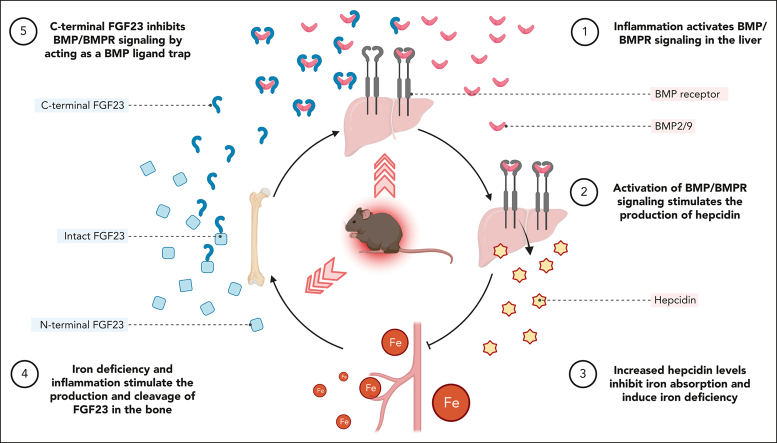
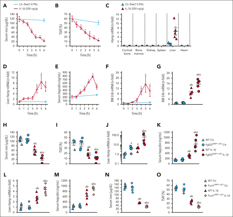
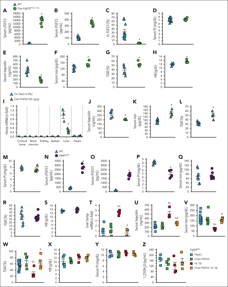
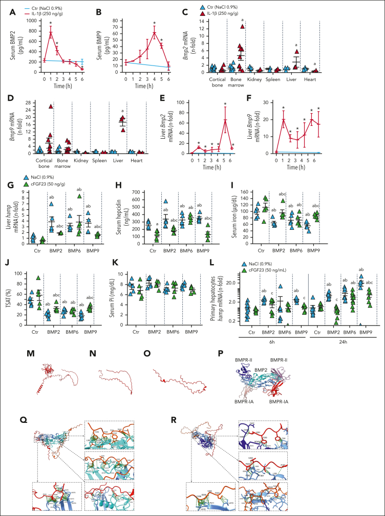
Inflammation leads to functional iron deficiency by increasing the expression of the hepatic iron regulatory peptide hepcidin. Inflammation also stimulates fibroblast growth factor 23 (FGF23) production by increasing both Fgf23 transcription and FGF23 cleavage, which paradoxically leads to excess in C-terminal FGF23 peptides (Cter-FGF23), rather than intact FGF23 (iFGF23) hormone. We determined that the major source of Cter-FGF23 is osteocytes and investigated whether Cter-FGF23 peptides play a direct role in the regulation of hepcidin and iron metabolism in response to acute inflammation. Mice harboring an osteocyte-specific deletion of Fgf23 showed a ∼90% reduction in Cter-FGF23 levels during acute inflammation. Reduction in Cter-FGF23 led to a further decrease in circulating iron in inflamed mice owing to excessive hepcidin production. We observed similar results in mice showing impaired FGF23 cleavage owing to osteocyte-specific deletion of Furin. We next showed that Cter-FGF23 peptides bind members of the bone morphogenetic protein (BMP) family, BMP2 and BMP9, which are established inducers of hepcidin. Coadministration of Cter-FGF23 and BMP2 or BMP9 prevented the increase in Hamp messenger RNA and circulating hepcidin levels induced by BMP2/9, resulting in normal serum iron levels. Finally, injection of Cter-FGF23 in inflamed Fgf23KO mice and genetic overexpression of Cter-Fgf23 in wild type mice also resulted in lower hepcidin and higher circulating iron levels. In conclusion, during inflammation, bone is the major source of Cter-FGF23 secretion, and independently of iFGF23, Cter-FGF23 reduces BMP-induced hepcidin secretion in the liver.


Conflict of interest statement

Conflict-of-interest disclosure: V.D. received research funding from Akebia and has received research funding from Vifor Pharma and consulting honoraria from Keryx Biopharmaceuticals, Vifor Pharma, Luitpold, and Amgen, outside of the submitted work. The remaining authors declare no competing financial interests.

Figures

None
Graphical abstract
Figure 1.
Figure 1.
Acute inflammation increases bone FGF23 production. (A) Fgf23 promoter activity in MC3T3-E1 osteoblasts in response to escalating doses of IL-1β and calcium used as positive control. Serum (B) cFGF23, (C) iFGF23, (D) i/cFGF23, and (E) bone Fgf23 mRNA expression in 6-week-old WT mice, 6 hours after administration of a single dose of IL-1β. n ≥ 3 per group, P < .05 vs (a) 0 (Ctr), (b) 5, (c) 10 ng/mL, and (d) 50 ng/g IL-1β.
Figure 2.
Figure 2.
Deletion of Fgf23 in osteocytes reduces circulating FGF23 in acute inflammation. Serum (A) cFGF23, (B) iFGF23, (C) i/cFGF23, (D) phosphate, (E) 1,25OH2D, bone (F) Fgf23 mRNA, and (G) FGF23 protein expression in 6-week-old WT and Fgf23Dmp1-cKO mice 6 hours after administration of a single dose of 250 ng/g IL-1β. n ≥ 5 per group, P < .05 vs (a) WT Ctr, (b) Fgf23Dmp1-cKO Ctr (c) WT IL-1β. (H) Fgf23 mRNA levels and secreted (I) cFGF23 and (J) iFGF23 in primary osteoblast cultures cultured for 3 weeks in osteoblastic medium and treated with saline or 10 ng/mL of IL-1β for 6 hours. n ≥ 3 per group, P < .05 vs (a) WT Ctr, (b) Fgf23Dmp1-cKO Ctr (c) WT IL-1β.
Figure 3.
Figure 3.
FGF23 cleavage by furin in osteocytes contributes to a time dependent secretion of FGF23 cleaved peptides. (A) Cortical bone Fgf23 mRNA, serum (B) cFGF23, (C) iFGF23, (D) i/cFGF23, (E) IP/WB of circulating FGF23 peptides, and cortical bone mRNA expression of (F) Galnt3, (G) Fam20c and (H) Furin in 6-week-old WT mice up to 6 hours after administration of a single dose of 250 ng/g IL-1β. Supernatant (I) cFGF23, (J) iFGF23 and (K) i/cFGF23 in 24 hours cultures of bone explants from Fgf23KO mice treated with recombinant murine iFGF23 (10 ng/mL) and IL-1β (10 ng/mL) and furin inhibitor (0 or 15 μg/mL). Serum (L) cFGF23, (M) iFGF23, (N) i/cFGF23, (O) bone Fgf23 mRNA, (P) serum phosphate, and (Q) 1,25OH2D in 6-week-old WT and FurinDmp1-cKO mice 6 hours after administration of a single dose of 250 ng/g IL-1β. n ≥ 3 per group, P < .05 vs (∗) Ctr, (a) Fgf23KO + FGF23, (b) Fgf23KO + FGF23 + IL-1β, (c) WT Ctr, (d) FurinDmp1-cKO Ctr, (e) WT IL-1β.
Figure 4.
Figure 4.
Impaired FGF23 cleavage aggravates functional iron deficiency in acute inflammation. Time course of serum (A) iron and (B) TSAT, (C) Hamp mRNA expression in selected organs, (D) liver Hamp mRNA expression, (E) serum hepcidin, and (F) bone marrow Erfe mRNA expression in 6-week-old WT mice, 6 hours after administration of a single dose of 250 ng/g IL-1β. (G) bone marrow Erfe mRNA expression and serum (H) iron and (I) TSAT, (J) liver Hamp mRNA expression and serum (K) hepcidin in 6-week-old WT and Fgf23Dmp1-cKO mice 6 hours after administration of a single dose of 250 ng/g IL-1β. (L) Liver Hamp mRNA expression, serum (M) hepcidin, (N) iron and (O) TSAT in 6-week-old WT and FurinDmp1-cKO mice 6 hours after administration of a single dose of 250 ng/g IL-1β. n ≥ 5 per group. P < .05 vs (∗) Ctr, (a) WT Ctr, (b) cKO Ctr, (c) WT IL-1β.
Figure 5.
Figure 5.
Cter-FGF23 peptides directly regulate iron metabolism in mice. Serum (A) cFGF23, (B) iFGF23, (C) i/cFGF23 ratio, (D) phosphate, (E) hepcidin, (F) iron, (G) TSAT, and (H) Hb in 6-week-old WT and Cter-Fgf23Dmp1-cTg mice. Liver (I) Hamp mRNA expression, serum (J) hepcidin, (K) iron and (L) TSAT and (M) phosphate in 6-week-old WT mice 6 hours after administration of a single dose of 50 ng/g Cter-FGF23. Serum (N) cFGF23, (O) iFGF23, (P) phosphate, (Q) iron, (R) TSAT, and (S) Hb in 6-week-old WT and Dmp1KO mice. Liver (T) Hamp mRNA, serum (U) hepcidin, (V) iron and (W) TSAT, (X) Hb, (Y) phosphate and (Z) 1,25OH2D in 6-week-old FGF23KO mice 6 hours after administration of a single dose of Cter-FGF23 (50 ng/g), IL-1β (250 ng/g), or both. n ≥ 5 per group. P < .05 vs (a) WT/Ctr, (b) Fgf23KO + NaCl, (c) Fgf23KO + Cter-FGF23, (d) Fgf23KO + IL-1β.
Figure 6.
Figure 6.
Cter-FGF23 peptides antagonize BMP2/9-induced hepcidin secretion. Time course of serum (A) BMP2 and (B) BMP9, mRNA expression of (C) Bmp2 and (D) Bmp9 in selected organs and time course mRNA expression of liver (E) Bmp2 and (F) Bmp9 in 6-week-old WT mice, 6 hours after administration of a single dose of 250 ng/g IL-1β. Liver (G) Hamp mRNA expression and serum (H) hepcidin, (I) iron and (J) TSAT and (K) phosphate in 6-week-old WT mice 6 hours after administration of a single dose of 50 ng/g Cter-FGF23, 10 ng/g BMP (2, 6 or 9), or both. n ≥ 5 per group. Expression of (L) Hamp mRNA in primary hepatocytes treated with Cter-FGF23 (50 ng/mL), 10 ng/mL BMP (2, 6 or 9) or both for 6 and 24 hours. n ≥ 5 per group. P < .05 vs (∗) Ctr T0, (a) Ctr NaCl, (b) Ctr Cter-FGF23, (c) BMP (2, 6, 9). (M) iFGF23 predicted 3D structure, (N) Cter-FGF23 isolated from panel M, (O) predicted AlphaFold2 Cter-FGF23 structure (model 0), (P) BMP2 dimer (blue) binding BMPR-II (purple) and BMPR-IA (red), (Q) Interaction of 2 molecules of Cter-FGF23 with a dimeric BMP2 at the BMPR-II site, (R) Interaction of 2 molecules of Cter-FGF23 with a dimeric BMP9.

Comment in

  • Matryoshka hormones.
    Wolf M. Wolf M. Blood. 2023 Jul 6;142(1):7-9. doi: 10.1182/blood.2023020770. Blood. 2023. PMID: 37410509 No abstract available.

References

    1. Liu S, Zhou J, Tang W, Jiang X, Rowe DW, Quarles LD. Pathogenic role of Fgf23 in Hyp mice. Am J Physiol Endocrinol Metab. 2006;291(1):E38–E49. - PubMed
    1. Feng JQ, Ward LM, Liu S, et al. Loss of DMP1 causes rickets and osteomalacia and identifies a role for osteocytes in mineral metabolism. Nat Genet. 2006;38(11):1310–1315. - PMC - PubMed
    1. Pereira RC, Juppner H, Azucena-Serrano CE, Yadin O, Salusky IB, Wesseling-Perry K. Patterns of FGF-23, DMP1, and MEPE expression in patients with chronic kidney disease. Bone. 2009;45(6):1161–1168. - PMC - PubMed
    1. Riminucci M, Collins MT, Fedarko NS, et al. FGF-23 in fibrous dysplasia of bone and its relationship to renal phosphate wasting. J Clin Invest. 2003;112(5):683–692. - PMC - PubMed
    1. Bonewald LF, Wacker MJ. FGF23 production by osteocytes. Pediatr Nephrol. 2013;28(4):563–568. - PMC - PubMed

Publication types