Observational Study
. 2023 Jul:127:43-53.
doi: 10.1016/j.neurobiolaging.2023.03.008. Epub 2023 Mar 21.

Associations of physical function and body mass index with functional brain networks in community-dwelling older adults

Affiliations
Observational Study

Associations of physical function and body mass index with functional brain networks in community-dwelling older adults

Paul J Laurienti et al. Neurobiol Aging. 2023 Jul.

Abstract

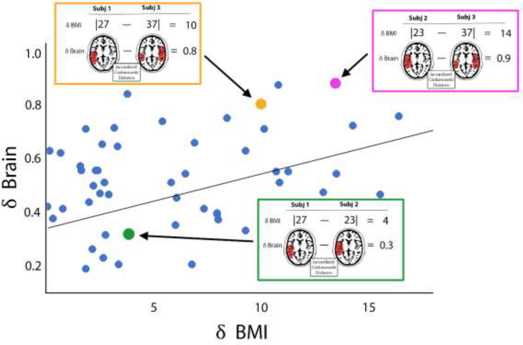
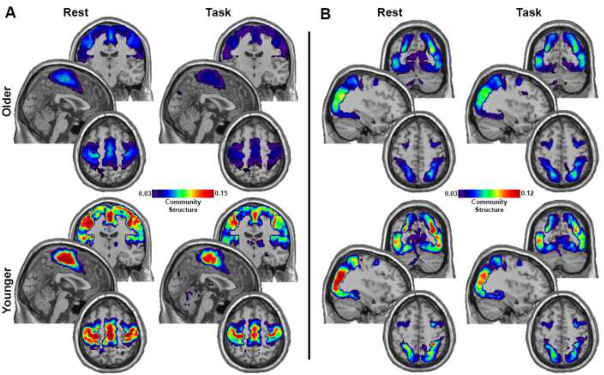
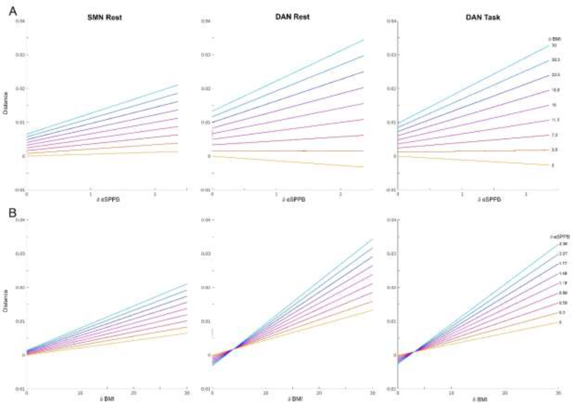
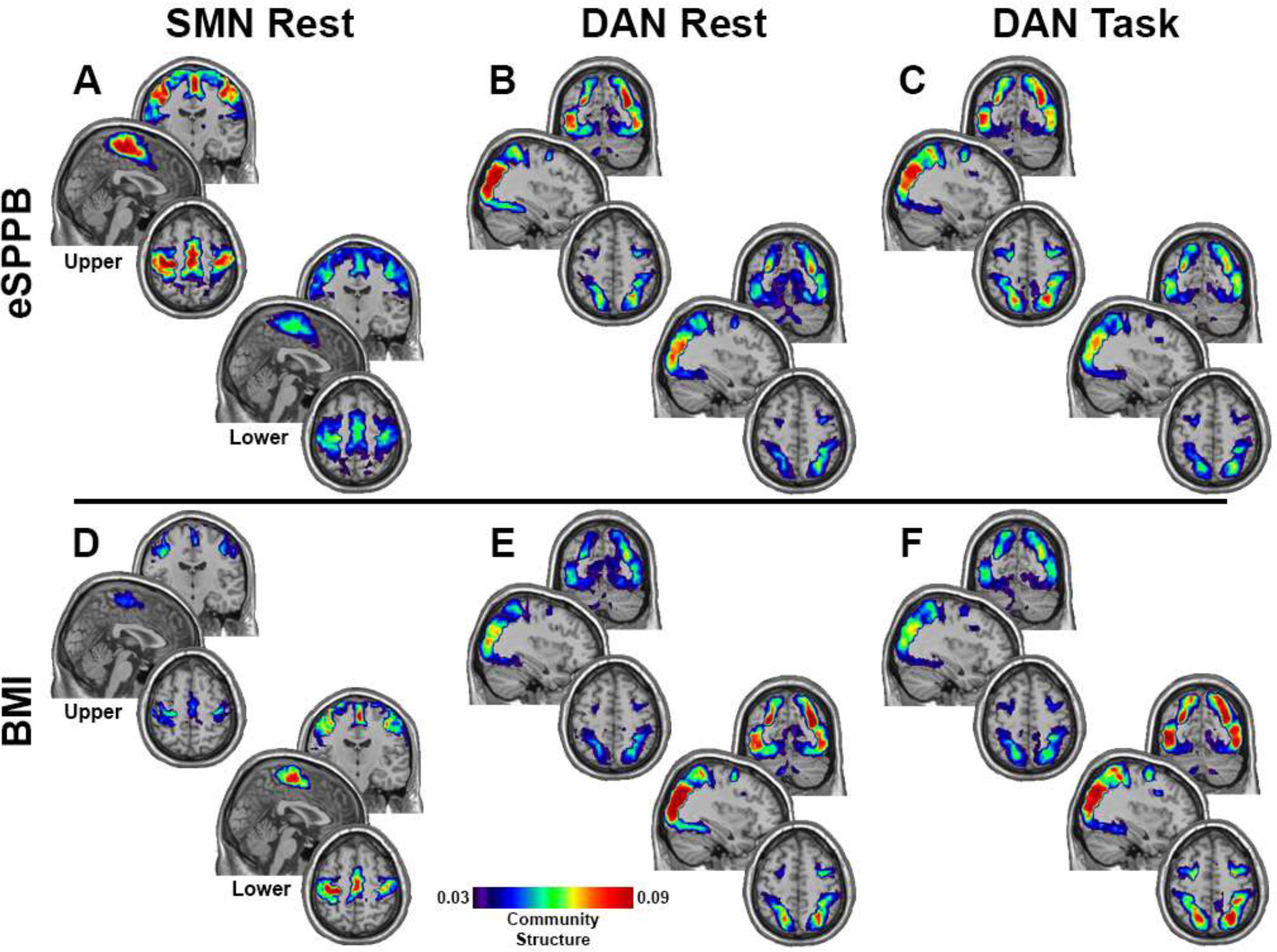
Deficits in physical function that occur with aging contribute to declines in quality of life and increased mortality. There has been a growing interest in examining associations between physical function and neurobiology. Whereas high levels of white matter disease have been found in individuals with mobility impairments in structural brain studies, much less is known about the relationship between physical function and functional brain networks. Even less is known about the association between modifiable risk factors such as body mass index (BMI) and functional brain networks. The current study examined baseline functional brain networks in 192 individuals from the Brain Networks and mobility (B-NET) study, an ongoing longitudinal, observational study in community-dwelling adults aged 70 and older. Physical function and BMI were found to be associated with sensorimotor and dorsal attention network connectivity. There was a synergistic interaction such that high physical function and low BMI were associated with the highest network integrity. White matter disease did not modify these relationships. Future work is needed to understand the causal direction of these relationships.

Keywords: Aging; Body mass index; Brain network; Connectome; Physical function.


Conflict of interest statement

Declarations of interest: none

Figures

Figure 1.
Figure 1.
Schematic cartoon depicting the distance regression method. In this cartoon, the brain maps and BMI from three simulated participants from a larger group are shown. Each participant is compared to each other participant using distance measures. For numeric variables (the independent variables in this study), the absolute difference between participant is computed. This distance is the X value in the regression model. For the brain maps of network community structure (the dependent variable in this study), the weighted Jaccard distance is computed. This metric assesses spatial similarity of the two brain maps. The Jaccard distance (δBrain) is the Y variable in the regression. Note that subject 1 and subject 2 both have low BMIs, the δBMI is low (green box). They also have very similar brain maps resulting in low δBrain. When these two participants are compared to subject 3 with a higher BMI (red and blue boxes), the δBMI values are also higher. Since BMI and brain network organization are correlated in this cartoon example, the higher δBMIs are associated with larger δBrain values. Once the distances between all participants pairs is computed (simulated blue data points), a regression is performed. This cartoon shows a simple linear regression but the reality is that all data points for an individual participant compared to each of the remaining participants are correlated. Thus, the regression model is performed using an F test with individual level effects (ILE). This more complex model is not easily depicted in cartoon form, but the premise of the distance regression is the same.
Figure 2.
Figure 2.
Community structure maps for the younger and older adults. A) Maps for the SMN. B) Maps for the DAN. Each map is an average across all participants in each group for the condition (Rest and Task) and network (SMN and DAN). Brain regions with hotter colors were more frequently part of the community across participants, indicating greater spatial consistency. The color scale represents the average SI value across each group/condition and has been scaled the min and max and applies to all images within each figure section (A and B). Each image collage contains a coronal slice at the top (y: SMN=−15, DAN=−70), a sagittal slice in the middle (x: SMN=2, DAN 36), and an axial slice on the bottom (z: SMN=58, DAN= 54).
Figure 3.
Figure 3.
Plots showing how δeSPPB and δBMI interact in their effects on community structure. A) The colored lines represent the relationships between eSPPB distances (δeSPPB) on the x-axis and community structure distances on the y-axis for ten evenly spaced, discrete BMI distances (δBMI). The bottom yellow line represents the relationship between δeSPPB and community structure distance when the BMI distance is 0 and the top light blue line shows a δBMI of 30 kg/m2. The y-intercept shows the effect of δBMI when eSPPB scores are the same (δeSPPB=0). Note that there are systematic increases in community structure distance with increases in δBMI even between individuals with the comparable eSPPB scores. B) Colored lines represent the relationship between δBMI between participants on the x-axis and community structure distance on the y-axis for ten evenly spaced, discrete δeSPPB values. Similarly to panel A, the bottom yellow line represents the relationship between δBMI and community structure distance when there is no difference in eSPPB between participants and the y-intercept shows the effect of δeSPPB when BMI scores are the same. For the SMN at rest there are no meaningful effects of eSPPB in individuals with the same BMI. For the DAN at rest and during task, there is actually a decrease, albeit quite small, in distance with greater δeSPPB in individuals with the same BMI.
Figure 4.
Figure 4.
Community structure maps for the groups in the upper and lower tertiles (N=54 in each group) of eSPPB and BMI. For eSPPB, the upper tertile had greater community spatial overlap across participants for all three conditions/networks (A-C). BMI exhibited the opposite relationship with community structure with the lower tertile having the higher spatial consistency (D-F). Brain regions with hotter colors were more frequently part of the community across participants, indicating greater spatial consistency. The color bar applies to all images and is scaled as in Figure 2. Slice locations are the same as in Figure 2.

References

    1. Albert SM, Bear-Lehman J and Anderson SJ (2015). “Declines in mobility and changes in performance in the instrumental activities of daily living among mildly disabled community-dwelling older adults.” J Gerontol A Biol Sci Med Sci 70(1): 71–77. DOI: 10.1093/gerona/glu088. - DOI - PMC - PubMed
    1. Avants BB, Epstein CL, Grossman M and Gee JC (2008). “Symmetric diffeomorphic image registration with cross-correlation: evaluating automated labeling of elderly and neurodegenerative brain.” Med Image Anal 12(1): 26–41. DOI: 10.1016/j.media.2007.06.004. - DOI - PMC - PubMed
    1. Benjamini Y and Hochberg Y (2000). “On the adaptive control of the false discovery fate in multiple testing with independent statistics.” Journal of Educational and Behavioral Statistics 25(1): 60–83. DOI: 10.3102/10769986025001060. - DOI
    1. Birn RM, Molloy EK, Patriat R, Parker T, Meier TB, Kirk GR, Nair VA, Meyerand ME and Prabhakaran V (2013). “The effect of scan length on the reliability of resting-state fMRI connectivity estimates.” Neuroimage 83: 550–558. DOI: 10.1016/j.neuroimage.2013.05.099. - DOI - PMC - PubMed
    1. Carson N, Leach L and Murphy KJ (2018). “A re-examination of Montreal Cognitive Assessment (MoCA) cutoff scores.” Int J Geriatr Psychiatry 33(2): 379–388. DOI: 10.1002/gps.4756. - DOI - PubMed

Publication types