An official website of the United States government
The .gov means it’s official.
Federal government websites often end in .gov or .mil. Before
sharing sensitive information, make sure you’re on a federal
government site.
The site is secure.
The https:// ensures that you are connecting to the
official website and that any information you provide is encrypted
and transmitted securely.
1 Division of Pulmonary, Critical Care, and Sleep Medicine, National Jewish Health, 1400 Jackson St, Denver, CO 80206, USA; Department of Medicine, National Jewish Health, 1400 Jackson St, Denver, CO 80206, USA. Electronic address: koslowm@njhealth.org.
2 Department of Radiology, National Jewish Health, 1400 Jackson St, Denver, CO 80206, USA.
3 Department of Pathology, University of Colorado School of Medicine Anschutz Medical Campus, 13001 E 17th Pl, Aurora, CO 80045, USA; Division of Pathology, Department of Medicine, National Jewish Health, Denver, CO, USA.
4 Department of Medicine, National Jewish Health, 1400 Jackson St, Denver, CO 80206, USA.
5 Department of Medicine, National Jewish Health, 1400 Jackson St, Denver, CO 80206, USA; Department of Pediatrics, National Jewish Health; Department of Immunology and Genomic Medicine, National Jewish Health.
Cysts and cavities in the lung are commonly encountered on chest imaging. It is necessary to distinguish thin-walled lung cysts (≤2 mm) from cavities and characterize their distribution as focal or multifocal versus diffuse. Focal cavitary lesions are often caused by inflammatory, infectious, or neoplastic processes in contrast to diffuse cystic lung diseases. An algorithmic approach to diffuse cystic lung disease can help narrow the differential diagnosis, and additional testing such as skin biopsy, serum biomarkers, and genetic testing can be confirmatory. An accurate diagnosis is essential for the management and disease surveillance of extrapulmonary complications.
A. Axial CT image shows a moderate right chylous effusion and thin-walled cysts of variable sizes evenly distributed throughout the lungs. B. CXR shows a resolved effusion following 4 months of medical therapy. C. Coronal CT image in a different patient shows bilateral fat-containing renal angiomyolipomas (arrows). Metal artifact in the right kidney is from arterial embolization coils. D. Coronal CT in a different patient shows a large tubular and lobulated fluid attenuating structure in the retroperitoneum (arrows) compatible with retroperitoneal lymphangeioleiomyoma which resolved with prolonged therapy (not shown). A large right chylous effusion is also present.
Figure 2.. Histopathologic findings of LAM.
A …
Figure 2.. Histopathologic findings of LAM.
A . H&E stained section at low power shows…
Figure 2.. Histopathologic findings of LAM.
A. H&E stained section at low power shows a cystic structure with a nodule of LAM cells protruding into the cyst space in a polypoid manner. B. High power image of the LAM nodule demonstrating clusters of spindle shaped cells growing in a haphazard manner. C. HMB45 immunohistochemical stain of LAM cells shows patchy cytoplasmic staining. D. SMA (smooth muscle active) immunohistochemistry stain highlights the nodules of smooth muscle cells within the wall of a cyst in LAM. (Images courtesy of Dr. Carlyne D. Cool and Dr. Steve D. Groshong, Division of Pathology, National Jewish Health).
Figure 3.. Characteristic findings of Birt-Hogg-Dube syndrome.
A . Axial CT shows elliptical thin-walled pulmonary…
Figure 3.. Characteristic findings of Birt-Hogg-Dube syndrome.
A. Axial CT shows elliptical thin-walled pulmonary cysts predominating in the paracardiac regions of the lower lungs. B. Subpleural cystic structure lined by bland alveolar cells without atypical morphology. C. White/gray papules on midface, forehead and post-auricular of a patient with Birt-Hogg-Dube syndrome characteristic of fibrofolliculomas or similar lesions (e.g., acrochordons). (Fig 3B, courtesy of Dr. Carlyne D. Cool, Division of Pathology, National Jewish Health).
Figure 4.. Algorithm to reliably differentiate BHD…
Figure 4.. Algorithm to reliably differentiate BHD from other cystic lung diseases (AJR 2019; 212:…
Figure 4.. Algorithm to reliably differentiate BHD from other cystic lung diseases (AJR 2019; 212: 1260–1264.
Figure 5.
A . Coronal CT in…
Figure 5.
A . Coronal CT in a patient with Sjogren’s syndrome shows multiple lower…
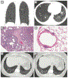
Figure 5.
A. Coronal CT in a patient with Sjogren’s syndrome shows multiple lower lung predominant thin-walled cysts with peribronchovascular predominance, compatible with LIP. B. Axial CT through the lower lungs in a patient with biopsy-proven LIP and amyloidosis in Sjogren’s syndrome shows a combination of irregular nodules, cysts and ground glass opacities. C. Low power image demonstrating diffuse interstitial infiltrate by mature lymphocytes and plasma cells. Cysts are seen adjacent to airways. D. Lymphoid follicles with germinal centers in a patient with Sjogren’s syndrome and cystic lung disease. E, F. CT scans two years apart in a patient with LIP and Sjogren syndrome show increased ground glass abnormality and septal thickening indicating progression of lymphocytic infiltrates. BAL demonstrated 60% lymphocytic predominance without monoclonality or infection. (Images C, D, images courtesy of Dr. Carlyne D. Cool, Division of Pathology, National Jewish Health).
Figure 6.
A . Axial CT image…
Figure 6.
A . Axial CT image of thin-walled pulmonary cysts and nodules, some calcified,…
Figure 6.
A. Axial CT image of thin-walled pulmonary cysts and nodules, some calcified, in a patient with biopsy-proved LIP and amyloidosis. B. Axial CT image of diffuse lung cysts in the mid and upper lung zones in a patient with advanced LCDD of the kidney secondary to multiple myeloma C. Nodular amyloidosis characterized by intraalveolar and interstitial deposits of amorphous eosinophilic material. D. Congo red stain highlights the amyloid material. (Image C, courtesy of Dr. Carlyne D. Cool, Division of Pathology, National Jewish Health. Image D, with permission, Rosane Duarte Achcar MD, FCAP, FASCP, Steve D. Groshong MD, PhD, Carlyne D. Cool MD, Differential Diagnoses in Surgical Pathology: Pulmonary Pathology, copyright 2017 Wolters Kluwer Health, Inc.
Figure 7.
A,B Coronal CT images in…
Figure 7.
A,B Coronal CT images in a patient with pLCH show characteristic irregular cysts…
Figure 7.
A,B Coronal CT images in a patient with pLCH show characteristic irregular cysts with mid to upper zone predominance, relative sparing of the costophrenic sulci, and scattered small nodules. C. Bronchiolocentric stellate (star-like) scars develop with aging of the lesions and depletion of the Langerhans cells. D. Immunohistochemical stain for CD1a highlights the Langerhans cells. (Images C, D courtesy of Dr. Steve D. Groshong, Division of Pathology, National Jewish Health).
Figure 8.. An algorithmic approach to the…
Figure 8.. An algorithmic approach to the diagnosis of diffuse cystic lung disease.
Modified from…
Figure 8.. An algorithmic approach to the diagnosis of diffuse cystic lung disease.
Hansell DM, Bankier AA, MacMahon H, McLoud TC, Muller NL, Remy J. Fleischner Society: glossary of terms for thoracic imaging. Radiology. Mar 2008;246(3):697–722. doi:10.1148/radiol.2462070712
-
DOI
-
PubMed
Woodring JH, Fried AM. Significance of wall thickness in solitary cavities of the lung: a follow-up study. AJR Am J Roentgenol. Mar 1983;140(3):473–4. doi:10.2214/ajr.140.3.473
-
DOI
-
PubMed
Moss J, Avila NA, Barnes PM, et al. Prevalence and clinical characteristics of lymphangioleiomyomatosis (LAM) in patients with tuberous sclerosis complex. Am J Respir Crit Care Med. Aug 15 2001;164(4):669–71. doi:10.1164/ajrccm.164.4.2101154
-
DOI
-
PubMed
Ryu JH, Moss J, Beck GJ, et al. The NHLBI lymphangioleiomyomatosis registry: characteristics of 230 patients at enrollment. Am J Respir Crit Care Med. Jan 1 2006;173(1):105–11. doi:10.1164/rccm.200409-1298OC
-
DOI
-
PMC
-
PubMed
Han MK, Tyburczy ME, Darling TN, et al. Apparent Sporadic Lymphangioleiomyomatosis in a Man as a Result of Extreme Mosaicism for a TSC2 Mutation. Ann Am Thorac Soc. Jul 2017;14(7):1227–1229. doi:10.1513/AnnalsATS.201703-229LE
-
DOI
-
PMC
-
PubMed