Review
. 2023 Jul;29(7):898-957.
doi: 10.1261/rna.079620.123. Epub 2023 Apr 13.

The life and times of a tRNA

Affiliations
Review

The life and times of a tRNA

Eric M Phizicky et al. RNA. 2023 Jul.

Abstract

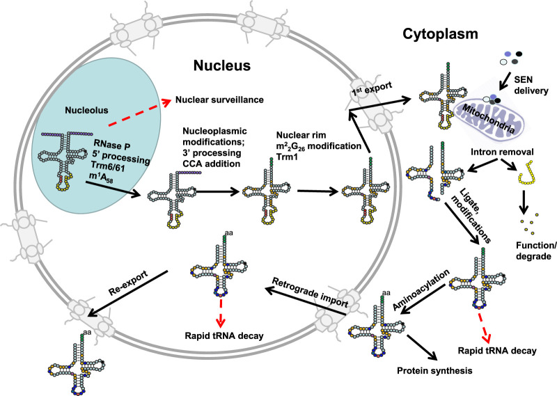
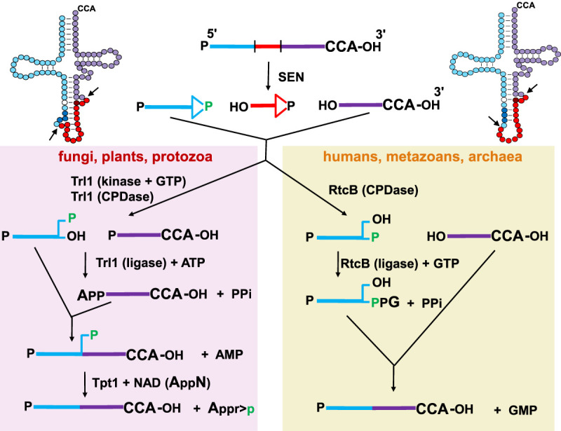
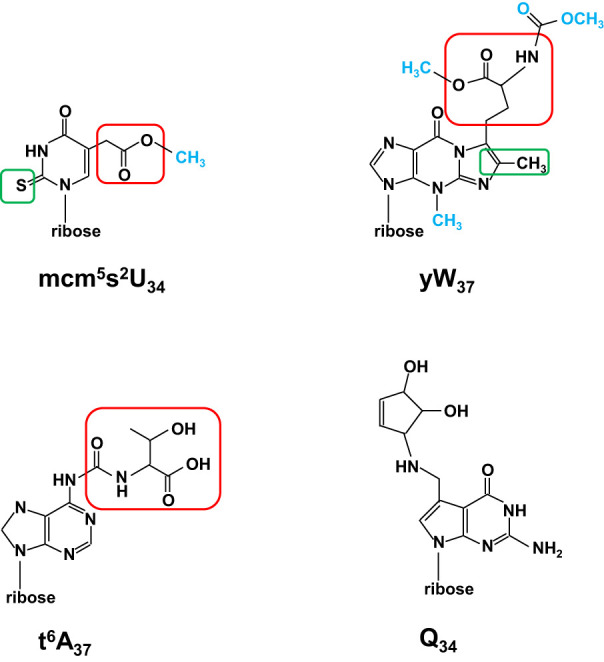
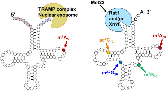
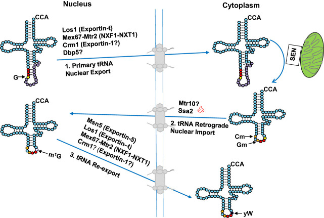
The study of eukaryotic tRNA processing has given rise to an explosion of new information and insights in the last several years. We now have unprecedented knowledge of each step in the tRNA processing pathway, revealing unexpected twists in biochemical pathways, multiple new connections with regulatory pathways, and numerous biological effects of defects in processing steps that have profound consequences throughout eukaryotes, leading to growth phenotypes in the yeast Saccharomyces cerevisiae and to neurological and other disorders in humans. This review highlights seminal new results within the pathways that comprise the life of a tRNA, from its birth after transcription until its death by decay. We focus on new findings and revelations in each step of the pathway including the end-processing and splicing steps, many of the numerous modifications throughout the main body and anticodon loop of tRNA that are so crucial for tRNA function, the intricate tRNA trafficking pathways, and the quality control decay pathways, as well as the biogenesis and biology of tRNA-derived fragments. We also describe the many interactions of these pathways with signaling and other pathways in the cell.

Keywords: decay; modification; splicing; tRNA; tRNA-derived fragments.


Figures

FIGURE 1.
FIGURE 1.
Schematic of tRNA biogenesis, subcellular dynamics, and quality control turnover pathways in S. cerevisiae. tRNAs are transcribed in the nucleolus where the 5′ leader (left purple circles) of the initial transcript is removed by RNase P and likely where m1A58 is modified (black circle) by Trm6/61. About half of the known modifications (examples, orange circles) occur in the nucleoplasm where 3′ CCA nucleotides (green circles) are also added. Dimethylation of G26 (magenta circle) is catalyzed by Trm1, which is located on the inner nuclear membrane, prior to nuclear export of the end-matured, partially processed, intron-containing (yellow circles) pre-tRNAs; end-processed, partially modified tRNAs encoded by genes lacking introns are also exported to the cytoplasm. Introns are removed on the mitochondrial cytoplasmic surface. After/during splicing, additional modifications are added in the cytoplasm (examples, blue circles), and the freed introns are destroyed. Processed/modified cytoplasmic tRNAs return to the nucleoplasm via retrograde tRNA nuclear import and under stress conditions accumulate there; in favorable conditions the tRNAs return to the cytoplasm via reexport where they participate in protein synthesis. There are quality control steps, indicated by red dashed arrows, that destroy tRNAs that have not undergone the canonical (black arrows) steps appropriately. Further details of the cell biology and quality control pathways are provided in the text and Figures 7 and 8.
FIGURE 2.
FIGURE 2.
tRNA structure. A schematic of tRNA structure. tRNA is shown in its usual secondary structure, with colored circles representing nucleotides in and adjacent to the acceptor stem (pink), D stem–loop (green), anticodon stem–loop (red), variable arm (aqua) and T-stem–loop (gray), and lines representing base pairs. The 3′ CCA residues N74–N76 are shown in dark pink, and the anticodon residues N34–N36 are dark red. Outer disks of circles are colored to indicate common tertiary interactions, as first detailed for tRNAPhe from yeast (Kim et al. 1974b) (8–14, dark pink; 9–12–23, yellow; 13–22–46, red; 15–48, purple; 18–55, green; 19–56, blue; 26–44, light gray; 54–58, dark gray). Note that different tRNA species can have a D-stem with only 3 bp, a D-loop of variable length, a variable arm with 4 nt or a longer variable arm comprising a stem–loop. Note that tRNA residues are numbered so as to conserve constant numbering of major structural and functional elements, with the anticodon as N34–N36 and the CCA end as N74–N76 (Sprinzl et al. 1998). To this end, additional residues in the D-loop and variable arm have specialized names, and missing residues in some tRNA species are designated by gaps in the numbering for the appropriate residues. On the right is the corresponding crystal structure of tRNAPhe (1EHZ) (Shi and Moore 2000), with residues colored to match the schematic.
FIGURE 3.
FIGURE 3.
A schematic of modifications and the corresponding genes found in cytoplasmic tRNA in S. cerevisiae. The tRNA secondary structure has gray circles indicating residues without known modifications and blue numbered circles indicating residues with modifications, for each of which the boxed text indicates the corresponding modification and the required gene products. a and b represent nucleotides N20a and N20b, which are found in some tRNAs. Names in all caps (ELP+, NCS+, KEOPS+) refer to the main text for the corresponding genes involved in modification. Conventional abbreviations are used; they are described in the Modomics database (https://genesilico.pl/modomics/) (Boccaletto et al. 2022). (Ψ) pseudouridine, (Am) 2′-O-methyladenosine, (Cm) 2′-O-methylcytidine, (m1G) 1-methylguanosine, (m2G) 2-methylguanosine, (ac4C) 4-acetylcytidine, (D) dihydrouridine, (Gm) 2′-O-methylguanosine, (m2,2G) N2,N2-dimethylguanosine, (m3C) 3-methylcytidine, (I) inosine, (m5C) 5-methylcytidine, (mcm5U) 5-methoxycarbonylmethyluridine, (mcm5s2U) 5-methoxycarbonylmethyl-2-thiouridine, (ncm5U) 5-carbamoylmethyluridine, (ncm5Um) 5-carbamoylmethyl-2′-O-methyluridine, (m1I) 1-methylinosine, (i6A) N6-isopentenyl adenosine, (yW) wybutosine, (t6A) N6-threonylcarbamoyladenosine, (ct6A) cyclic form of t6A, (Um) 2′-O-methyluridine, (m7G) 7-methylguanosine, (rT) ribothymidine, [Ar(p)] 2′-O-ribosyladenosine (phosphate).
FIGURE 4.
FIGURE 4.
Effect of lack of tRNA modifications in S. cerevisiae and humans. (A) Prominent phenotypes resulting from mutations in S. cerevisiae modification genes. 5-FUs, sensitivity to 5-fluorouracil; ts, temperature sensitivity. (B) Prominent diseases and disorders resulting from mutations in human modification genes. (ID) Intellectual disability.
FIGURE 5.
FIGURE 5.
Schematic of tRNA splicing pathways in different eukaryotes. (Top left and right) A typical unspliced pre-tRNA is shown in its accepted secondary structure, with the intron residues indicated by red circles except for the antepenultimate intron residue (dark red); residues N1–N37 of the 5′ exon indicated by light blue circles, except for N32 (white) and anticodon residues N34–N36, (dark blue); and residues N38–N73 indicated by purple residues. The antepenultimate intron residue pairs with N32 in the pre-tRNA. Arrows indicate sites of endonucleolytic cleavage of the pre-tRNA by the SEN/TSEN splicing complex. (Top left) Canonical pre-tRNA with a well-defined BHL motif. (Top right) One of several pre-tRNAs with a slightly different BHL motif. (Top center) A typical unspliced pre-tRNA is shown in linear form with the 5′ exon in blue, the intron in red, and the 3′ exon in purple. Endonucleolytic cleavage of the pre-tRNA results in formation of a 2′–3′-cyclic phosphate at the 3′ end of both the 5′ exon and and the intron, leaving a 5′-OH at the 5′ end of both the 3′ exon and the intron. (Left panel) In fungi, plants, and protozoa, the RNA 5′-kinase activity of the ligase Trl1 phosphorylates the 5′-OH end of the 3′-half-molecule using GTP, and the cyclic phosphodiesterase (CPDase) activity of Trl1 opens the 2′–3′ cyclic phosphate to a 2′-phosphate (green). Then the ligase activity of Trl1 joins the half-molecules by adenylylation of the 5′-phosphate of the 3′ exon and ligation to the 3′-OH of the 5′ exon, leaving a 2′ phosphate (green) at the splice junction. This 2′-phosphate is subsequently transferred to NAD by the 2′-phosphotransferase (Tpt1). (Right panel) In humans and metazoans, as well as in some archaea, the CPDase activity of the ligase RtcB opens the 2′–3′ cyclic phosphate of the 5′ exon to form a 3′-phosphate (green). Then, the ligase activity of RtcB joins the half-molecules by guanylylation of the 3′-phosphate of the 5′ exon and ligation to the 5′-OH of the 3′ exon, releasing GMP.
FIGURE 6.
FIGURE 6.
Schematic of complex modifications. All modifications are shown as nucleosides. (Top left) The mcm5s2U34 modification. The schematic is shown with the 2-thio moiety s2 boxed in green, the 5-carboxymethyl moiety cm5 boxed in red, and the terminal methyl group colored blue. In ncm5U, the terminal methyl group would be an amino group, and the sulfur in the 2-thio moiety would be an oxygen. (Top right) The yW37 modification. The schematic is shown with the methyl/methylene residues added to m1G to form the additional ring of imG14 boxed in green, the α-amino-α-carboxypropyl group added from S-adenosylmethionine boxed in red, and other added groups colored blue. (Bottom left) The t6A37 modification. The schematic is shown with the threonylcarbamoyl group boxed in red. (Bottom right) The Q34 modification.
FIGURE 7.
FIGURE 7.
Two different tRNA decay pathways in S. cerevisiae. (Left) A pre-tRNAiMet molecule is depicted in the typical secondary structure shortly after transcription, with uncolored circles representing tRNA residues, pale red circles representing the 5′ leader nucleotides, pale blue circles representing the 3′ trailer nucleotides, and a bright red circle indicating the site for m1A58 modification. A pre-tRNAiMet lacking m1A58 is targeted for decay by the nucelar surveillance pathway in S. cerevisiae, involving oligoadenylation of the pre-tRNA by Trf4 of the TRAMP complex, and then 3′–5′ exonucleolytic degradation of the pre-tRNA by Rrp6 and Rrp44 of the nuclear exosome. Spliced leader-containing pre-tRNAs are also targeted for decay by the nuclear surveillance pathway (Kramer and Hopper 2013; Chatterjee et al. 2022). (Right) A mature tRNA with a CCA end is depicted in its typical secondary structure, with residues that are normally modified to form ac4C12 in yellow, m2,2G26 in blue, m7G46 in green, and m1A58 in red. Specific mature tRNAs lacking one of these modifications are targeted for decay by the rapid tRNA decay pathway in S. cerevisiae, involving 5′–3′ exonucleolytic decay of the tRNA by Rat1 and Xrn1 in the nucleus and cytoplasm, respectively, both of which are inhibited by pAp, which accumulates in met22Δ mutants.
FIGURE 8.
FIGURE 8.
Bidirectional tRNA trafficking between the nucleus and cytoplasm and generation of tRNAPhe yW37 in S. cerevisiae. Step 1. Upon 5′ and 3′ processing and addition of several nucleoside modifications to newly transcribed intron-containing tRNAs, Los1, Mex67–Mtr2, and Crm1 escort the end-processed, partially modified intron-containing tRNAs to the cytoplasm via the primary tRNA nuclear export step. The tRNAs are then spliced on the mitochondrial outer membrane. Numerous additional nucleoside modifications also occur in the cytoplasm after splicing. Cm32 and Gm34 (orange circles) modifications added in the cytoplasm are important for yW biogenesis. Step 2. Spliced, modified tRNAs are returned to the nucleus via the tRNA retrograde nuclear import step. Mtr10 functions indirectly in tRNA nuclear import both constitutively and upon amino acid deprivation (red symbol), whereas Ssa2 functions only upon amino acid deprivation. tRNAPhe imported into the nucleus is further modified at G37 (yellow circle) to m1G37 (empty colored circle). Step 3. Msn5, Los1, Mex67–Mtr2, and pehaps also Crm1, escort the imported tRNAs back to the cytoplasm via the tRNA reexport step. Once reexported to the cytoplasm, tRNAPhe m1G37 is further modified to yW (black circle). Red circles indicate anticodon nucleotides 34, 35, and 36.
FIGURE 9.
FIGURE 9.
Biogenesis of tRNA fragments. (A) tRNA fragments generated from pre-tRNAs prior to 5′ leader and 3′ trailer removal. Green font and bracket indicate tRNA fragments derived from 3′ trailers upon endonucleolytic cleavage by RNase Z. Blue bracket demarcates region of fragments resulting from cleavage of 5′ leader-containing pre-tRNAs in the ACL. (B) 5′ (left blue bracket) or 3′ (right blue bracket) ∼ half size tRNA fragments generated upon cleavage of mature tRNAs in the ACL. (C) 5′ (left puple bracket) or 3′ (right purple bracket) ∼ ¼ size tRNA framents resulting from endonucleolytic cleavage of mature tRNAs in the D- or T-loops, respectively. Black, blue, and purple fonts near brackets indicate the various names of the tRNA fragments; blue font nomenclature is used in this review. Red font refers to the proposed future systematic nomenclature for tRNA fragments. Arcs indicate the possible locations of loop cleavages. Names below each arc refer to the various endonucleases implicated in cleavages. Angiogenin (also refered to as ANG) is a vertebrate RNase A-like enzyme, and RNase L is an interferon induced 2′–5′ oligoadenylate synthetase-dependent RNase. Rny1 is a yeast T2-like endonuclease; Rnt2 and RNS1, RNS2, and RNS3 are plant T2-like endonucleases. Metazoan Dicer and plant Dicer-like DCLs are RNase III-like enzymes also functioning in pre-miRNA biogenesis.

Similar articles

Cited by

References

    1. Abad MG, Rao BS, Jackman JE. 2010. Template-dependent 3′–5′ nucleotide addition is a shared feature of tRNAHis guanylyltransferase enzymes from multiple domains of life. Proc Natl Acad Sci 107: 674–679. 10.1073/pnas.0910961107 - DOI - PMC - PubMed
    1. Abad MG, Long Y, Willcox A, Gott JM, Gray MW, Jackman JE. 2011. A role for tRNAHis guanylyltransferase (Thg1)-like proteins from Dictyostelium discoideum in mitochondrial 5′-tRNA editing. RNA 17: 613–623. 10.1261/rna.2517111 - DOI - PMC - PubMed
    1. Abbasi-Moheb L, Mertel S, Gonsior M, Nouri-Vahid L, Kahrizi K, Cirak S, Wieczorek D, Motazacker MM, Esmaeeli-Nieh S, Cremer K, et al. 2012. Mutations in NSUN2 cause autosomal-recessive intellectual disability. Am J Hum Genet 90: 847–855. 10.1016/j.ajhg.2012.03.021 - DOI - PMC - PubMed
    1. Abdel-Fattah W, Jablonowski D, Di Santo R, Thuring KL, Scheidt V, Hammermeister A, Ten Have S, Helm M, Schaffrath R, Stark MJ. 2015. Phosphorylation of Elp1 by Hrr25 is required for elongator-dependent tRNA modification in yeast. PLoS Genet 11: e1004931. 10.1371/journal.pgen.1004931 - DOI - PMC - PubMed
    1. Abdelrahman HA, Al-Shamsi AM, Ali BR, Al-Gazali L. 2018. A null variant in PUS3 confirms its involvement in intellectual disability and further delineates the associated neurodevelopmental disease. Clin Genet 94: 586–587. 10.1111/cge.13443 - DOI - PubMed

LinkOut - more resources