

Characterizing the lens regeneration process in Pleurodeles waltl
- PMID: 37055300
- PMCID: PMC10493237
- DOI: 10.1016/j.diff.2023.02.003
Characterizing the lens regeneration process in Pleurodeles waltl
Abstract
Background: Aging and regeneration are heavily linked processes. While it is generally accepted that regenerative capacity declines with age, some vertebrates, such as newts, can bypass the deleterious effects of aging and successfully regenerate a lens throughout their lifetime.
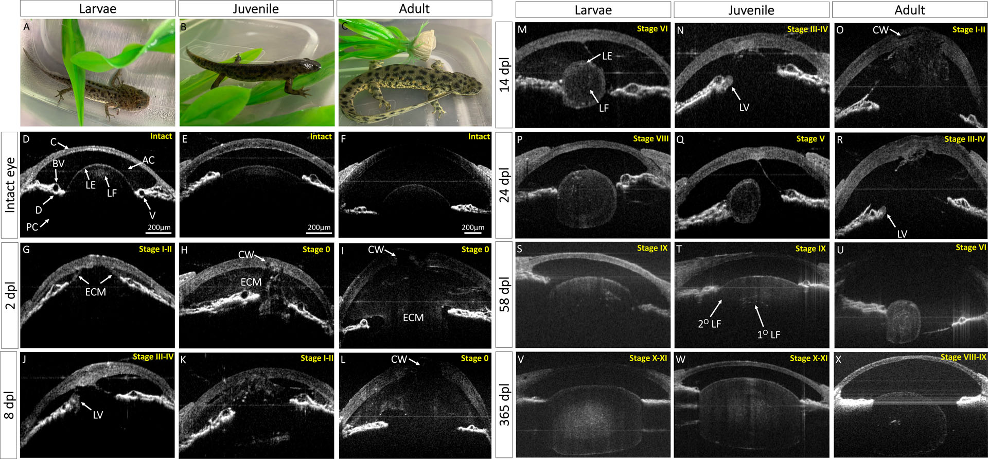
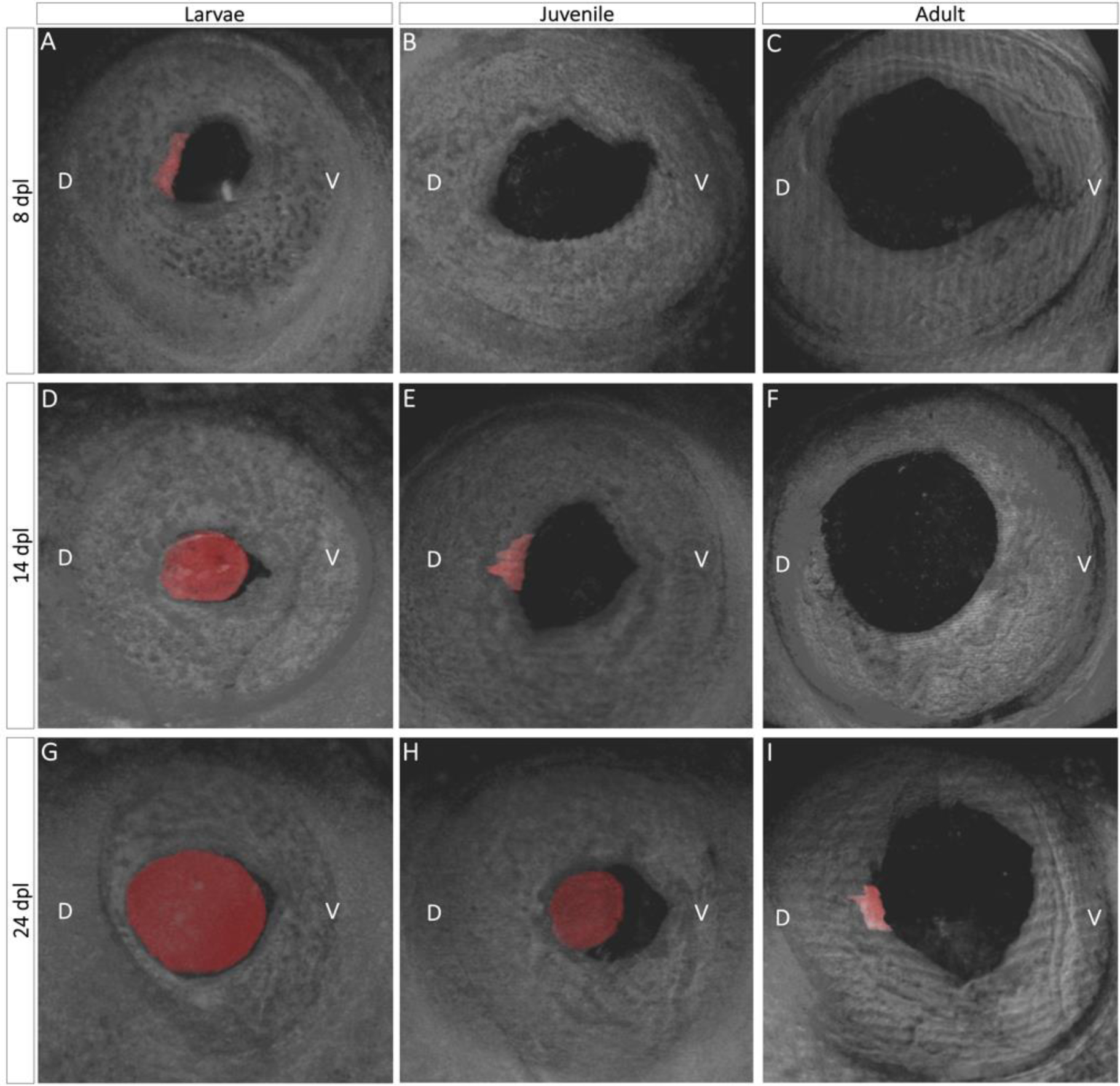
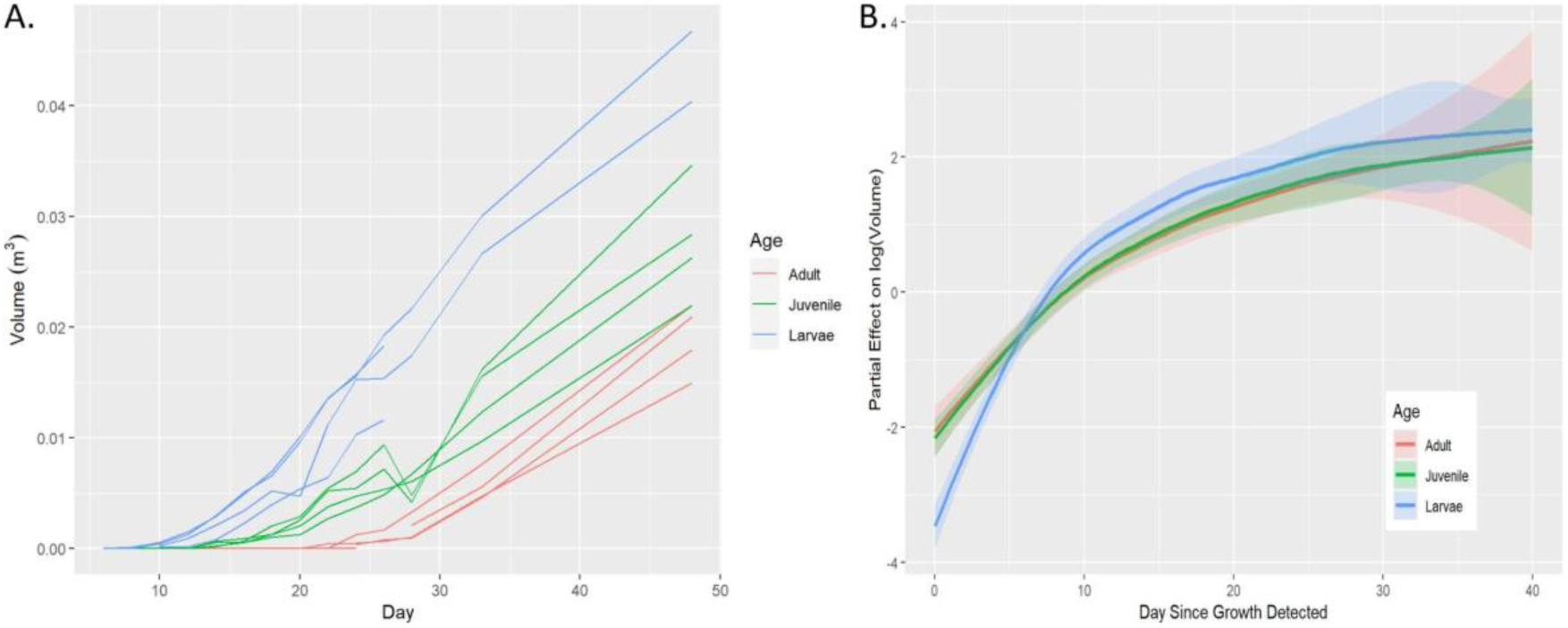
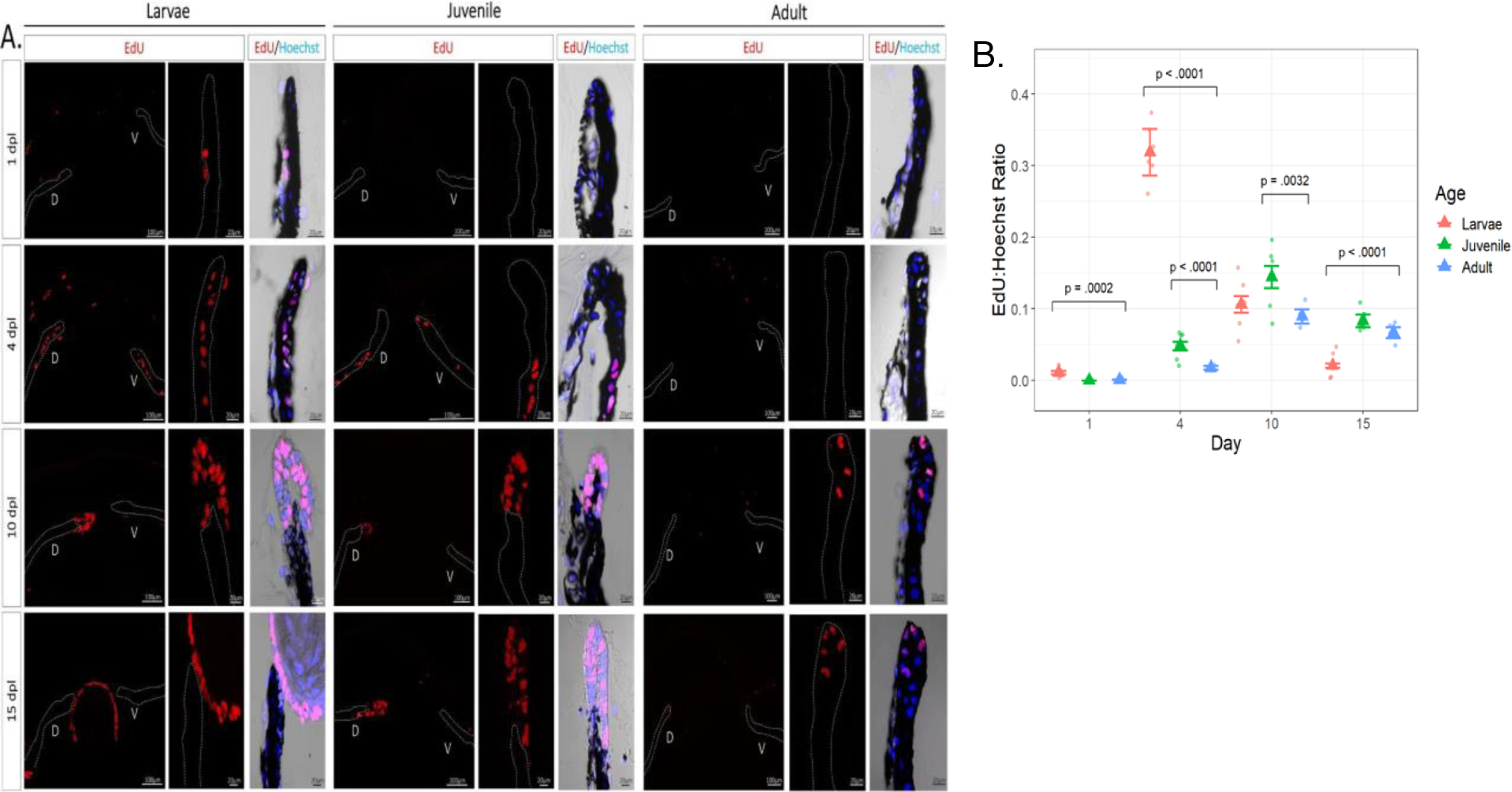
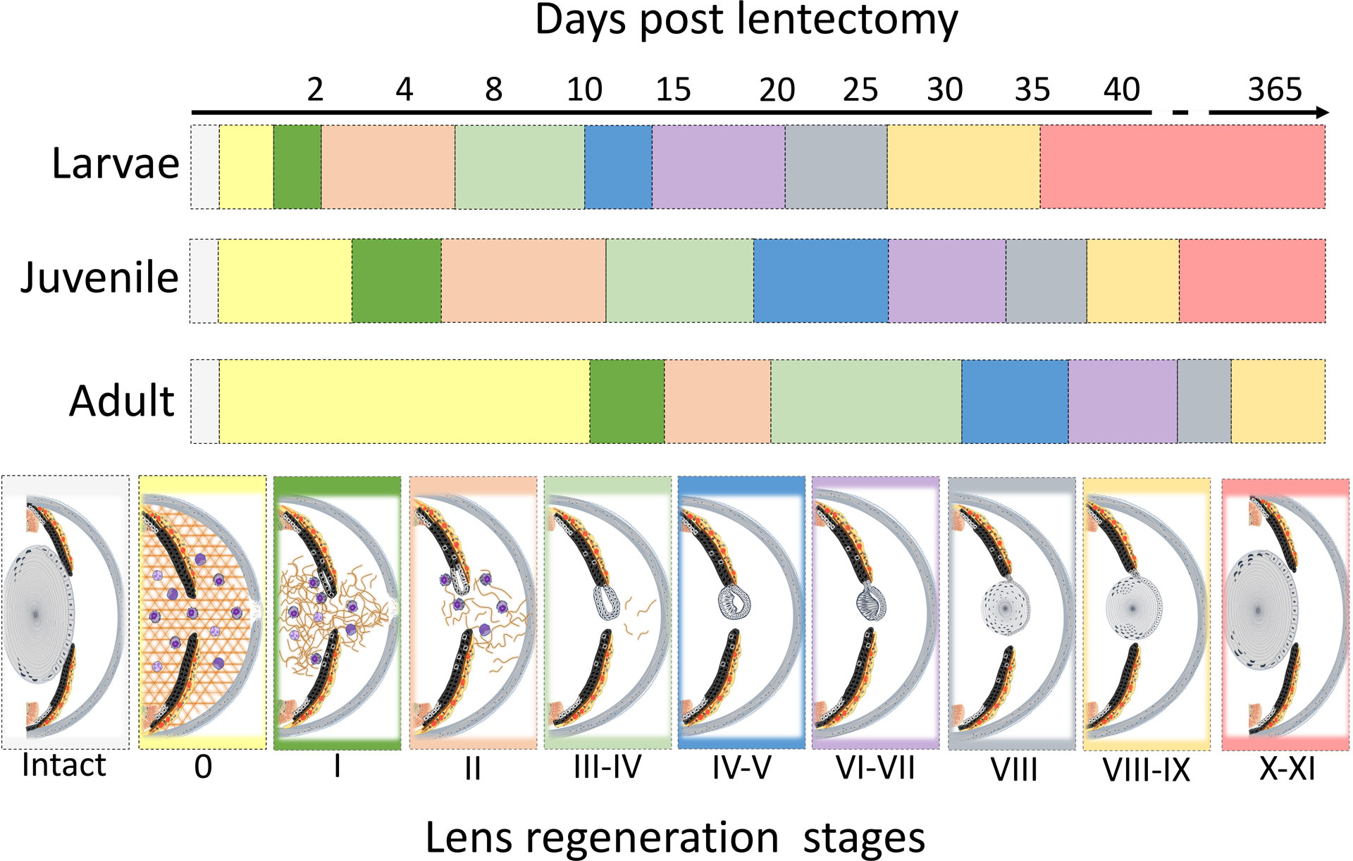
Results: Here, we used Spectral-Domain Optical Coherence Tomography (SD-OCT) to monitor the lens regeneration process of larvae, juvenile, and adult newts. While all three life stages were able to regenerate a lens through transdifferentiation of the dorsal iris pigment epithelial cells (iPECs), an age-related change in the kinetics of the regeneration process was observed. Consistent with these findings, iPECs from older animals exhibited a delay in cell cycle re-entry. Furthermore, it was observed that clearance of the extracellular matrix (ECM) was delayed in older organisms.
Conclusions: Collectively, our results suggest that although lens regeneration capacity does not decline throughout the lifespan of newts, the intrinsic and extrinsic cellular changes associated with aging alter the kinetics of this process. By understanding how these changes affect lens regeneration in newts, we can gain important insights for restoring the age-related regeneration decline observed in most vertebrates.
Keywords: Aging; Cell cycle; ECM remodeling; Lens regeneration; SD-OCT; iPECs.
Copyright © 2023 The Authors. Published by Elsevier B.V. All rights reserved.
Conflict of interest statement
Declaration of competing interest None.
Figures

Similar articles
-
Macrophages modulate fibrosis during newt lens regeneration.Stem Cell Res Ther. 2024 May 14;15(1):141. doi: 10.1186/s13287-024-03740-1. Stem Cell Res Ther. 2024. PMID: 38745238 Free PMC article.
-
The Black Book of Psychotropic Dosing and Monitoring.Psychopharmacol Bull. 2024 Jul 8;54(3):8-59. Psychopharmacol Bull. 2024. PMID: 38993656 Free PMC article. Review.
-
Short-Term Memory Impairment.2024 Jun 8. In: StatPearls [Internet]. Treasure Island (FL): StatPearls Publishing; 2025 Jan–. 2024 Jun 8. In: StatPearls [Internet]. Treasure Island (FL): StatPearls Publishing; 2025 Jan–. PMID: 31424720 Free Books & Documents.
-
"It Was Like the Final Piece in the Puzzle for Me": A Qualitative Study on the Experiences of Autistic Women Initially Diagnosed with Borderline Personality Disorder.Autism Adulthood. 2024 Dec 2;6(4):428-437. doi: 10.1089/aut.2023.0031. eCollection 2024 Dec. Autism Adulthood. 2024. PMID: 40018060
-
Signs and symptoms to determine if a patient presenting in primary care or hospital outpatient settings has COVID-19.Cochrane Database Syst Rev. 2022 May 20;5(5):CD013665. doi: 10.1002/14651858.CD013665.pub3. Cochrane Database Syst Rev. 2022. PMID: 35593186 Free PMC article.
Cited by
-
OCTSharp: an open-source and real-time OCT imaging software based on C.Biomed Opt Express. 2023 Oct 31;14(11):6060-6071. doi: 10.1364/BOE.505308. eCollection 2023 Nov 1. Biomed Opt Express. 2023. PMID: 38021120 Free PMC article.
-
Macrophages modulate fibrosis during newt lens regeneration.Stem Cell Res Ther. 2024 May 14;15(1):141. doi: 10.1186/s13287-024-03740-1. Stem Cell Res Ther. 2024. PMID: 38745238 Free PMC article.
-
Epigenetic Modifications in the Retinal Pigment Epithelium of the Eye During RPE-Related Regeneration or Retinal Diseases in Vertebrates.Biomedicines. 2025 Jun 25;13(7):1552. doi: 10.3390/biomedicines13071552. Biomedicines. 2025. PMID: 40722628 Free PMC article. Review.
-
Macrophages modulate fibrosis during newt lens regeneration.Res Sq [Preprint]. 2023 Nov 25:rs.3.rs-3603645. doi: 10.21203/rs.3.rs-3603645/v1. Res Sq. 2023. Update in: Stem Cell Res Ther. 2024 May 14;15(1):141. doi: 10.1186/s13287-024-03740-1. PMID: 38045376 Free PMC article. Updated. Preprint.
References
-
- Benjamini Y, & Hochberg Y, 1995. Controlling the false discovery rate: a practical and powerful approach to multiple testing. Journal of the Royal statistical society: series B (Methodological) 57(1), 289–300.
-
- Brockes JP, Kumar A, 2008. Comparative aspects of animal regeneration. Annu Rev Cell Dev Biol 24, 525–549. - PubMed
-
- Carpaij OA, Goorsenberg AWM, d’Hooghe JNS, de Bruin DM, van den Elzen RM, Nawijn MC, Annema JT, van den Berge M, Bonta PI, Burgess JK, 2020. Optical Coherence Tomography Intensity Correlates with Extracellular Matrix Components in the Airway Wall. Am J Respir Crit Care Med 202, 762–766. - PubMed
Publication types
MeSH terms
Grants and funding
LinkOut - more resources
Full Text Sources
