

Complosome - the intracellular complement system
- PMID: 37055581
- PMCID: PMC10100629
- DOI: 10.1038/s41581-023-00704-1
Complosome - the intracellular complement system
Abstract
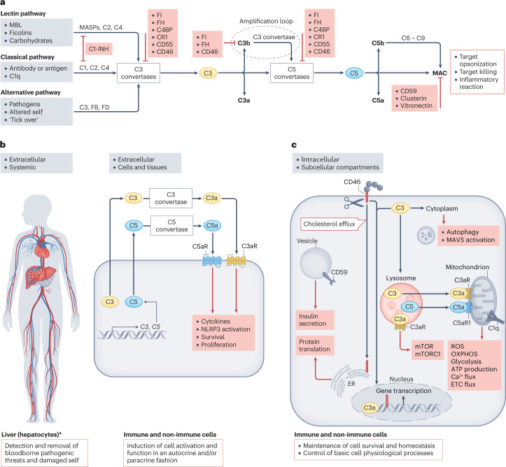
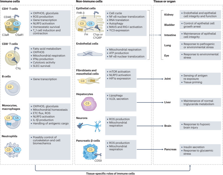
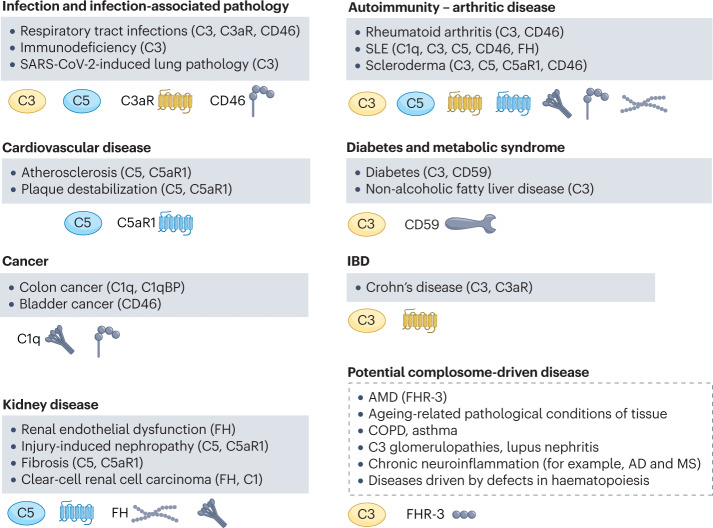
The complement system is a recognized pillar of host defence against infection and noxious self-derived antigens. Complement is traditionally known as a serum-effective system, whereby the liver expresses and secretes most complement components, which participate in the detection of bloodborne pathogens and drive an inflammatory reaction to safely remove the microbial or antigenic threat. However, perturbations in normal complement function can cause severe disease and, for reasons that are currently not fully understood, the kidney is particularly vulnerable to dysregulated complement activity. Novel insights into complement biology have identified cell-autonomous and intracellularly active complement - the complosome - as an unexpected central orchestrator of normal cell physiology. For example, the complosome controls mitochondrial activity, glycolysis, oxidative phosphorylation, cell survival and gene regulation in innate and adaptive immune cells, and in non-immune cells, such as fibroblasts and endothelial and epithelial cells. These unanticipated complosome contributions to basic cell physiological pathways make it a novel and central player in the control of cell homeostasis and effector responses. This discovery, together with the realization that an increasing number of human diseases involve complement perturbations, has renewed interest in the complement system and its therapeutic targeting. Here, we summarize the current knowledge about the complosome across healthy cells and tissues, highlight contributions from dysregulated complosome activities to human disease and discuss potential therapeutic implications.
© 2023. This is a U.S. Government work and not under copyright protection in the US; foreign copyright protection may apply.
Conflict of interest statement
The authors declare no competing interests.
Figures
Similar articles
-
Complement, complosome, and complotype: A perspective.Eur J Immunol. 2023 Dec;53(12):e2250042. doi: 10.1002/eji.202250042. Epub 2023 May 11. Eur J Immunol. 2023. PMID: 37120820 Free PMC article. Review.
-
Complement's favourite organelle-Mitochondria?Br J Pharmacol. 2021 Jul;178(14):2771-2785. doi: 10.1111/bph.15238. Epub 2020 Sep 28. Br J Pharmacol. 2021. PMID: 32840864 Free PMC article. Review.
-
Complement and human T cell metabolism: Location, location, location.Immunol Rev. 2020 May;295(1):68-81. doi: 10.1111/imr.12852. Epub 2020 Mar 12. Immunol Rev. 2020. PMID: 32166778 Free PMC article. Review.
-
The role of the complosome in health and disease.Front Immunol. 2023 Mar 10;14:1146167. doi: 10.3389/fimmu.2023.1146167. eCollection 2023. Front Immunol. 2023. PMID: 36969185 Free PMC article. Review.
-
Complement Has Brains-Do Intracellular Complement and Immunometabolism Cooperate in Tissue Homeostasis and Behavior?Front Immunol. 2021 Feb 25;12:629986. doi: 10.3389/fimmu.2021.629986. eCollection 2021. Front Immunol. 2021. PMID: 33717157 Free PMC article. Review.
Cited by
-
Immunity and Coagulation in COVID-19.Int J Mol Sci. 2024 Oct 19;25(20):11267. doi: 10.3390/ijms252011267. Int J Mol Sci. 2024. PMID: 39457048 Free PMC article. Review.
-
Structural biology of complement receptors.Front Immunol. 2023 Sep 11;14:1239146. doi: 10.3389/fimmu.2023.1239146. eCollection 2023. Front Immunol. 2023. PMID: 37753090 Free PMC article. Review.
-
Senescent fibro-adipogenic progenitors are potential drivers of pathology in inclusion body myositis.Acta Neuropathol. 2023 Nov;146(5):725-745. doi: 10.1007/s00401-023-02637-2. Epub 2023 Sep 29. Acta Neuropathol. 2023. PMID: 37773216 Free PMC article.
-
Canonical and non-canonical roles of complement in atherosclerosis.Nat Rev Cardiol. 2024 Nov;21(11):743-761. doi: 10.1038/s41569-024-01016-y. Epub 2024 Apr 10. Nat Rev Cardiol. 2024. PMID: 38600367 Review.
-
High-fat diet induces pre-eclampsia through dampening cell-autonomous C3 in trophoblasts.Commun Biol. 2025 Jun 6;8(1):879. doi: 10.1038/s42003-025-08298-z. Commun Biol. 2025. PMID: 40481230 Free PMC article.
References
-
- Bordet JGO. Sur l’existence de substances sensibilisatrices dans la plupart des serums antimicrobiens. Ann. De. l’Inst. Pasteur. 1901;15:289–303.
Publication types
MeSH terms
Substances
Grants and funding
LinkOut - more resources
Full Text Sources
