Review
. 2023 Jun;28(5-6):754-768.
doi: 10.1007/s10495-023-01837-1. Epub 2023 Apr 13.

Apoptotic Janus-faced mycotoxins against thoracal and breast metastases

Affiliations
Review

Apoptotic Janus-faced mycotoxins against thoracal and breast metastases

Gaspar Banfalvi. Apoptosis. 2023 Jun.

Abstract

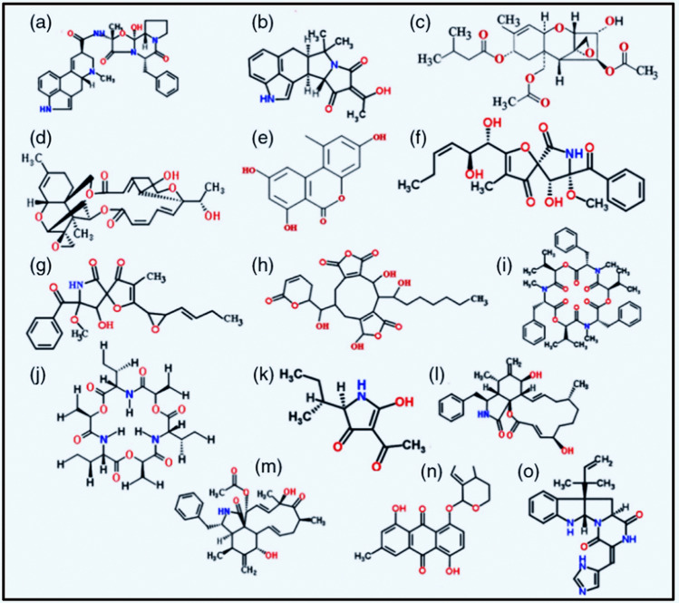
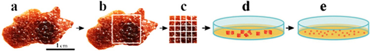
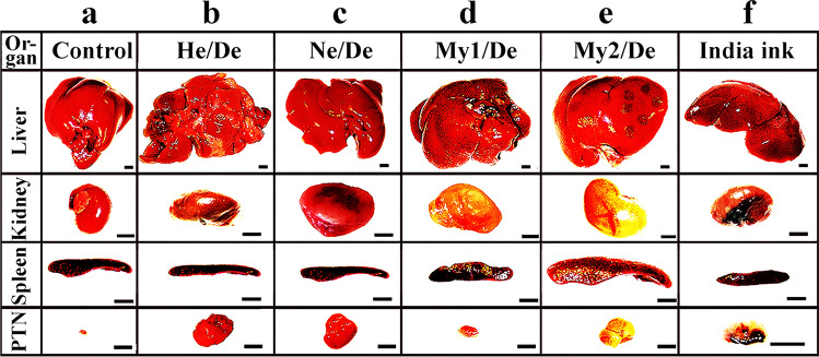
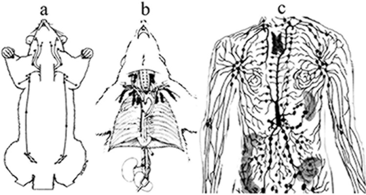
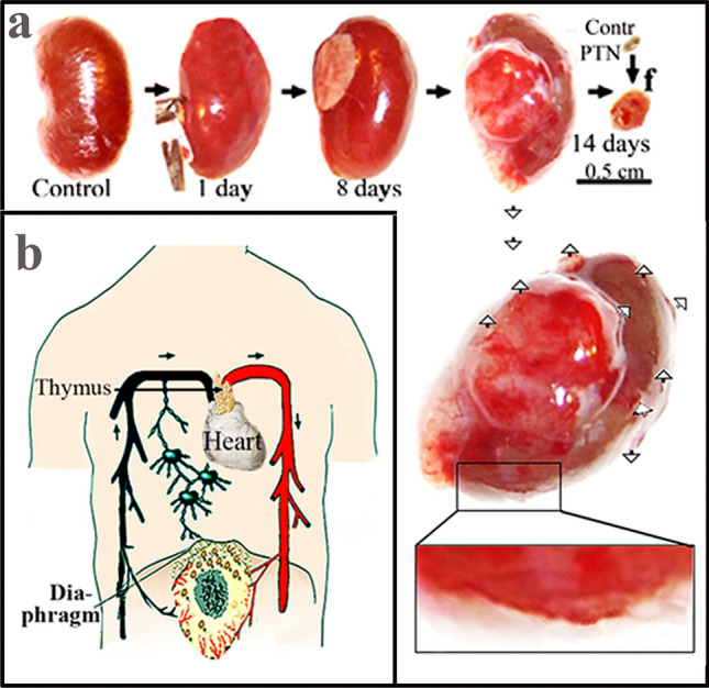
Abdominal organs (liver, kidney, spleen) are frequent targets of cancer cell invasion but their primary tumours are less known for their metastatic potential to other organs e.g. to the breast. Despite the known connection of the pathogenesis from breast cancer to liver metastasis, the study of the spread in the opposite direction has been neglected. The notion that breast cancer could be a metastasis besides being a primary tumour is based on rodents' tumour models upon implantation of tumour cells under the capsule of the kidney or under the Glisson's capsule of the liver of rats and mice. Tumour cells develop into a primary tumour at the site of subcutaneous implantation. The metastatic process starts with peripheral disruptions of blood vessels near the surface of primary tumours. Tumour cells released into the abdomen cross the apertures of the diaphragm, enter the thoracal lymph nodes and accumulate in parathymic lymph nodes. Abdominal colloidal carbon particles injected into the abdomen faithfully mimicked the migration of tumour cells and deposited in parathymic lymph nodes (PTNs). An explanation is provided why the connection between abdominal tumours and mammary tumours escaped attention, notably, parathymic lymph nodes in humans were referred to as internal mammary or parasternal lymph nodes. The apoptotic effect of Janus-faced cytotoxins is suggested to provide a new approach against the spread of abdominal primary tumours, and metastatic development.

Keywords: Abdominal tumours; Antitumour agents; Carcinogens; Double-edged molecules; Metastatic spread to PTNS; Metastatic tumour models; Prevention of breast cancer.


Conflict of interest statement

The author has declared that no conflict of interest exists.

Figures

Fig. 1
Fig. 1
Chemical structures of selected mycotoxins with strong toxic effects and anticancer potential. a Ergotamine, b cyclopiazonic acid, c T-2 toxin, d satratoxin H, e alternariol,f pseurotin, g synerazol, h rubratoxin, i beauvericin, j enniatin, k tenuazonic acid,l cytochalasin B, m cytochalasin C, n MT81, and o roquefortine of beneficial low toxicity. Permitted by [22]
Fig. 2
Fig. 2
Establishment of rat hepatocarcinoma tumour cell line (HeDe). Under strictly sterile conditions a Hepatocarcinoma tumour of the rat was surgically removed, b designated part of the tumour (boxed), c was minced into 2 × 2x2 mm pieces, d and digested with elastase medium, filtered through sterile gauze, suspended in RPMI 1640 medium. After overnight incubation at 37 °C in a CO2 incubator, the non-adherent cells were discarded and the adherent cells were subcultured. e After 20 subcultures the new hepatocarcinoma He/De cell line was established. The same procedure was applied when other (Ne/De, My1/De and My2/De) cell lines were isolated [40]
Fig. 3
Fig. 3
Primary tumours spread to distant organs. The location of the primary tumours in major human organs is indicated by concentric rings
Fig. 4
Fig. 4
Primary liver tumours spread to other organs. a Organs of rats before implanting tumour cells into the liver. b-e Implantation of tumour or cells inducing abdominal primary tumours or f) colloidal particles. Solid tumour (HeDe, NeDe) cells, (My1De or My2De) (106) were placed under the capsule of the left kidney or the Glisson’s capsule of rats. Two weeks after HeDe or NeDe and four weeks after myeloid leukaemia cell implantation animals were euthanized. The liver, the impacted left kidney, spleen and parathymic lymph nodes (PTNs) were removed surgically postmortem. a Control: liver, right kidney, spleen and parathymic lymph nodes 14 days after subrenal implantation of saline. b Liver hepatocarcinoma formation after HeDe cell (106) implantation under the Glisson’s capsule. Bottom panel: enlargement of parathymic lymph nodes (PTNs). c Nephroblastoma formation in the kidney after subrenal implantation of NeDe cells (106) into the liver. Bottom panel: Enlargement of PTNs. d Liver, kidney, spleen, PTN enlargement upon subrenal implantation of My1De cells (106). e Subrenal implantation of My2De cells (106). f Implantation under the renal capsule of 0.1 ml 0.1% India ink and appearance 24 h after administration in the cortical region of parathymic lymph nodes. Abbreviations: LN, lymph node; PTN, parathymic lymph node. Bar, 0.5 cm each
Fig. 5
Fig. 5
Tumour models to follow the metastatic migration of tumour cells from primary tumours to PTNs Heterotopic rat model. Growth and metastatic spread of abdominal primary tumour upon heterotopic implantation of hepatocarcinoma (HeDe) rat tumour cells (106) under the subrenal capsule of rats. a Haemorrhagic ruptures in the primary kidney tumour two weeks after implantation. Ruptures are indicated by the arrowheads. The outer part of the primary tumours is seen in the upper right corner of f panel. b Hematoxylin–eosin staining of a tissue section near the surface of the tumour. c. Hematoxylin–eosin staining of control PTNs.d) Haematoxylin–eosin staining of PTNs two weeks after subrenal implantation of HeDe cells
Fig. 6
Fig. 6
Tissue distribution of radioactivity after i.v. administration of 18FDG in Ne/De and He/De tumour-bearing rats. Metastatic potential of Ne/De and He/De cells, plasma, muscle, tumour, paratumourlymph nodes, thymus, liver and kidney expressed as differential absorption ratio (DAR). With permission [37]
Fig. 7
Fig. 7
Comparison of lymph nodes in the thoracic region. a The low number and small size of thoracic lymph nodes explain why opossums are inefficient against tuberculosis. b The lymphatic system of rats is resistant to tubercle bacilli. c The lymphatic system of the human thorax is more resistant to tuberculosis than marsupials but less developed than the upper thoracal lymph nodes of rats. With permission [56]
Fig. 8
Fig. 8
Blood-borne and lymphatic phases of metastasis. Phase (1). Hematogeneous growth of primary tumour cells (a). Phase (2) Hematogeneous growth of primary tumour and release of tumour cells into the abdominal cavity, and passage through the diaphragm (b). Phase (3) Lymphatic migration of tumour cells from the thoracal capillaries to the blood circulation at the left subclavian and internal jugular veins (b). Phase (4) Generalization of metastasis through blood circulation (b)

Similar articles

Cited by

  • Molecular docking and antitumor evaluation of liposomal nanoformulations containing citrinin.
    de Menezes APM, Moura MLV, de Oliveira Filho JWG, do Nascimento MLLB, Gonçalves JCR, Sobral MV, Marques KKG, da Silva FCC, De Freitas GBL, Silva VC, Coêlho SGC, Gualter MP, Negreiros HA, do Lago JPAD, de Sousa IGB, Rolim HML, de Castro E Sousa JM. de Menezes APM, et al. Naunyn Schmiedebergs Arch Pharmacol. 2025 May 1. doi: 10.1007/s00210-025-04201-z. Online ahead of print. Naunyn Schmiedebergs Arch Pharmacol. 2025. PMID: 40310529

References

    1. Salehi B, Mishra AP, Nigam M, Sener B, Kilic M, Sharifi-Rad M, Fokou PVT, Martins N, Sharifi-Rad J. Resveratrol: a double-edged sword in health benefits. Biomedicines. 2018;6:91. doi: 10.3390/biomedicines6030091. - DOI - PMC - PubMed
    1. Renaud S, de Lorgeril M. Wine, alcohol, platelets, and the French paradox for coronary heart disease. Lancet. 1992;339:1523–1526. doi: 10.1016/0140-6736(92)91277-f. - DOI - PubMed
    1. Malir F, Ostry V, Pfohl-Leszkowicz A, Novotna E, Ochratoxin A. Developmental and reproductive toxicity. an overview. Birth Defects Res B Dev Reprod Toxicol. 2013;98:493–502. doi: 10.1002/bdrb.21091. - DOI - PubMed
    1. Wang JS, Groopman JD. DNA damage by mycotoxins. Mutat Res. 1999;424:167–181. doi: 10.1016/S0027-5107(99)00017-2. - DOI - PubMed
    1. Dornetshuber R, Heffeter P, Kamyar MR, Peterbauer T, Berger W. Lemmens-Gruber R (2007) Enniatin exerts p53-dependent cytostatic and p53-independent cytotoxic activities against human cancer cells. Chem Res Toxicol. 2007;20:465–473. doi: 10.1016/s0027-5107(99)00017-2. - DOI - PubMed

Publication types