

Epidemiological profile of microbial keratitis in Alexandria-Egypt a 5 years retrospective study
- PMID: 37055636
- PMCID: PMC10102273
- DOI: 10.1186/s12348-023-00332-7
Epidemiological profile of microbial keratitis in Alexandria-Egypt a 5 years retrospective study
Abstract
Objective: To evaluate the epidemiologic profile of microbial keratitis in Alexandria- Egypt, with special emphasis on risk factors, visual outcome and microbiological results.
Methods: This retrospective study reviewed files of patients treated for microbial keratitis during a period of 5 years at Alexandria Ophthalmology Hospital Cornea Clinic, Alexandria- Egypt, between February 2017 and June 2022. The patients were evaluated for the risk factors e.g., trauma, eyelid disorders, co-morbidities, and contact lens use. They were also evaluated for their clinical picture, the identified microorganisms, visual outcomes, and complications. Non-microbial keratitis and incomplete files were excluded from the study.
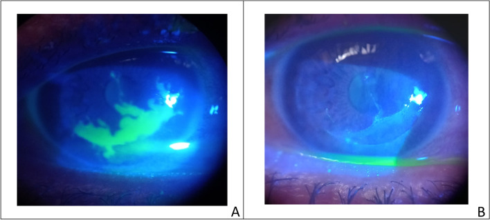
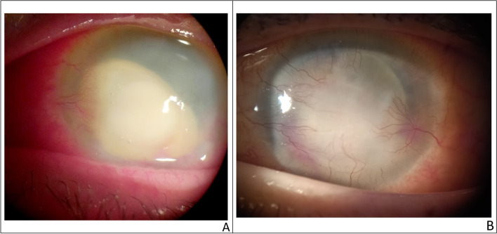
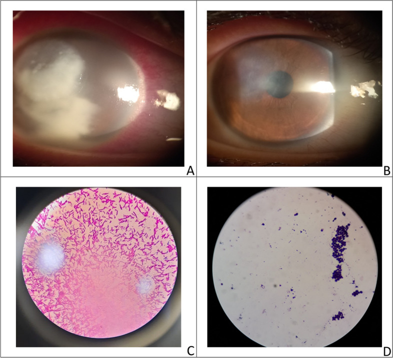
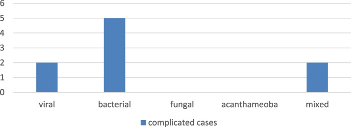
Results: A total of 284 patients were diagnosed as microbial keratitis in our study. Viral keratitis was the most common cause of microbial keratitis (n = 118 (41.55%)), followed by bacterial keratitis (n = 77 (27.11%)), mixed keratitis (n = 51 (17.96%)), acanthamoeba keratitis (n = 22 (7.75%)) and the least cause was fungal keratitis (n = 16 (5.63%)). Trauma was the most common risk factor for microbial keratitis (29.2%). Fungal keratitis had a statistically significant association with trauma (p < 0.001), while the use of contact lenses had a statistically significant association with Acanthamoeba keratitis (p < 0.001). The percentage of culture-positive results in our study was 76.8%. Gram-positive bacteria were the most frequently isolated bacterial isolate (n = 25 (36.2%)), while filamentous fungi were the most frequently isolated fungi (n = 13(18.8%)). After treatment, there was a significant increase in the mean visual acuity among all groups; it was significantly higher in Acanthamoeba keratitis group with a mean difference of 0.262 ± 0.161 (p = 0.003).
Conclusion: Viral keratitis followed by bacterial keratitis were the most frequent etiologic agents causing microbial keratitis found in our study. Although trauma was the most frequent risk factor for microbial keratitis, contact lens wear was found an important preventable risk factor for microbial keratitis in young patients. Performing culture properly whenever indicated before starting antimicrobial treatment increased the cultures' positive results.
Keywords: Acanthamoeba; Contact lens; Filamentous fungi; Gram-positive bacteria; Microbial keratitis/risk factors; Viral keratitis, Microorganisms.
© 2023. The Author(s).
Conflict of interest statement
The authors declare no competing interests.
Figures


References
LinkOut - more resources
Full Text Sources
