. 2023 Apr 13;15(1):24.
doi: 10.1186/s13073-023-01175-6.

Stalled oligodendrocyte differentiation in IDH-mutant gliomas

Affiliations

Stalled oligodendrocyte differentiation in IDH-mutant gliomas

Yanfei Wei et al. Genome Med. .

Abstract

Background: Roughly 50% of adult gliomas harbor isocitrate dehydrogenase (IDH) mutations. According to the 2021 WHO classification guideline, these gliomas are diagnosed as astrocytomas, harboring no 1p19q co-deletion, or oligodendrogliomas, harboring 1p19q co-deletion. Recent studies report that IDH-mutant gliomas share a common developmental hierarchy. However, the neural lineages and differentiation stages in IDH-mutant gliomas remain inadequately characterized.

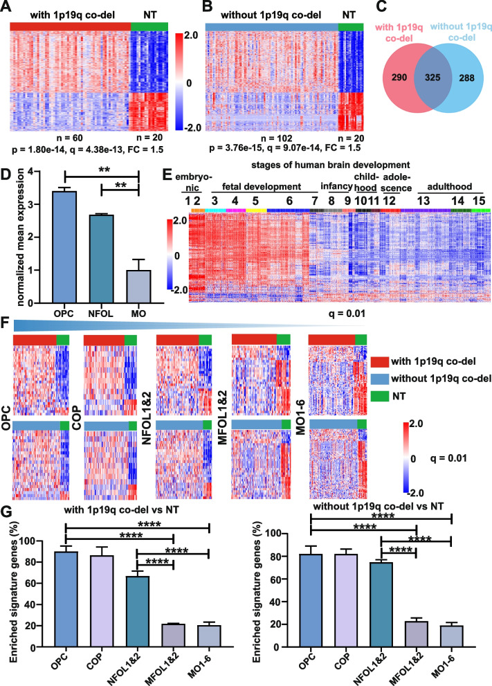
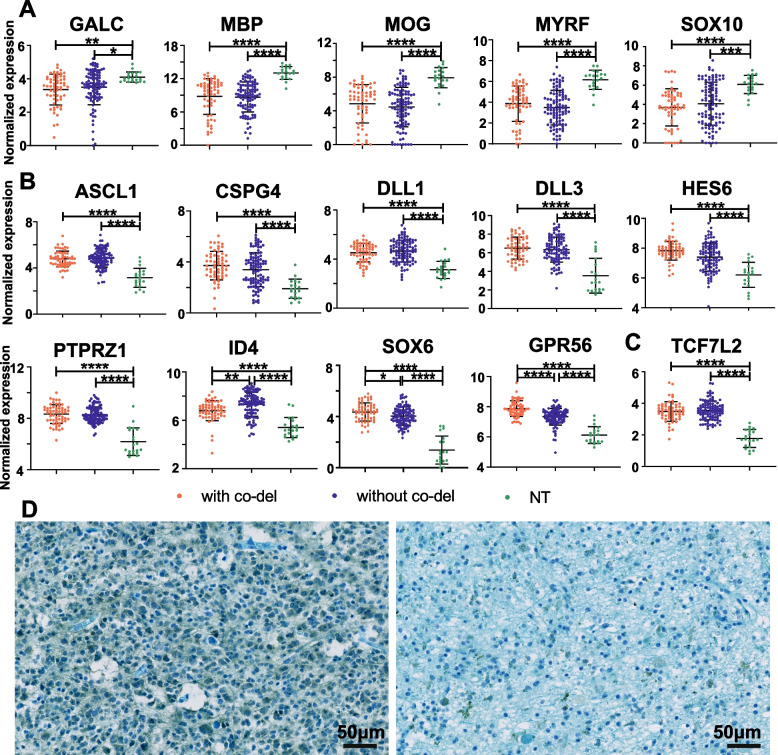
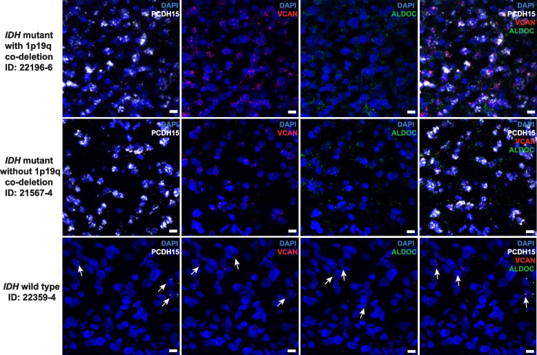
Methods: Using bulk transcriptomes and single-cell transcriptomes, we identified genes enriched in IDH-mutant gliomas with or without 1p19q co-deletion, we also assessed the expression pattern of stage-specific signatures and key regulators of oligodendrocyte lineage differentiation. We compared the expression of oligodendrocyte lineage stage-specific markers between quiescent and proliferating malignant single cells. The gene expression profiles were validated using RNAscope analysis and myelin staining and were further substantiated using data of DNA methylation and single-cell ATAC-seq. As a control, we assessed the expression pattern of astrocyte lineage markers.

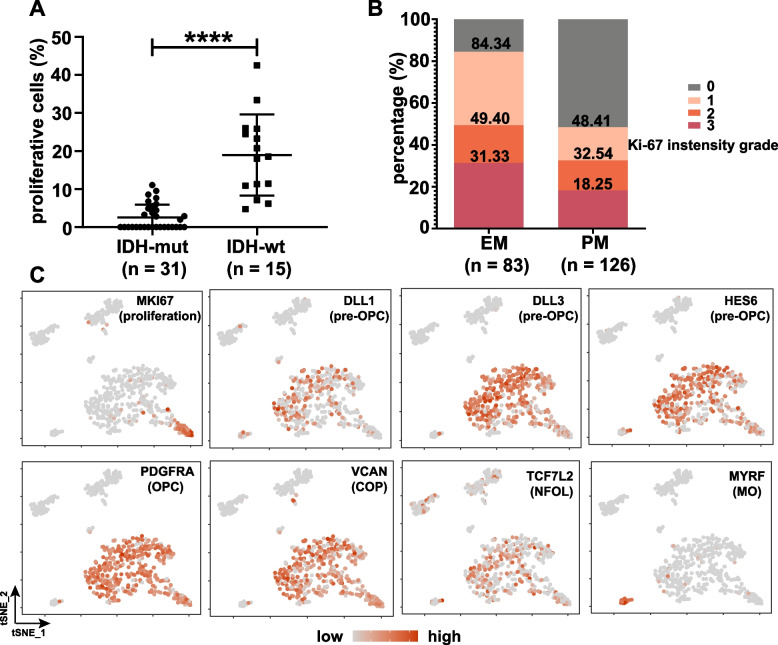
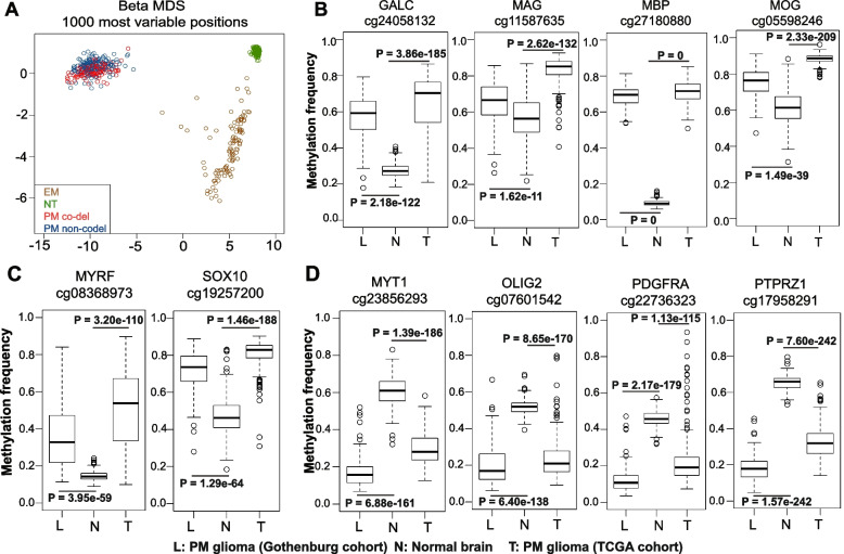
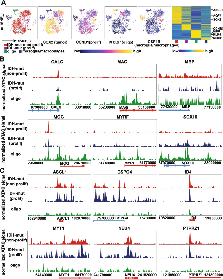
Results: Genes concordantly enriched in both subtypes of IDH-mutant gliomas are upregulated in oligodendrocyte progenitor cells (OPC). Signatures of early stages of oligodendrocyte lineage and key regulators of OPC specification and maintenance are enriched in all IDH-mutant gliomas. In contrast, signature of myelin-forming oligodendrocytes, myelination regulators, and myelin components are significantly down-regulated or absent in IDH-mutant gliomas. Further, single-cell transcriptomes of IDH-mutant gliomas are similar to OPC and differentiation-committed oligodendrocyte progenitors, but not to myelinating oligodendrocyte. Most IDH-mutant glioma cells are quiescent; quiescent cells and proliferating cells resemble the same differentiation stage of oligodendrocyte lineage. Mirroring the gene expression profiles along the oligodendrocyte lineage, analyses of DNA methylation and single-cell ATAC-seq data demonstrate that genes of myelination regulators and myelin components are hypermethylated and show inaccessible chromatin status, whereas regulators of OPC specification and maintenance are hypomethylated and show open chromatin status. Markers of astrocyte precursors are not enriched in IDH-mutant gliomas.

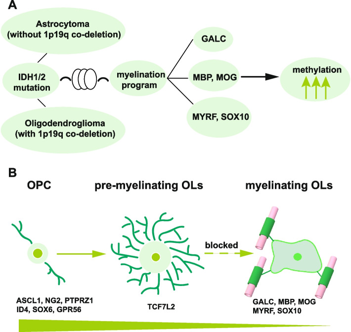
Conclusions: Our studies show that despite differences in clinical manifestation and genomic alterations, all IDH-mutant gliomas resemble early stages of oligodendrocyte lineage and are stalled in oligodendrocyte differentiation due to blocked myelination program. These findings provide a framework to accommodate biological features and therapy development for IDH-mutant gliomas.


Conflict of interest statement

The authors declare that they have no competing interests.

Figures

Fig. 1
Fig. 1
Declining expression of NFOL and MFOL signatures in PM gliomas from the CGGA dataset. A, B Heatmaps of differential gene expression profile between PM gliomas and NT brain tissues at the indicated p and q values with a fold change (FC) at 1.5. Genes concordantly enriched in both PM glioma subtypes (C) show upregulated expression in OPC and NFOL of mouse oligodendrocyte lineage as analyzed in GSE52564 [30] (D), and in early stages of human brain development as analyzed in GSE25219 [31] (E). F Drastically declining expression in the gene expression signatures of NFOL and MFOL in PM gliomas with (upper) or without (lower) 1p19q co-deletion. The same color codes were used in A, B, C, and F. Data shown in A to F were derived from CGGA dataset. G Percentages of the members of the indicated stage-specific signature gene sets enriched in PM gliomas from CGGA, GSE4290, and Rembrandt datasets; mean and standard deviation are shown. NT: non-tumor brain tissues, OPC: oligodendrocyte progenitor cell, COP: differentiation-committed oligodendrocyte precursors, NFOL: newly formed oligodendrocyte, MFOL: myeline-forming oligodendrocyte, MO: mature oligodendrocyte. **p < 0.01; ****p < 0.0001
Fig. 2
Fig. 2
Suppressed myelination program in PM gliomas from the CGGA dataset. A Under expression in PM gliomas of the MO marker GALC/O1, myelin components (MBP, MOG), and myelination regulators MYRF/C11ORF9. B Sustained expression of stage-specific markers of pre-OPC and OPC in PM gliomas. C Sustained expression of stage-specific marker of NFOL in PM gliomas. D Co-staining with LFB which labels myelin structures, and with the anti-IDH1 R132H antibody which stains IDH1 mutant cells, of a representative PM glioma with 1p19q co-deletion is shown for tumor (left) and NT (right) regions. Myelin fiber is absent in tumor regions with strong cytosolic staining of the IDH1 R132H mutant protein
Fig. 3
Fig. 3
Uniform expression of OPC and COP markers in individual PM glioma cells. AC Results of single-cell RNA-seq analysis of a representative PM glioma with 1p19q co-deletion from CGGA (sample ID: sc15). t-SNE plot and cell numbers of the cell populations identified. B Heatmap of the top 200 most differentially expressed genes across the cell populations; lineage-specific hallmarks are shown. C Inferred chromosome CNVs in the OPC/COP-like cells with non-tumor cells as the control. D Concordant expression of SOX2 and early oligodendrocytic lineage markers but sporadic expression of astrocytic lineage markers in representative PM glioma samples with (MGH54, sc16 and sc15) or without 1p19q co-deletion (sc5, MGH43 and MGH45). O: oligodendroglioma grade 2, AO: anaplastic oligodendroglioma, OA: oligoastrocytoma grade 2, A:  astrocytoma grade 2, AA: anaplastic astrocytoma, O/C: OPC/COP-like cells, PLR: proliferating cells, APC: astrocyte precursor cells
Fig. 4
Fig. 4
Expression of OPC and COP markers in individual PM glioma cells validated with RNAscope analysis. Cells from IDH-mutant/PM gliomas with or without 1p19q co-deletion uniformly co-expressed markers of OPC (PCDH15, white) and COP (VCAN, red); the expression of the astrocytic marker ALDOC (green) was sporadic. PCDH15 and VCAN were however sporadically expressed in IDH-wild-type/EM gliomas. RNAscope images of representative samples are shown, scale bar: 10 μm
Fig. 5
Fig. 5
Common differentiation stages in proliferating and non-proliferating PM glioma cells. A Frequencies of proliferating cells in single-cell RNA-seq data from the PM and EM gliomas analyzed. B Ki-67 staining of the CGGA cohort. C Expression of cell proliferation or oligodendrocyte lineage stage-specific markers overlaid on the t-SNE map for one representative sample (MGH45)
Fig. 6
Fig. 6
Suppressed myelination program in PM gliomas due to IDH-mutation induced DNA hypermethylation. A Multidimensional scaling (MDS) plot of the top 1000 most variable CpG sites in PM gliomas, EM gliomas, and normal brain tissues. B Hypermethylation of the MO marker GALC and the myelin components MAG, MBP, and MOG in PM gliomas. C Hypermethylation of MYRF/C11orf9 and SOX10 in PM gliomas. D Hypomethylation of OPC regulators MYT1, OLIG2, PDGFRA, and PTPRZ1 in PM gliomas. L, N, and T indicate PM glioma samples from Gothenburg, normal brain samples, and PM glioma samples from TCGA, respectively
Fig. 7
Fig. 7
Chromatin status of key members of myelination program and OPC specification and maintenance in IDH-mutant gliomas. A Chromatin status-based identification of cell populations and the chromatin status of canonical markers across the respective cell populations identified. t-SNE plots and heatmap are shown. B Inaccessible chromatin status of oligodendrocyte marker, myelin components, and myelination-regulatory genes in both proliferating and non-proliferating IDH-mutant glioma cells. C Open chromatin status in genes involved in OPC specification and maintenance in both proliferating and non-proliferating IDH-mutant glioma cells. IDH-mut: IDH-mutant, non-prolif: non-proliferating cells, prolif: proliferating cells, oligo: oligodendrocytes
Fig. 8
Fig. 8
Schematic depiction of differentiation blockage of IDH-mutant gliomas. A, B IDH-mutant astrocytomas and oligodendrogliomas are both blocked at the premyelination stage due to hypermethylation and down-regulated expression of myelination regulators and myelin components. OPC: oligodendrocyte progenitor cell, OLs: oligodendrocytes

References

    1. Ostrom QT, Bauchet L, Davis FG, Deltour I, Fisher JL, Langer CE, Pekmezci M, Schwartzbaum JA, Turner MC, Walsh KM, et al. The epidemiology of glioma in adults: a "state of the science" review. Neuro Oncol. 2014;16:896–913. doi: 10.1093/neuonc/nou087. - DOI - PMC - PubMed
    1. Stupp R, Hegi ME, Mason WP, van den Bent MJ, Taphoorn MJ, Janzer RC, Ludwin SK, Allgeier A, Fisher B, Belanger K, et al. Effects of radiotherapy with concomitant and adjuvant temozolomide versus radiotherapy alone on survival in glioblastoma in a randomised phase III study: 5-year analysis of the EORTC-NCIC trial. Lancet Oncol. 2009;10:459–466. doi: 10.1016/S1470-2045(09)70025-7. - DOI - PubMed
    1. Laug D, Glasgow SM, Deneen B. A glial blueprint for gliomagenesis. Nat Rev Neurosci. 2018;19:393–403. doi: 10.1038/s41583-018-0014-3. - DOI - PMC - PubMed
    1. Louis DN, Perry A, Reifenberger G, von Deimling A, Figarella-Branger D, Cavenee WK, Ohgaki H, Wiestler OD, Kleihues P, Ellison DW. The 2016 world health organization classification of tumors of the central nervous system: a summary. Acta Neuropathol. 2016;131:803–820. doi: 10.1007/s00401-016-1545-1. - DOI - PubMed
    1. Weller M, van den Bent M, Preusser M, Le Rhun E, Tonn JC, Minniti G, Bendszus M, Balana C, Chinot O, Dirven L, et al. EANO guidelines on the diagnosis and treatment of diffuse gliomas of adulthood. Nat Rev Clin Oncol. 2021;18:170–186. doi: 10.1038/s41571-020-00447-z. - DOI - PMC - PubMed

Publication types