. 2023 Jul 5;31(7):2028-2041.
doi: 10.1016/j.ymthe.2023.04.005. Epub 2023 Apr 13.

Development of a translatable gene augmentation therapy for CNGB1-retinitis pigmentosa

Affiliations

Development of a translatable gene augmentation therapy for CNGB1-retinitis pigmentosa

Laurence M Occelli et al. Mol Ther. .

Abstract

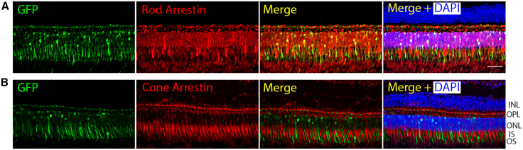
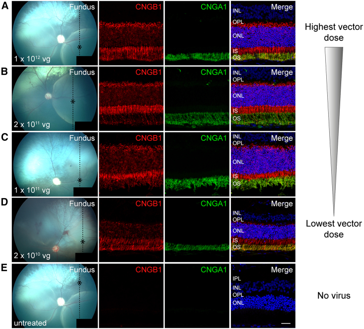
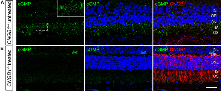
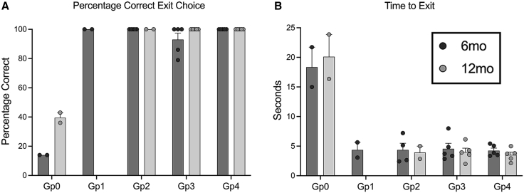
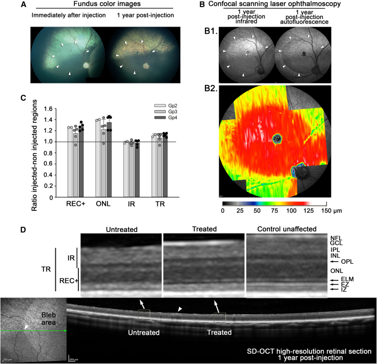
In this study, we investigate a gene augmentation therapy candidate for the treatment of retinitis pigmentosa (RP) due to cyclic nucleotide-gated channel beta 1 (CNGB1) mutations. We use an adeno-associated virus serotype 5 with transgene under control of a novel short human rhodopsin promoter. The promoter/capsid combination drives efficient expression of a reporter gene (AAV5-RHO-eGFP) exclusively in rod photoreceptors in primate, dog, and mouse following subretinal delivery. The therapeutic vector (AAV5-RHO-CNGB1) delivered to the subretinal space of CNGB1 mutant dogs restores rod-mediated retinal function (electroretinographic responses and vision) for at least 12 months post treatment. Immunohistochemistry shows human CNGB1 is expressed in rod photoreceptors in the treated regions as well as restoration of expression and trafficking of the endogenous alpha subunit of the rod CNG channel required for normal channel formation. The treatment reverses abnormal accumulation of the second messenger, cyclic guanosine monophosphate, which occurs in rod photoreceptors of CNGB1 mutant dogs, confirming formation of a functional CNG channel. In vivo imaging shows long-term preservation of retinal structure. In conclusion, this study establishes the long-term efficacy of subretinal delivery of AAV5-RHO-CNGB1 to rescue the disease phenotype in a canine model of CNGB1-RP, confirming its suitability for future clinical development.

Keywords: CNGB1; adeno-associated virus; dog; electroretinography; gene therapy; nonhuman primate; perifoveal chorioretinal atrophy; retinitis pigmentosa; short rhodopsin promoter; spectral domain optical coherence tomography.


Conflict of interest statement

Declaration of interests C.R.O. and A.F. are employees of Sanofi. S.M. is listed as inventor on the patent application WO2018172961A1 ‘‘Gene therapy for the treatment of cngb1-linked retinitis pigmentosa’’ and is co-founder of the gene therapy company ViGeneron GmbH.

Figures

None
Graphical abstract
Figure 1
Figure 1
Reporter gene expression in nonhuman primates following subretinal injection (A) Retinal section across injection bleb showing GFP expression (green) within the bleb. Blue is DAPI nuclear stain. Size bar, 100 μm. (B) Representative sections from blebs of the four dose groups (B1–B4). In each case, GFP expression (using anti-GFP antibody) is shown in green in the left panel, and GFP expression merged with DAPI (blue) is shown in the right panel. Doses (vector genomes) are indicated above each panel, and animal number, eye, and bleb imaged are indicated beside each panel. A dose effect is evident. Size bar, 50 μm. Key: INL, inner nuclear layer; OPL, outer plexiform layer; ONL, outer nuclear layer; IS, inner segment; OS, outer segment. (C) Comparative number of GFP expressing outer nuclear cell nuclei per unit length for each dose group, illustrating the dose effect. The color key indicates the individual animals.
Figure 2
Figure 2
Representative IHC of NHP retinal sections showing exclusive rod transduction (A) Panel illustrating the co-localization of GFP expression with rod arrestin. (B) Double labeling for GFP with cone arrestin showing that there is no detectable GFP expression in cone photoreceptors. Size bar, 50 μm. Key: INL, inner nuclear layer; OPL, outer plexiform layer; ONL, outer nuclear layer; IS, inner segment; OS, outer segment.
Figure 3
Figure 3
Montage showing retinal cross-sections from canine eyes from each treatment group from the highest to the lowest dose (A) Dose group 4 (17-011 OD), (B) dose group 3 (17-007 OD), (C) dose group 2 (17-040 OD), and (D) dose group 1 (18-076 OS). (E) Untreated portion of a treated eye (17-011 OD). The first column is a color fundus image taken immediately after subretinal injection. The dotted line shows the plane of the histological section and the asterisk the region of the section imaged. The first IHC column is labeled for CNGB1 (in red) and shows a dosage effect. The next column shows CNGA1 labeling (in green) also showing a dosage effect. Note that CNGA1 expression is dependent on CNGB1 expression. The final column is the merge. Size bar, 25 μm.
Figure 4
Figure 4
Gene augmentation therapy reverses disease-related cGMP accumulation (A) shows an untreated retinal area of a CNGB1 mutant dog (17-011 OD group 4). cGMP is in green with the insert showing a magnified view of the region in the white box. Note cGMP is detected in the inner and outer segment layers of the photoreceptors. (B) shows the same eye in a treated retinal region cGMP is green and CNGB1 red. Note the lack of accumulation of cGMP in the treated retinal region where CNGB1 labeling is present. Size bar, 25 μm.
Figure 5
Figure 5
Gene augmentation rescue of rod function (A) ERG dark-adapted luminance series from an untreated eye (17–042 OS 6 mo), an eye 6 months post injection (17-007 OD Gp3), and the same eye 12 months post injection. The red tracings indicate the ISCEV recommended ERG flash stimuli of 0.01, 3, and 10 cd‧s/m2. (B) Rod 5-Hz flicker (0.025 cd‧s/m2) results in the same eyes as in (A). (C–E) The mean (+/− SEM) dark-adapted ERG amplitudes for the ISCEV flash stimuli of 0.01, 3, and 10 cd‧s/m2 for the control eyes (Gp0) and the four dose groups (Gp1, Gp2, Gp3, Gp4). The 6-month post injection time point is in black, and the 12-month time point is in gray. Note that Gp1 was only maintained to 6 months post injection. There is no a-wave response at 0.01 cd‧/m2. (F) The mean (+/− SEM) 5-Hz rod flicker amplitudes for the same groups as in (C)–(E).
Figure 6
Figure 6
Vision testing. Scotopic vision of CNGB1 mutant dogs restored by gene augmentation therapy Vision testing outcome from four-choice vision testing device showing results under scotopic lighting levels (0.057 lux). (A) Shows mean (+/−SEM) percentage correct exit choices. (B) Mean (+/− SEM) time to exit. While the control dogs (GP0) fail to regularly choose the open exit and take a prolonged time to exit the device, dogs in all treated groups were able to identify the open exit accurately and rapidly exit the device. Gp1 dogs and two of the Gp2 dogs were only kept for 6 months so are not shown in the 12-month post-injection graphs.
Figure 7
Figure 7
Structural preservation in treated eye (A) Fundus photographs of Gp 3 (1E+12 Titer, 2E+11 Dose) dog 17-007 OD (right eye). The color fundus images show the injection bleb immediately after injection and then the fundus 1 year later. (B) B1. The cSLO images show infrared and autofluorescent imaging of the injected region. B2. A heatmap clearly shows the preservation of the treated retinal area, showing REC+ thickness. (C) REC+, ONL, IR, and TR thicknesses ratio of thickness in treated regions versus thickness in untreated regions at 12 months post injection for three doses/groups (mean ratio +/- SD). This shows preservation of the outer retina layers REC+ and ONL for all groups in the treated regions. (D) cSLO (left) and SD-OCT high-resolution retinal cross-section (right) images show the treated and untreated adjacent regions. The red arrow indicates the junction between treated and untreated regions. The two enlarged images above show high magnification of untreated and treated regions. Notice the thicker outer nuclear layer in the treated region and the improved definition of ELM, EZ, and IZ bands. TR, total retina; REC+, Receptor+; IR, inner retina; NFL, nerve fiber layer; GCL, ganglion cell layer; IPL, inner plexiform layer; INL, inner nuclear layer; OPL, outer plexiform layer; ONL, outer nuclear layer; ELM, external limiting membrane; EZ, ellipsoid zone; IZ, interdigitation zone.

References

    1. Russell S., Bennett J., Wellman J.A., Chung D.C., Yu Z.F., Tillman A., Wittes J., Pappas J., Elci O., McCague S., et al. Efficacy and safety of voretigene neparvovec (AAV2-hRPE65v2) in patients with RPE65-mediated inherited retinal dystrophy: a randomised, controlled, open-label, phase 3 trial. Lancet. 2017;390:849–860. - PMC - PubMed
    1. Georgiou M., Fujinami K., Michaelides M. Inherited retinal diseases: therapeutics, clinical trials and end points-A review. Clin. Exp. Ophthalmol. 2021;49:270–288. - PubMed
    1. Daiger S.P., Rossiter B.J.F., Greenberg J., Christoffels A., Hide W. Data services and software for identifying genes and mutations causing retinal degeneration. Invest. Ophthalmol. Vis. Sci. 1998;39:S295.
    1. Hartong D.T., Berson E.L., Dryja T.P. Retinitis pigmentosa. Lancet. 2006;368:1795–1809. - PubMed
    1. Bareil C., Hamel C.P., Delague V., Arnaud B., Demaille J., Claustres M. Segregation of a mutation in CNGB1 encoding the beta-subunit of the rod cGMP-gated channel in a family with autosomal recessive retinitis pigmentosa. Hum. Genet. 2001;108:328–334. - PubMed

Publication types

Substances

Supplementary concepts

LinkOut - more resources