. 2023 Oct;18(10):2246-2251.
doi: 10.4103/1673-5374.369114.

Exosomes from bone marrow mesenchymal stem cells are a potential treatment for ischemic stroke

Affiliations

Exosomes from bone marrow mesenchymal stem cells are a potential treatment for ischemic stroke

Chang Liu et al. Neural Regen Res. 2023 Oct.

Abstract

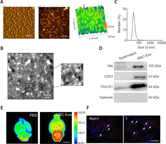
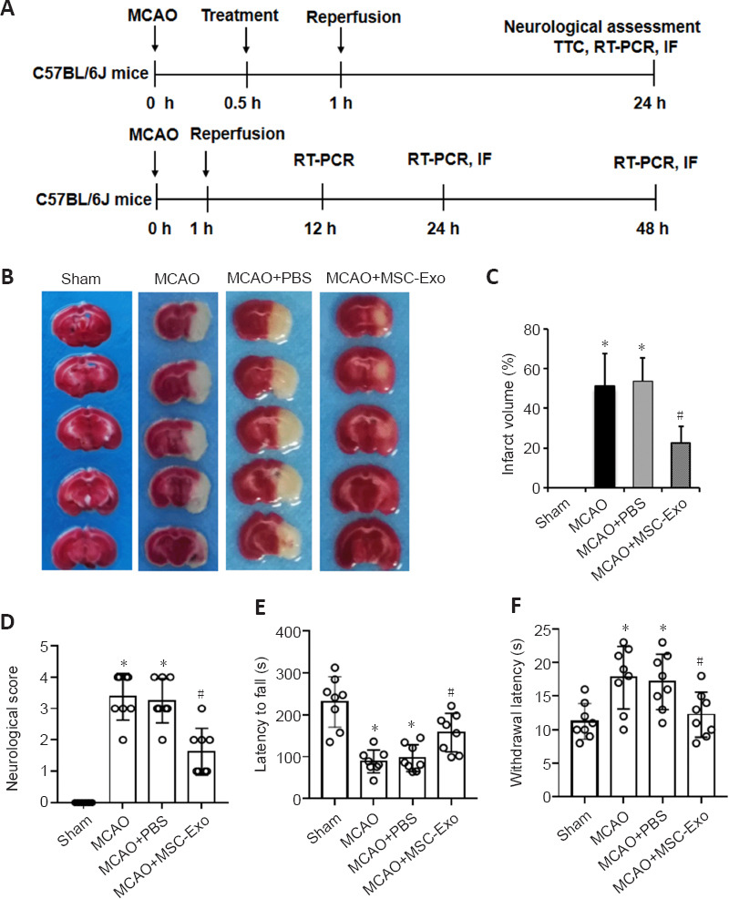
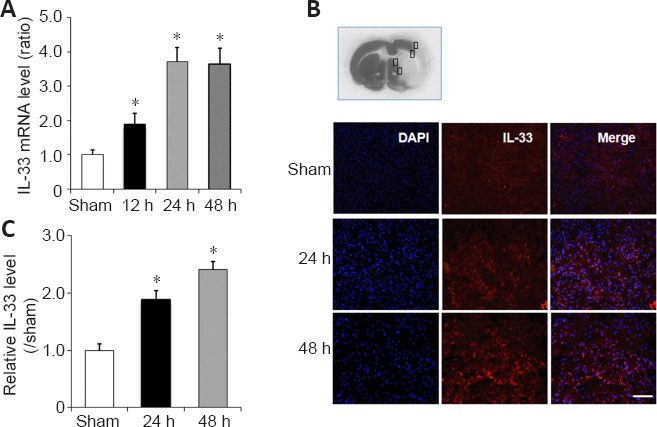
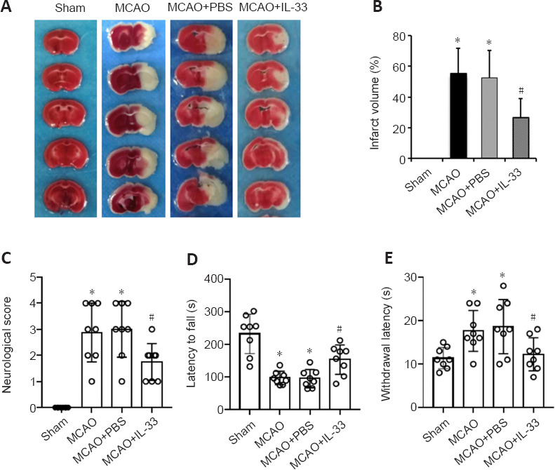
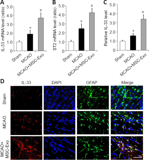
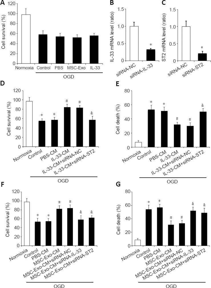
Exosomes derived from human bone marrow mesenchymal stem cells (MSC-Exo) are characterized by easy expansion and storage, low risk of tumor formation, low immunogenicity, and anti-inflammatory effects. The therapeutic effects of MSC-Exo on ischemic stroke have been widely explored. However, the underlying mechanism remains unclear. In this study, we established a mouse model of ischemic brain injury induced by occlusion of the middle cerebral artery using the thread bolt method and injected MSC-Exo into the tail vein. We found that administration of MSC-Exo reduced the volume of cerebral infarction in the ischemic brain injury mouse model, increased the levels of interleukin-33 (IL-33) and suppression of tumorigenicity 2 receptor (ST2) in the penumbra of cerebral infarction, and improved neurological function. In vitro results showed that astrocyte-conditioned medium of cells deprived of both oxygen and glucose, to simulate ischemia conditions, combined with MSC-Exo increased the survival rate of primary cortical neurons. However, after transfection by IL-33 siRNA or ST2 siRNA, the survival rate of primary cortical neurons was markedly decreased. These results indicated that MSC-Exo inhibited neuronal death induced by oxygen and glucose deprivation through the IL-33/ST2 signaling pathway in astrocytes. These findings suggest that MSC-Exo may reduce ischemia-induced brain injury through regulating the IL-33/ST2 signaling pathway. Therefore, MSC-Exo may be a potential therapeutic method for ischemic stroke.

Keywords: IL-33; ST2; astrocytes; bone marrow mesenchymal stem cells; brain injury; exosome; inflammation; ischemic stroke; neurological function; neuron.


Conflict of interest statement

None

Figures

Figure 1
Figure 1
Characterization of MSC-Exo. (A) Morphology of MSC-Exo was observed by atomic force microscopy. Panels on the left and center show peak force error images. The panel on the right shows 3D height sensor images. Scale bars: 2 µm. (B) Morphology of MSC-Exo (arrows) was observed by transmission electron microscopy. (C) Particle size distribution of MSC-Exo. (D) Western blot analysis of exosomal markers and calnexin. (E) Accumulation of PKH26-labeled MSC-Exo in mouse brain after MCAO. Fluorescence of PKH26-MSC-Exo was observed in both the ischemic hemisphere and contralateral hemisphere compared with PBS administration, and higher PKH26 fluorescence was seen in the ischemic cerebral hemisphere. Scale bar: 5 mm. Four mice were used. (F) Representative fluorescent images of PKH26-labeled MSC-Exo (red, some are indicated by arrows) at the ischemic brain region (I) and the non-ischemic brain region (Non-I). Nuclei were stained with DAPI (in blue). The fluorescence intensity of PKH26-MSC-Exo in the ischemic region was higher than that in the non-ischemic region. Scale bar: 100 µm. DAPI: 4′,6-Diamidino-2-phenylindole; I: ischemic brain region; MSC-Exo: exosomes made from bone marrow mesenchymal stem cells; Non-I: non-ischemic brain region; PBS: phosphate-buffered saline.
Figure 2
Figure 2
Effects of MSC-Exo on the infarct volume and neurological function of mice with ischemia-induced cerebral injury. (A) Experimental design for the animal study. (B) Representative images of TTC staining. MSC-Exo reduced the infarct volume (white) of ischemic mice compared with MCAO + PBS. (C) Percentage of infarct volume (n = 5 per group). (D) Longa 5-point score (n = 8 per group). (E) Latency in rotarod test (n = 8 per group). (F) Latency in hot plate test (n = 8 per group). Data are expressed as mean ± SD. *P < 0.05, vs. sham group; #P < 0.05, vs. MCAO + PBS group (C, E, F: one-way analysis of variance followed by the Tukey’s post hoc test; D: Kruskal-Wallis test). IF: Immunofluorescence; MCAO: middle cerebral artery occlusion; MSC-Exo: Exosomes made from bone marrow mesenchymal stem cells; PBS: phosphate-buffered saline; TTC: 2,3,5-triphenyltetrazolium chloride.
Figure 3
Figure 3
IL-33 expression level in the peri-infarct penumbra of mice with ischemic stroke. (A) RT-PCR results of Il-33 mRNA levels (normalized by levels in the sham group) in peri-infarct penumbra. (B) Representative immunofluorescence images of IL-33 (red) in the peri-infarct penumbra (corresponding to the box in the upper image). Nuclei were stained with DAPI (in blue). Immunofluorescence labeling revealed substantial IL-33 expression in the peri-infarct penumbra of mice 24 and 48 hours after reperfusion. Scale bar: 50 µm. (C) Quantitative analysis of immunofluorescence staining for IL-33 (normalized by levels in the sham group) in B. All values are expressed as mean ± SD (n = 5 for each group). *P < 0.05, vs. sham group (one-way analysis of variance followed by the Tukey’s post hoc test). DAPI: 4′,6-Diamidino-2-phenylindole; IL-33: interleukin-33; RT-PCR: reverse transcription-polymerase chain reaction.
Figure 4
Figure 4
Recombinant IL-33 alleviates the infarct volume and improves neurological functional outcomes in mice with ischemic stroke. (A) Representative images of TTC staining after 1-hour MCAO and 24-hour reperfusion. Recombinant IL-33 reduced the infarct volume (white) of ischemic mice compared with PBS administration. (B) Quantitative analysis of infarct volumes (n = 5 per group). (C) Longa 5-point scores (n = 8 per group). (D) Latency in rotarod test (n = 8 per group). (E) Latency in hot plate test (n = 8 per group). Data are expressed as mean ± SD. *P < 0.05, vs. sham group; #P < 0.05, vs. MCAO + PBS group (B, D, E: one-way analysis of variance followed by the Tukey’s post hoc test; C: Kruskal-Wallis test). IL-33: Interleukin-33; MCAO: middle cerebral artery occlusion; PBS: phosphate-buffered saline; TTC: 2,3,5-triphenyltetrazolium chloride.
Figure 5
Figure 5
MSC-Exo regulates the IL-33/ST2 pathway in the peri-infarct penumbra of mice. (A, B) mRNA expression of Il-33 (A) and St2 (B) (normalized by levels in the sham group) were determined by RT-PCR after 24 hours of reperfusion. (C) Quantitative analysis of immunopositivity for IL-33 (normalized by levels in the sham group). Data are expressed as mean ± SD (n = 5 per group). *P < 0.05, vs. sham group; #P < 0.05, vs. MCAO group (one-way analysis of variance followed by the Tukey’s post hoc test). (D) Representative images of immunofluorescence staining for IL-33 (red) and GFAP (green) in the peri-infarct penumbra of mice. The immunofluorescence intensity of IL-33 was higher in the MCAO group compared with the sham group. MSC-Exo treatment further increased IL-33 level in MCAO + MSC-Exo group compared with the MCAO group. Scale bar: 20 µm. MSC-Exo: Exosomes derived from bone marrow mesenchymal stem cells; IL-33: interleukin-33; MCAO: middle cerebral artery occlusion; RT-PCR: reverse transcription-polymerase chain reaction; ST2: suppression of tumorigenicity 2.
Figure 6
Figure 6
IL-33/ST2 is essential for MSC-Exo’s neuroprotective effects in vitro. (A) MTT assay after 3-hour OGD/24-hour reoxygenation. (B, C) Expression of IL-33 (B) and ST2 (C) (normalized by siRNA-NC group) were determined by quantitative reverse transcription-polymerase chain reaction. *P < 0.05, vs. siRNA-NC group (Student’s t-test). (D, E) The results of MTT (D) and LDH (E) assays showed IL-33-CM significantly increased neuronal survival, but ST2 siRNA could block the IL-33-condition medium-induced neuroprotective effect. (F, G) The results of MTT (F) and LDH (G) assays showed MSC-Exo-CM significantly increased neuronal survival, but IL-33 siRNA or ST2 siRNA could block the MSC-Exo-CM-induced neuroprotective effect. Data are expressed as mean ± SD (n = 5 per group). *P < 0.05, vs. normoxia group; #P < 0.05, vs. OGD group; &P < 0.05, vs. MSC-Exo-CM group (one-way analysis of variance followed by the Tukey’s post hoc test). CM: Conditioned medium; IL-33: interleukin-33; LDH: lactate dehydrogenase; MSC-Exo: exosomes derived from bone marrow mesenchymal stem cells; MTT: thiazolyl blue tetrazolium bromide; siRNA: small interfering RNA; ST2: suppression of tumorigenicity 2.

References

    1. Ali S, Huber M, Kollewe C, Bischoff SC, Falk W, Martin MU. IL-1 receptor accessory protein is essential for IL-33-induced activation of T lymphocytes and mast cells. Proc Natl Acad Sci U S A. 2007;104:18660–18665. - PMC - PubMed
    1. Bacova M, Bimbova K, Kisucka A, Lukacova N, Galik J. Epidural oscillating field stimulation increases axonal regenerative capacity and myelination after spinal cord trauma. Neural Regen Res. 2022;17:2730–2736. - PMC - PubMed
    1. Cayrol C, Girard JP. The IL-1-like cytokine IL-33 is inactivated after maturation by caspase-1. Proc Natl Acad Sci U S A. 2009;106:9021–9026. - PMC - PubMed
    1. Dinarello CA. An IL-1 family member requires caspase-1 processing and signals through the ST2 receptor. Immunity. 2005;23:461–462. - PubMed
    1. Doeppner TR, Herz J, Görgens A, Schlechter J, Ludwig AK, Radtke S, de Miroschedji K, Horn PA, Giebel B, Hermann DM. Extracellular vesicles improve post-stroke neuroregeneration and prevent postischemic immunosuppression. Stem Cells Transl Med. 2015;4:1131–1143. - PMC - PubMed