. 2023 Oct;18(10):2291-2300.
doi: 10.4103/1673-5374.368300.

Exosomes derived from human umbilical cord mesenchymal stem cells alleviate Parkinson's disease and neuronal damage through inhibition of microglia

Affiliations

Exosomes derived from human umbilical cord mesenchymal stem cells alleviate Parkinson's disease and neuronal damage through inhibition of microglia

Zhong-Xia Zhang et al. Neural Regen Res. 2023 Oct.

Abstract

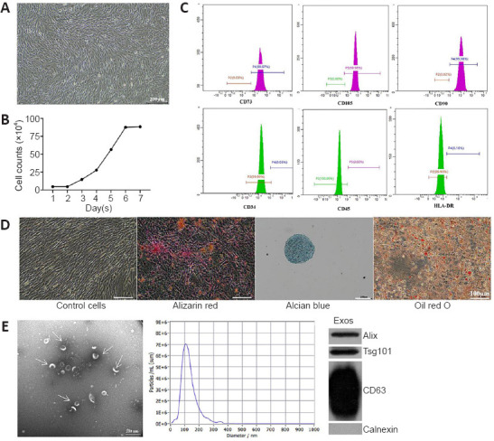
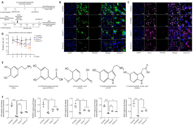
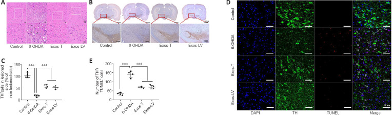
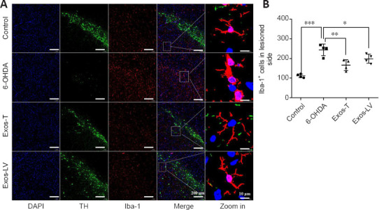
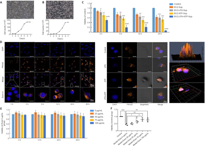
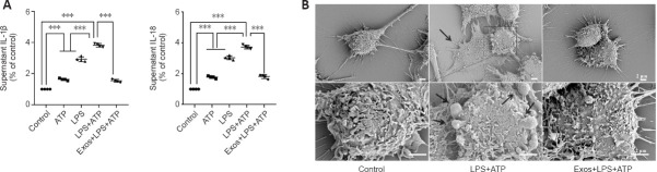
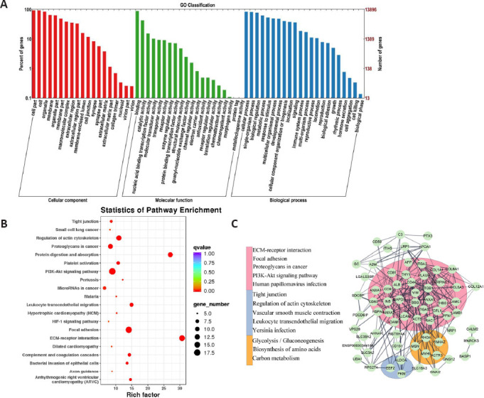
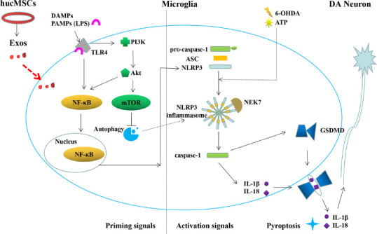
Microglia-mediated inflammatory responses have been shown to play a crucial role in Parkinson's disease. In addition, exosomes derived from mesenchymal stem cells have shown anti-inflammatory effects in the treatment of a variety of diseases. However, whether they can protect neurons in Parkinson's disease by inhibiting microglia-mediated inflammatory responses is not yet known. In this study, exosomes were isolated from human umbilical cord mesenchymal stem cells and injected into a 6-hydroxydopamine-induced rat model of Parkinson's disease. We found that the exosomes injected through the tail vein and lateral ventricle were absorbed by dopaminergic neurons and microglia on the affected side of the brain, where they repaired nigral-striatal dopamine system damage and inhibited microglial activation. Furthermore, in an in vitro cell model, pretreating lipopolysaccharide-stimulated BV2 cells with exosomes reduced interleukin-1β and interleukin-18 secretion, prevented the adoption of pyroptosis-associated morphology by BV2 cells, and increased the survival rate of SH-SY5Y cells. Potential targets for treatment with human umbilical cord mesenchymal stem cells and exosomes were further identified by high-throughput microRNA sequencing and protein spectrum sequencing. Our findings suggest that human umbilical cord mesenchymal stem cells and exosomes are a potential treatment for Parkinson's disease, and that their neuroprotective effects may be mediated by inhibition of excessive microglial proliferation.

Keywords: 6-hydroxydopamine; Parkinson’s disease; dopamine neurons; exosomes; inflammation; mesenchymal stem cells; microglia; pyroptosis.


Conflict of interest statement

None

Figures

Figure 1
Figure 1
Typical characteristics of hucMSCs and hucMSC-Exos. (A) Morphology of hucMSCs. Scale bar: 200 μm. (B) hucMSCs growth curve. The cells displayed a logarithmic growth followed by a static growth period, which is in line with the growth of stem cell populations. (C) hucMSCs surface antigens were assayed by flow cytometry. The cells were positive for the MSC-specific cell surface antigens CD73, CD105, and CD90 and negative for the hematopoietic stem cell antigens CD34, CD45, and HLA-DR. (D) Three-directional differentiation of hucMSCs. Adipogenic, osteogenic and chondrogenic differentiation were detected by oil red O, Alizarin red, and Alcian blue staining, respectively. Scale bars: 100 μm. (E) Typical characteristics of Exos: transmission electron microscope (TEM) image (left), particle size shown by nano-particle size tracking analysis (NTA) (middle), positive expression of Exos markers including Alix, Tsg 101, and CD63, and negative expression of organelle protein calnexin shown by western blotting (right). Scale bar: 200 nm. Exos: Exosomes; HLA: human leukocyte antigen; hucMSCs: human umbilical cord mesenchymal stem cells.
Figure 2
Figure 2
HucMSC-Exos co-localized with DA neurons and microglia in the lesioned substantia nigra improved behavior, and increased the concentrations of DA and its metabolites in lesioned striatum of PD model rats. (A) Schematic diagram of the in vivo experiment. (B) Colocalization of Exos (red) and DA neurons (green), identified by TH expression, in the lesioned substantia nigra (n = 3). (C) Colocalization of Exos (red) and microglia (pink), identified by Iba-1 expression, in the lesioned substantia nigra. Blue indicates DAPI-stained nuclei (n = 3). The arrow indicates Exos, and the box indicates the enlarged area. Scale bars: 20 μm in B and C. (D) Intraperitoneal injection of APO (0.5 mg/kg) induced rotation in rats. Rotation was assessed 21 days after 6-OHDA injection (before Exos injection, i.e., 0 week) and 2, 4, 6, and 8 weeks after Exos injection. 6-OHDA group: injection of 6-OHDA without Exos; control group: no injection of 6-OHDA or Exos; Exos-LV group: 6-OHDA + Exos injected into the lateral ventricle; Exos-T group: 6-OHDA + Exos injected into the tail vein. **P < 0.01, vs. 6-OHDA group before injection (0 week), #P < 0.01, vs. before injection (0 week) (n = 6; repeated measures analysis of variance followed by Tukey’s post hoc test). (E) The chemical structure of all analytes including DA, DOPAC, HVA, 5-HT, and HIAA. (F) The levels of the metabolites were assayed by HPLC-MS. All data are shown as the mean ± SD. *P < 0.05, **P < 0.01, ***P < 0.001 (n = 3; one-way analysis of variance followed by least significant difference test). Exos-T and Exos-LV indicate 6-OHDA-induced PD models treated with Exos injection through the tail or lateral ventricle, respectively. 6-OHDA: 6-Hydroxydopamine; APO: apomorphine; DA: dopamine; DAPI: 4’,6-diamidino-2-phenylindole; Exos: exosomes; HPLC-MS: High performance liquid chromatography-mass spectrometry; hucMSCs-Exos: human umbilical cord mesenchymal stem cells derived exosomes; Iba-1: Ionized calcium binding adapter molecule 1; PD: Parkinson’s disease; TH: tyrosine hydroxylase.
Figure 3
Figure 3
HucMSC-Exos reduces the damage to DA neuron in the substantia nigra of PD model rats. (A) HE staining. Arrows indicate areas where neuronal loss and morphological changes were observed. The rectangle indicates the location of the damage. Compared with the control group, cells in the 6-OHDA group were disarranged, some neurons were shrunk, and severe structural damage could be seen. These pathological changes were not apparent in the Exos-T and Exos-LV groups. Scale bars: 20 μm. (B) Representative TH immunohistochemical staining of the substantia nigra. The left side was the damaged side, and the right side was the undamaged side. Scale bars: 2000 μm in the first row, 200 μm in the second row. (C) TH+ cells. (D) TH (green) and TUNEL (red) immunoreactivity in neurons in the substantia nigra. Blue indicates DAPI-stained nuclei. Scale bars: 50 μm. (E) Quantification of TUNEL+ and TH+ cells. Arrows indicated neurons positive for TUNEL staining and TH expression, and the box represents the enlarged area. All data shown in C and E are shown as the mean ± SD. ***P < 0.001 (n = 4; one-way analysis of variance followed by the least significant difference test). Exos-T and Exos-LV indicate 6-OHDA-induced PD models treated with Exos injection through the tail or lateral ventricle, respectively. 6-OHDA: 6-Hydroxydopamine; DA: dopamine; DAPI: 4′,6-diamidino-2-phenylindole; Exos: exosomes; HE: hematoxylin-eosin; hucMSCs-Exos: human umbilical cord mesenchymal stem cells derived exosomes; PD: Parkinson’s disease; TH: tyrosine hydroxylase; TUNEL: terminal deoxynucleotidyl transferase mediated dUTP Nick-end labeling.
Figure 4
Figure 4
HucMSC-Exos reduces microglial activation in the substantia nigra of PD model rats. (A) Iba-1 (red) expression in the substantia nigra of rats from each group. Blue indicates DAPI-stained nuclei, and green (TH) indicates the substantia nigra region. Scale bars: 200 μm in the DAPI, TH, and Iba-1-stained images and the merge column and 10 μm in the last column. (B) Quantification of Iba-1 expression. The box indicates the enlarged area. All data are shown as the mean ± SD. *P < 0.05, **P < 0.01, ***P < 0.001 (n = 4, one-way analysis of variance followed by the least significant difference test). Exos-T and Exos-LV indicate the 6-OHDA-induced PD model treated with Exos injection through the tail or lateral ventricle, respectively. 6-OHDA: 6-Hydroxydopamine; DAPI: 4′,6-diamidino-2-phenylindole; Exos: exosomes; hucMSCs-Exos: human umbilical cord mesenchymal stem cells derived exosomes; Iba-1: ionized calcium binding adapter molecule-1; PD: Parkinson’s disease; TH: tyrosine hydroxylase.
Figure 5
Figure 5
HucMSC-Exos are taken up by BV2 cells and mitigate the deleterious effect on SH-SY5Y cell viability caused by growth in conditioned medium from BV2 cells stimulated with LPS. (A, B) Morphology and growth curves of SH-SY5Y and BV2 cells. (C) Conditioned medium from BV2 cells stimulated with LPS + ATP reduced SH-SY5Y cell viability. Scale bars: 200 μm. (D) Uptake of Exos by normal BV2 cells at different time periods (left) (scale bars: 100 μm in DAPI, PKH26, and merge rows and 20 μm in the zoomed in row) and by BV2 cells stimulated with LPS and ATP at 24 hours (middle) (scale bars: 20 μm), as determined by 2.5D (upper) and 3D (middle and bottom) confocal microscopy of Exos in normal BV2 cells at 3 hours (right). Bright field and scanning microscopy showed that Exos entered the BV2 cells. Blue indicates the nuclei, and orange indicates the Exos. The box indicates the enlarged area. (E) The concentration-time curve of the effect of Exos on BV2 cell viability. The cell viability did not change significantly (P > 0.05) when treated with 0 to 100 μg/mL Exos for 6, 12, 24, or 48 hours. (F) The effect of conditioned medium from BV2 cells pretreated with different concentrations of Exos for 6 hours and stimulated with LPS and ATP on SH-SY5Y cell viability. Cell viability was assessed by CCK-8 assay. Three wells were assayed for each group, and the experiment was repeated three times. All data were shown as the mean ± SD. *P < 0.05, **P < 0.01, ***P < 0.001, vs. control group, #P < 0.05, vs. BV-sup group (n = 3, one-way analysis of variance followed by the least significant difference test). ATP: Adenosine triphosphate; CCK8: Cell Counting Kit-8; DAPI: 4′,6-diamidino-2-phenylindole; Exos: exosomes; hucMSCs-Exos: human umbilical cord mesenchymal stem cells derived exosomes; LPS: lipopolysaccharide.
Figure 6
Figure 6
HucMSC-Exos decreases IL-1β and IL-18 secretion by BV2 cells and prevents the adoption of pyroptosis-associated morphology by BV2 cells. (A) The effect of Exos on IL-1β and IL-18 secretion by BV2 cells was assayed by enzyme-linked immunosorbent assay. (B) The effect of Exos on BV2 cell morphology as observed by scanning electron microscopy. The arrow indicates pyroptotic cells. Scale bars: 2 μm. All data are shown by mean ± SD. ***P < 0.001 (n = 4; one-way analysis of variance followed by least significant difference test). ATP: Adenosine triphosphate; ELISA: Enzyme-linked immunosorbent assay; Exos: exosomes; hucMSCs-Exos: human umbilical cord mesenchymal stem cells derived exosomes; IL-18: interleukin-18; IL-1β: interleukin-1β; LPS: lipopolysaccharide.Ibeatem exerchici andi conse velessi id esequi dist mo et re pro temporum quidipsam et
Figure 7
Figure 7
MiRNA sequencing, protein spectrum sequencing, and bioinformatics analysis of Exos. (A) GO annotation of miRNA target genes. (B) Bubble chart of KEGG analysis of the top 100 proteins. (C) PPI network constructed from the top 100 proteins. Nodes represented proteins, lines represented correlation, different colors represented different subsets, and the bar on the left side shows the top five pathways in each subset identified by the KEGG enrichment analysis. Exos: Exosomes; GO: Gene Ontology; KEGG: Kyoto Encyclopedia of Genes and Genomes; miRNA: microRNAs; PPI: protein protein interaction.
Figure 8
Figure 8
Microglia pyroptosis and the therapeutic mechanism of hucMSC-Exos in PD. Stimulation of TLR4 with LPS activates the intracellular PI3K/Akt pathway, which then activates NF-κB and mTOR, accelerating NLRP3 inflammatory corpuscle aggregation and pyroptosis. hucMSC-Exos may inhibit pyroptosis through the TLR4/NF-κB and PI3K/AKT pathway. hucMSCs-Exos: Human umbilical cord mesenchymal stem cells-exosomes; LPS: Lipopolysaccharide; mTOR: mechanistic target of rapamycin; NLRP3: NOD-like receptor thermal protein domain associated protein 3; PD: Parkinson’s disease; TLR4: Toll-like receptor 4.

References

    1. Adams B, Nunes JM, Page MJ, Roberts T, Carr J, Nell TA, Kell DB, Pretorius E. Parkinson's disease: a systemic inflammatory disease accompanied by bacterial inflammagens. Front Aging Neurosci. 2019;11:210. - PMC - PubMed
    1. Ahmed S, Kwatra M, Ranjan Panda S, Murty USN, Naidu VGM. Andrographolide suppresses NLRP3 inflammasome activation in microglia through induction of parkin-mediated mitophagy in in-vitro and in-vivo models of Parkinson disease. Brain Behav Immun. 2021;91:142–158. - PubMed
    1. Aliotta JM, Pereira M, Wen S, Dooner MS, Del Tatto M, Papa E, Goldberg LR, Baird GL, Ventetuolo CE, Quesenberry PJ, Klinger JR. Exosomes induce and reverse monocrotaline-induced pulmonary hypertension in mice. Cardiovasc Res. 2016;110:319–330. - PMC - PubMed
    1. Avram CM, Brumbach BH, Hiller AL. A report of tamoxifen and Parkinson's disease in a US population and a review of the literature. Mov Disord. 2021;36:1238–1242. - PubMed
    1. Balasingam V, Tejada-Berges T, Wright E, Bouckova R, Yong VW. Reactive astrogliosis in the neonatal mouse brain and its modulation by cytokines. J Neurosci. 1994;14:846–856. - PMC - PubMed