Review
. 2022 Aug 11:25:500-526.
doi: 10.1016/j.bioactmat.2022.07.022. eCollection 2023 Jul.

Advances in extracellular vesicle functionalization strategies for tissue regeneration

Affiliations
Review

Advances in extracellular vesicle functionalization strategies for tissue regeneration

Dandan Zheng et al. Bioact Mater. .

Abstract

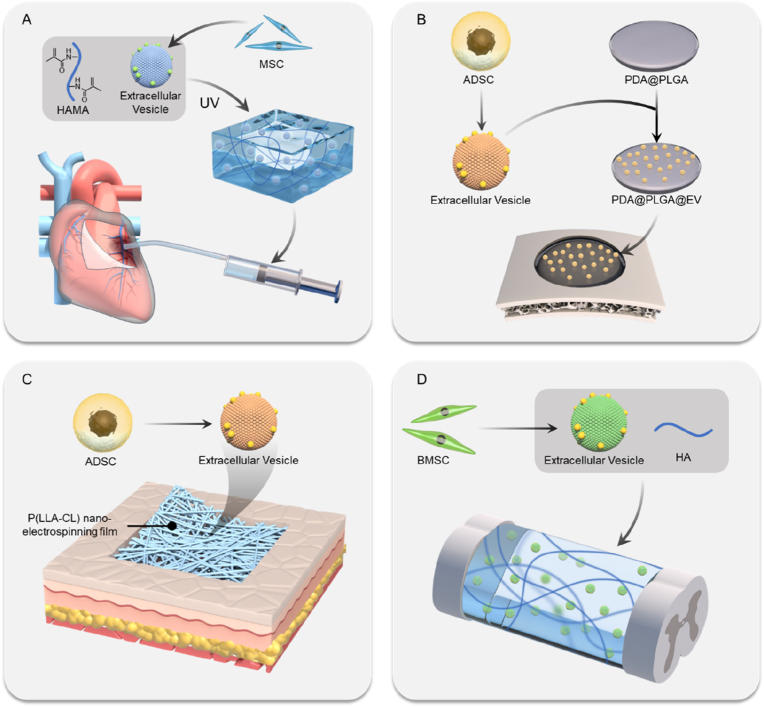
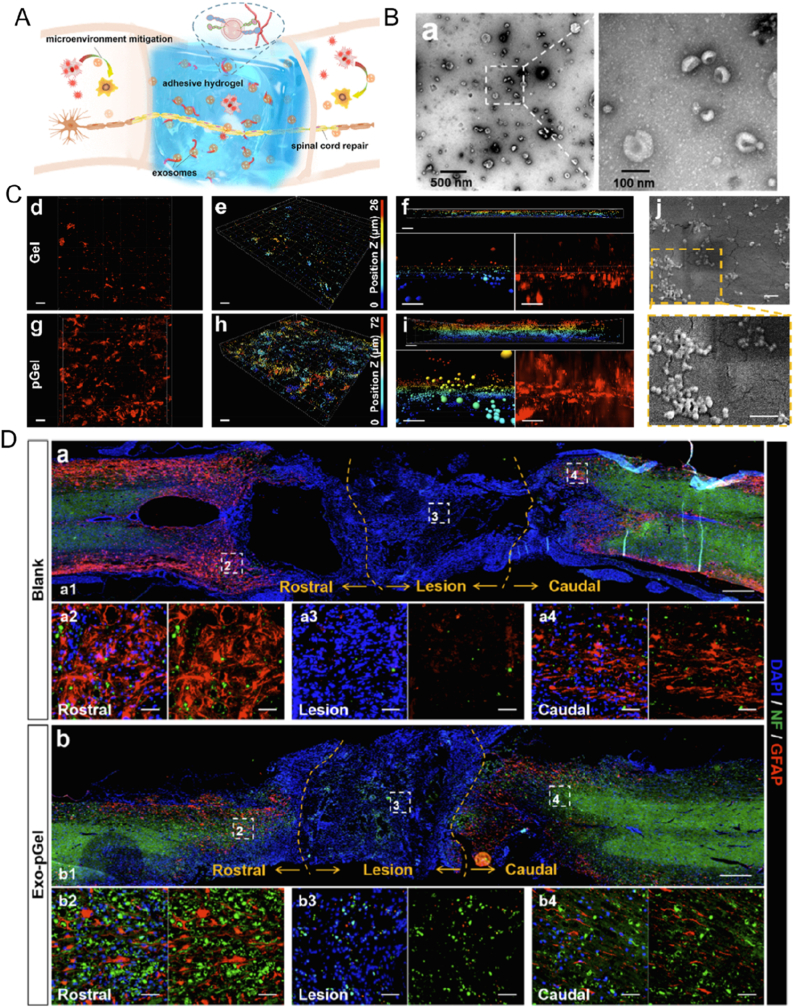
Extracellular vesicles (EVs) are nano-scale vesicles derived by cell secretion with unique advantages such as promoting cell proliferation, anti-inflammation, promoting blood vessels and regulating cell differentiation, which benefit their wide applications in regenerative medicine. However, the in vivo therapeutic effect of EVs still greatly restricted by several obstacles, including the off-targetability, rapid blood clearance, and undesired release. To address these issues, biomedical engineering techniques are vastly explored. This review summarizes different strategies to enhance EV functions from the perspective of drug loading, modification, and combination of biomaterials, and emphatically introduces the latest developments of functionalized EV-loaded biomaterials in different diseases, including cardio-vascular system diseases, osteochondral disorders, wound healing, nerve injuries. Challenges and future directions of EVs are also discussed.

Keywords: Biomaterials; Drug delivery; Extracellular vesicle; Modification; Regenerative medicine.


Conflict of interest statement

The authors declare that they have no known competing financial interests or personal relationships that could have appeared to influence the work reported in this paper.

Figures

Image 1
Graphical abstract
Fig. 1
Fig. 1
Schematic summary of EV-based treatment for regenerative medicine. (A) EVs with different origins bring bioactive molecules, including nucleic acids, proteins, lipids or membrane receptors, to target cells and exert multiple biological functions. (B) After surface modification, drug loading and combination with biomaterials, EVs can be used for treating degenerative diseases in different organs.
Fig. 2
Fig. 2
The major bioactive components of MSC-EVs. (A) A protein network analysis using proteomics illustrated candidate proteins and associated pathways involved in the therapeutic action of MSCs-derived EVs [16]. (B) The sequencing of EV miRNA showed that 23 miRNAs account for 79.1% of total miRNAs presented in MSC-EVs. (C)The predicted biological functions related to miRNA found in MSC-EVs [20]. Reproduced with permission from Ref. [16], copyright 2012, American Chemical Society; ref [20], copyright 2018, Springer Nature.
Fig. 3
Fig. 3
Characterization and application of the p-EVs. (A) Scheme of the preparation of p-EVs. PRP, platelet-rich plasma; RBCs, red blood cells. Morphology of the platelet (B) and p-EVs (C) observed by transmission electron microscopy (TEM). Scale bars, 1 μm. (D) SDS-PAGE of platelet lysate and p-EVs with Coomassie brilliant blue staining showing a different protein distribution between platelet and p-EVs. (E) Western blot analysis showed a high expression of CD41 in both the platelet lysate and p-EVs [53]. (F) p-EVs demonstrated increased therapeutic efficacy compared with PRP in a rat skin defect model. SAH, sodium alginate hydrogel [8]. Reproduced with permission from Ref. [53]. copyright 2020, Elsevier B.V.; ref [8]. copyright 2017, Ivyspring International Publisher.
Fig. 4
Fig. 4
Schematics illustrate the commonly used strategies of drug loading and modification of EVs. For drug-loading, the methods divided into drug incubation, parent cell engineering, and membrane punching such as freeze-thaw methos, electroporation or ultrasound. For modification, there are major two types of methods depending on whether the modification occur pre-isolation and post-isolation of EVs.
Fig. 5
Fig. 5
Engineering of light responsive “exosomes for protein loading via optically reversible protein-protein interactions” (EXPLORs) drug loading system. (A) Schematic diagram of EXPLOR technology. Cargo protein was linked to CRY2 protein and CIBN protein was linked to CD9 membrane marker. When light was applied, CRY2 was photoactivated and conjugated to CIBN, which produced a CD9-CIBN-CRY2-cargo protein complex. This complex was then loaded into EVs via the translocation of CD9 marker. After cargo was loaded, the closed light dissociated cargo from CD9 and cargo can be released in target cells. (B) HEK293T cells were transiently transfected with CIBN-EGFP-CD9 and mCherry-CRY2 expression vectors. It can be observed that when the blue light was switched on, the location of CRY2 overlapped with CIBN, indicating photoactivated conjugation between CRY2 and CIBN. Scale bars, 20 mm. (C) Brain sections of EXPLOR-injected transgenic imaged by fluorescence microscopy. The loading of Cre protein using EXPLOR system was verified using a genetically modified mice. The successful delivery of Cre activated EYFP in mice brains while non-Cre-loaded EXPLOR injected mice failed to induce fluorescence. Green: eNpHR3.0-EYFP, blue: cell nuclei. Scale bars, 500 mm. Hip, hippocampus; Th, thalamus. (D) Immunohistochemistry staining results showed that the activated EYFP were mainly located in neurons, indicating that this EV mainly targeted neurons [118]. Reproduced with permission from Ref. [118]. copyright 2016, Springer Nature.
Fig. 6
Fig. 6
Delivery of siRNA to the mouse brain by genetic engineering modified EVs. (A) Schematic diagram of production, collection and re-injection targeted self-exos for siRNA delivery. Targeting peptide was conjugated to the EV-specific marker in dendritic cells via transfection. In this way, the targeting peptide was modified on the surface of EVs with high modification efficacy. (B) Western blotting analysis showed the successful loading of targeted protein into DCs and EVs. The untransfected DCs and EVs were set as the control group, FLAG meant DCs transfected with FLAG-Lamp2b and derivative EVs. (C) TEM analysis of RVG-EVs. (D) Fluorescent images showed that different peptide-loaded exosomes can both show high targeting efficiency. siRNA + LP: siRNA delivered with lipofectamine 2000; RVG exos: RVG (Neuro2A targeting) peptide modified exosomes; MSP exos: MSP (C2C12 targeting) peptide modified exosomes. The top two images were Neuro2A cells and bottom two images were C2C12 cells. (E) Distribution of Cy3-GAPDH siRNA in the brain, indicating the successful delivery of siRNA to the brain using modified exosome [73]. Reproduced with permission from Ref. [73]. Copyright 2011, Springer Nature.
Fig. 7
Fig. 7
Surface modification of Evs via micelle-mediated physical modification and chemical modification. (A) TEM showed successful engineering of nanobody on the surface of EVs using post-insertion technique. (B) High magnification TEM image showing the nanobody on the membrane of EV-EGa1-PEG. (C) Plasma concentration of DiR-labeled Neuro2A EVs before and after post-insertion with nanobody-PEG micelles. The circulation time of PEG-modified EVs was significantly increased compared to non-modified EVs [150]. (D) Schematic illustration of modifying SDF-1 and DA7R on the EVs using copper-free click chemistry. DBCO was introduced to the surface of EVs using the covalent bond between NHS-PEG4-DBCO and the amino groups in EVs' membrane proteins. Then azide functionalized SDF-1 and DA7R peptide were reacted with DBCO. (E) Confocal fluorescence images to indicate the successful modification of Cy5.5-labeled SDF-1, FITC-labeled DA7R in the Dil-red-labeled-EVs. (F) IVIS images of brains from stroke model mice showed superior delivery and targeting ability of Dual-EVs. (G) Fluorescent images of harvested brain sections stained with the DCX antibody (green) indicated the migrating immature neurons, demonstrating that the Dual-EVs enabled to promote neurogenesis in the ischemic stroke mice model. DAPI (blue): cell nuclei [162]. Reproduced with permission from Ref. [150]. Copyright 2016, Elsevier B.V; ref [162], copyright 2022, Elsevier B.V.
Fig. 8
Fig. 8
Schematic diagram of biomaterials-based EVs therapy in tissue regeneration. The EVs are loaded in different biomaterials including natural or synthetic materials to exert their therapeutic functions in (A) cardiovascular system diseases (B) osteochondral disorders (C) wound healing and (D) nerve injuries.
Fig. 9
Fig. 9
The arrest and local delivery of in vivo circulating EVs via antibody-modified magnetic nanoparticles. (A) Scheme of the vesicle-shuttle approach. Characterization of GMNPEC-EVs, including TEM imaging (B), representative fluorescent images (C) and TEM analysis of released or control exosomes (D) Anti-CD63 and anti-MLC antibodies were conjugated to magnetic beads via acid-responsive bond. Magnetic beads were able to capture circulating EVs during their traveling towards heart. Anti-MLC antibody can guide magnetic beads to injured myocytes and release EVs in the acid environment. (E) Schematic demonstrated the animal experiment was designed to assess the capacity of GMNPEC to arrest EVs in vivo in a MI rat model. (F) Representative fluorescent images revealed that GMNPEC administration potentiated angiogenesis at the infarction border area 4 weeks post-treatment. (G) Masson's trichrome staining of papillary of the infarcted heart in different groups, illustrating superior regeneration ability provided by this vesicle-shuttle approach [9]. Reprinted with permission from Ref. [9]. Copyright 2020, Springer Nature.
Fig. 10
Fig. 10
circRNA3503-EVs loaded PLEL hydrogel for the osteoarthritis therapy. (A) Schematic illustration of the combinatorial strategy of SMSC-sEVs and sleep-related circRNA3503. CircRNA3503 was loaded into EVs by overexpressing circRNA3503 in parent cells. (B) Morphology of sEVs was observed by SEM (left) and TEM (right) (C) Schematic diagram illustrated the therapeutic mechanism of circRNA3503-OE-sEVs. (D) PAGE analysis indicating the successful loading of circRNA3503 into sEVs. (E) Safranin O, Alcian blue and Toluidine blue assays of in-vitro cartilage degeneration model treated with different formulations at day 21 [204]. Reprinted with permission from Ref. [204]. Copyright 2021, Elsevier B.V.
Fig. 11
Fig. 11
Combination of exosomes with anti-bacterial hydrogels for diabetic wound healing. (A) Schematic diagram displaying the potential treatment procedure of FEP@exo in skin reconstruction. (B) Photographs of the adhesive property of FEP dressing. (C) Cell migration assay via Transwell showed enhanced fibroblast migration in FEP@exo group after 48 h coculture. (D) FEP@exo promoted in vitro tube formation of HUVECs. (E) Representative images of wound healing progress in model mice with different therapeutic treatments [236]. Reproudced with permission of ref [236]. Copyright 2019, American Chemical Society.
Fig. 12
Fig. 12
MSC-EVs immobilized in adhesive hydrogel for SCI. (A) Scheme for Exo-pGel therapeutic procedures. (B) TEM showed typical exosomes morphology. (C) DiI-labeled exo in blank HA hydrogel (Gel) and (d–f) peptide-modified HA hydrogel (pGel) (g–i) observed via z-stack method using confocal laser scanning microscopy and SEM (j). The depth of exosome was marked using different colors. (D) Representative images revealing neurofilaments (NFs, green) and glial fibrillary acidic protein (GFAP, red) staining in blank (a) and Exo-pGel (b) groups of spinal-cord injured rat. The lesion size and cell density was significantly increased in Exo-pGel group [249]. Reproudced with permission of ref [249]. Copyright 2020, American Chemical Society.

References

    1. Kalluri R., LeBleu V.S. The biology, function, and biomedical applications of exosomes. Science. 2020;367(6478) - PMC - PubMed
    1. Wu P., Zhang B., Ocansey D.K.W., Xu W., Qian H. Extracellular vesicles: a bright star of nanomedicine. Biomaterials. 2021;269 - PubMed
    1. Phinney D.G., Pittenger M.F. Concise review: MSC-derived exosomes for cell-free therapy. Stem Cell. 2017;35(4):851–858. - PubMed
    1. Fu P., Zhang J., Li H., Mak M., Xu W., Tao Z. Extracellular vesicles as delivery systems at nano-/micro-scale. Adv. Drug Deliv. Rev. 2021 - PMC - PubMed
    1. Aliotta J.M., Pereira M., Wen S., Dooner M.S., Del Tatto M., Papa E., Goldberg L.R., Baird G.L., Ventetuolo C.E., Quesenberry P.J., Klinger J.R. Exosomes induce and reverse monocrotaline-induced pulmonary hypertension in mice. Cardiovasc. Res. 2016;110(3):319–330. - PMC - PubMed

LinkOut - more resources