. 2023 Mar 17;5(2):fcad072.
doi: 10.1093/braincomms/fcad072. eCollection 2023.

A potential protective role of the nuclear receptor-related factor 1 (Nurr1) in multiple sclerosis motor cortex: a neuropathological study

Affiliations

A potential protective role of the nuclear receptor-related factor 1 (Nurr1) in multiple sclerosis motor cortex: a neuropathological study

Jonathan Pansieri et al. Brain Commun. .

Abstract

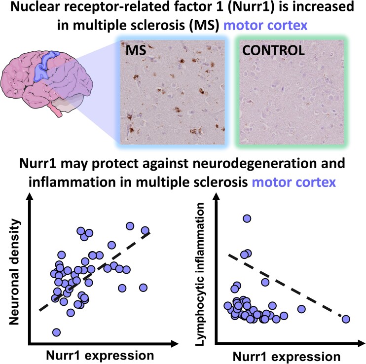
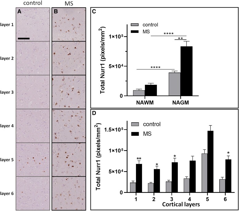
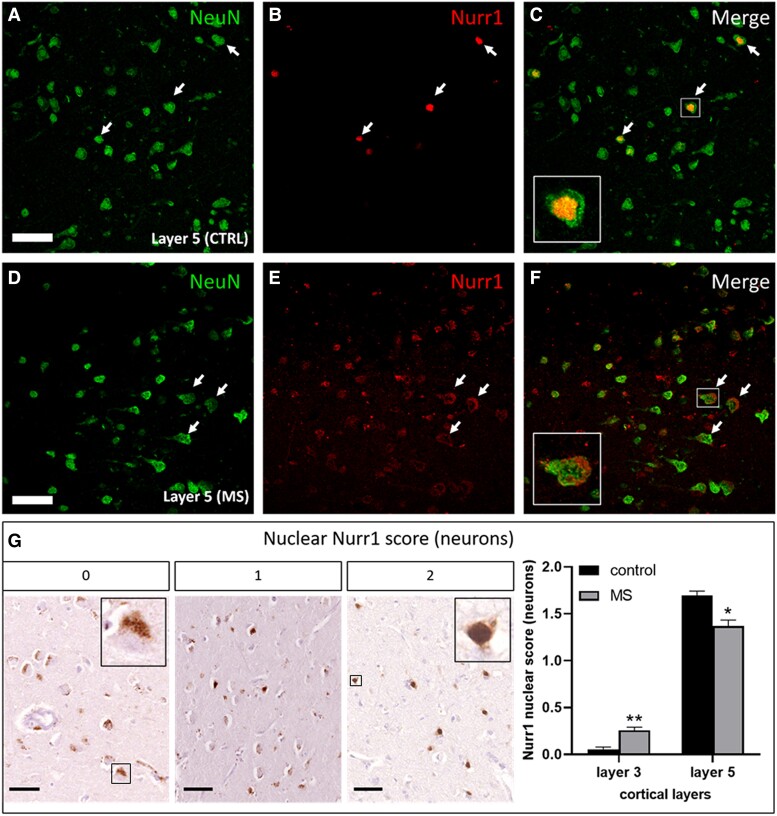
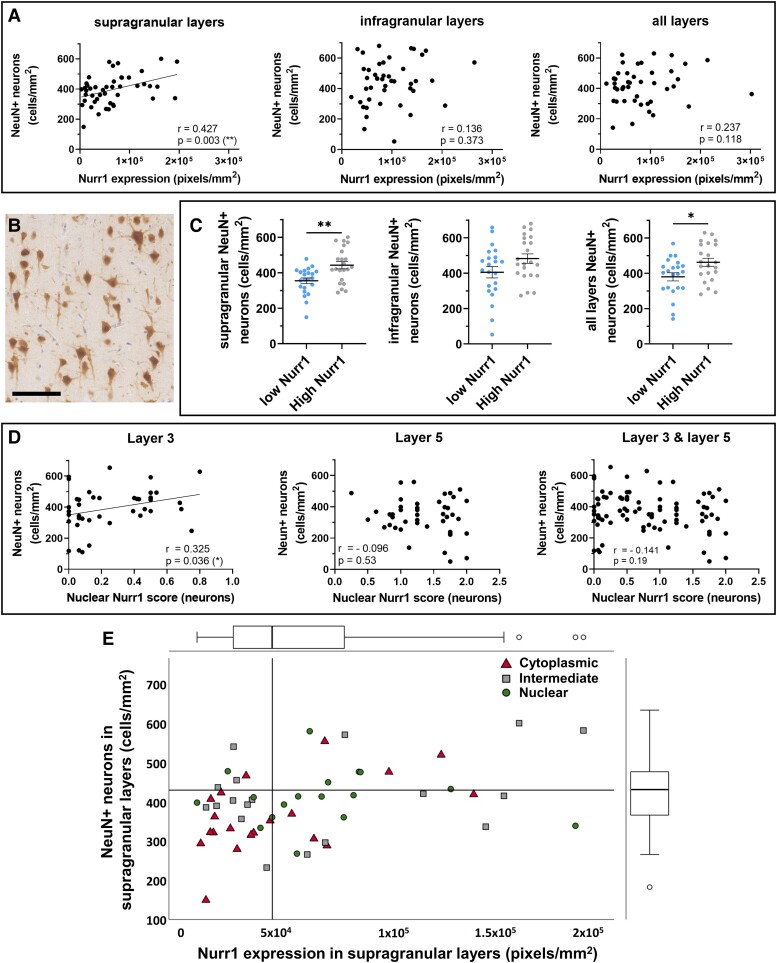
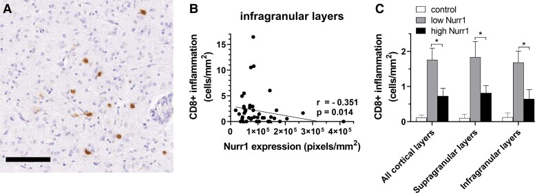
Cerebral cortical inflammation and neurodegeneration are hallmark pathological features of multiple sclerosis that contribute to irreversible neurological disability. While the reason for nerve cell death is unknown, the pathogenic inflammatory role of infiltrating lymphocytes is likely an important contributor. The nuclear receptor-related factor 1 counteracts inflammation in animal models of multiple sclerosis, and protects against neuronal loss in other neurodegenerative disorders, but its expression in post-mortem multiple sclerosis tissue is not known. This study aims to investigate the nuclear receptor-related factor 1 expression in multiple sclerosis motor cortex and evaluate its relationship with motor cortical pathology. To accomplish this, an autopsy cohort of pathologically confirmed multiple sclerosis (n = 46), and control (n = 11) cases was used, where the nuclear receptor-related factor 1 expression was related to neuronal and lymphocytic densities. Motor cortical nuclear receptor-related factor 1 was overexpressed in multiple sclerosis compared to control cases. Increased nuclear receptor-related factor 1 expression positively associated with neuronal densities, especially when present in nucleus of neurons, and associated with decreased CD8+ cytotoxic lymphocyte density. Our findings expand the current knowledge on nuclear receptor-related factor 1 in neurological diseases, and support the hypothesis that nuclear receptor-related factor 1 may play a dual neuroprotective role in multiple sclerosis by influencing inflammatory and neurodegenerative processes. Future studies elucidating the influence of nuclear receptor-related factor 1 on these processes in multiple sclerosis may cast light onto novel targets that may be modulated to alter clinical outcome.

Keywords: Nurr1; lymphocyte inflammation; multiple sclerosis; neuronal loss.


Conflict of interest statement

The authors report no competing interests.

Figures

Graphical Abstract
Graphical Abstract
Figure 1
Figure 1
Nurr1 expression in multiple sclerosis and control motor cortex. Nurr1 expression in the six cortical layers of (A) control and (B) multiple sclerosis cases revealed by DAB immunostaining. Quantitation of Nurr1 expression using a chromogen extraction method, comparing (C) normal-appearing white matter and normal-appearing grey matter and (D) each cortical layer, in control and multiple sclerosis cases. Mann-Whitney tests, data presented as mean ± SEM. *P < 0.05; **P < 0.01; ****P < 0.0001; scale bar 100 μm.
Figure 2
Figure 2
Representative NeuN+ and Nurr1+ neurons and semi-quantitative scoring on Nurr1 neuronal localisation. Confocal images of layer 5 pyramidal neurons in (AC) control and (DF) multiple sclerosis cases are labelled for NeuN (NeuN; A, D) and Nurr1 (Nurr1; B, E) at 400 × magnification, and representative merge image (C, F). Nuclear and cytoplasmic Nurr1 are illustrated in the inserts (arrows and inserts in C and F, respectively; scale bar 50 µm). (G) We devised a semiquantitative score for the localisation of Nurr1 in cytoplasm and nucleus of neurons in layer 3 and layer 5. For each FOV analysed, Nurr1 expression in each compartment was scored on a scale of 0 (mostly cytoplasmic), 1 (cytoplasmic/nuclear) and 2 (mostly nuclear). Cytoplasmic Nurr1 is greater in layer 3 of controls compared with multiple sclerosis cases, while nuclear Nurr1 is greater in layer 5 of controls compared with multiple sclerosis cases (Mann-Whitney tests). Cytoplasmic and nuclear Nurr1 are illustrated in the inserts of score 0 and score 1, respectively. Data presented as mean ± SEM. *P < 0.05; **P < 0.01; scale bar 100 μm.
Figure 3
Figure 3
Relationships between Nurr1 expression and neuronal localisation with neuronal density in multiple sclerosis. Nurr1 expression is positively correlated with (A) NeuN+ neuronal density, which is restricted to supragranular layers in multiple sclerosis (Spearman rank-correlation coefficients). (B) Representative labelling of NeuN+ neurons using DAB immunostaining in supragranular layer 3 in multiple sclerosis (scale bar 100 μm). (C) Further assessment shows an increase in NeuN+ neuronal density in the multiple sclerosis subgroup with high levels of Nurr1, restricted to supragranular layers (Mann-Whitney tests) (D) Nurr1 preferential localisation in nucleus is positively correlated with NeuN+ neuronal densities and restricted to superficial layer 3 (Spearman rank-correlation coefficients). (E) Nurr1 expression and sub-cellular localisation interact in predicting NeuN+ neuronal densities, as cases with low NeuN+ neuron densities (below median, horizontal line) tend to have both low Nurr1 expression (below median, vertical line) and a cytoplasmic Nurr1 expression (Generalised estimating equation model). The scatterplot shows the relationship between Nurr1 expression and NeuN+ neuronal density. Dots are classified based on the Nurr1 localisation score as predominant cytoplasmic (higher tertiles), intermediate or nuclear Nurr1 expression (bottom tertiles). Distribution of Nurr1 expression (on top) and NeuN+ neuronal densities (on the right) are represented with box plots. Data presented as mean ± SEM. *P < 0.05; **P < 0.01; scale bar 100 μm.
Figure 4
Figure 4
Relationships between Nurr1 expression and CD8+ lymphocyte density. (A) Representative labelling of CD8+ cells using DAB immunostaining. (B) A negative correlation between Nurr1 expression and CD8+ lymphocytes is restricted to infragranular layers in multiple sclerosis cohort (Spearman rank-correlation coefficients). (C) Further assessment shows a significant increase in CD8+ lymphocytes in the cases lying at the lowest extreme of Nurr1 expression (n = 20) compared with the cases with the highest extreme of Nurr1 expression throughout all cortical layers (Mann-Whitney tests). *P < 0.05; scale bar 100 μm.

Similar articles

Cited by

References

    1. Goldenberg MM. Multiple sclerosis review. P T. 2012;37(3):175–184. - PMC - PubMed
    1. Dutta R, Trapp BD. Mechanisms of neuronal dysfunction and degeneration in multiple sclerosis. Progr Neurobiol. 2011;93(1):1–12. - PMC - PubMed
    1. Fletcher JM, Lalor SJ, Sweeney CM, Tubridy N, Mills KHG. T cells in multiple sclerosis and experimental autoimmune encephalomyelitis. Clin Exp Immunol. 2010;162(1):1–11. - PMC - PubMed
    1. Compston A, Coles A. Multiple sclerosis. Lancet. 2008;372(9648):1502–1517. - PubMed
    1. Miranda BRD, Popichak KA, Hammond SL, et al. . The Nurr1 activator 1,1-bis(3′-indolyl)-1-(p-chlorophenyl)methane blocks inflammatory gene expression in BV-2 microglial cells by inhibiting nuclear factor κB. Mol Pharmacol. 2015;87(6):1021–1034. - PMC - PubMed

LinkOut - more resources