. 2023 Mar 15;5(8):2251-2260.
doi: 10.1039/d3na00014a. eCollection 2023 Apr 11.

Sortase A transpeptidation produces seamless, unbranched biotinylated nanobodies for multivalent and multifunctional applications

Affiliations

Sortase A transpeptidation produces seamless, unbranched biotinylated nanobodies for multivalent and multifunctional applications

Eugene M Obeng et al. Nanoscale Adv. .

Abstract

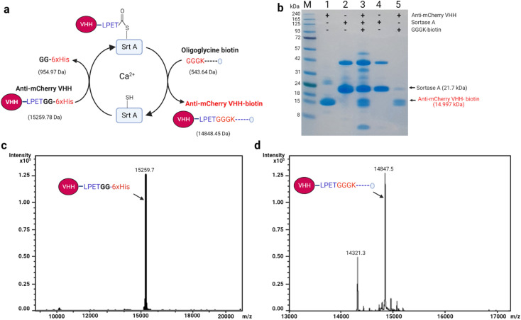
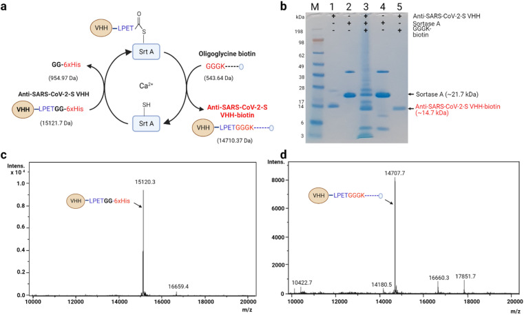
Exploitation of the biotin-streptavidin interaction for advanced protein engineering is used in many bio-nanotechnology applications. As such, researchers have used diverse techniques involving chemical and enzyme reactions to conjugate biotin to biomolecules of interest for subsequent docking onto streptavidin-associated molecules. Unfortunately, the biotin-streptavidin interaction is susceptible to steric hindrance and conformational malformation, leading to random orientations that ultimately impair the function of the displayed biomolecule. To minimize steric conflicts, we employ sortase A transpeptidation to produce quantitative, seamless, and unbranched nanobody-biotin conjugates for efficient display on streptavidin-associated nanoparticles. We further characterize the protein-nanoparticle complex and demonstrate its usefulness in optical microscopy and multivalent severe acute respiratory syndrome coronavirus (SARS-CoV-2) antigen interaction. The approach reported here provides a template for making novel multivalent and multifunctional protein complexes for avidity-inspired technologies.


Conflict of interest statement

The authors declare that they have no known competing financial interests or personal relationships that could have appeared to influence the work reported in this paper.

Figures

Fig. 1
Fig. 1. Schematic representation of biotinylation approaches. (a) Conventional chemical biotinylation approaches include reactions that form stable amide and thiol–maleimide bonds. These methods are often non-quantitative, non-homogeneous and could produce unwanted aggregates due to their penchant for variable labelling. (b) A two-dimensional schematic description of protein–particle homogeneity levels for common chemical and sortase A biotinylation approaches. The uniformity in biotin–streptavidin geometry and directionality determines the extent of aggregation. (c) Sortase A assisted biotinylation approach. The Srt A enzyme recognizes and cleaves the LPETG recognition motif on a molecule of interest after which it seamlessly transpeptidates an oligoglycine-biotin to the C-terminal of the molecule of interest. VHH: nanobody; DOL: degree of labelling.
Fig. 2
Fig. 2. Seamless biotinylation of anti-mCherry VHH via Srt A transpeptidation. (a) Schematic description of anti-mCherry VHH biotinylation. Anti-mCherry VHH (with Srt A recognition sequence, LPETG; 30 μM), Srt A enzyme (90 μM) and GGGK-biotin peptide (2 mM) were mixed in reaction buffer (40 mM Tris–HCl, 150 mM NaCl, and 10 mM CaCl2) and incubated at 42 °C for 4 h with shaking. The biotinylated anti-mCherry VHH was separated from the other components via a Ni2+-affinity purification approach. The estimated molecular weight changes (predicted with https://web.expasy.org/compute_pi/) are indicated in brackets. (b) SDS-PAGE analysis of the anti-mCherry VHH transpeptidation process. The last lane shows the state of the purified anti-mCherry VHH-biotin albeit with a little Srt A carryover. The higher molecular weight band (∼40 kDa) in the Srt A only lane is possibly an E. coli-associated protein that co-purifies with the Srt A enzyme. M: molecular weight marker; (1) anti-mCherry VHH; (2) Srt A protein; (3) reaction mixture containing anti-mCherry VHH, Srt A and GGGK-biotin; (4) separated Srt A (end of reaction); (5) biotinylated anti-mCherry VHH (with some cleaved but unbiotinylated forms). (c) Sample ESI spectra of unmodified anti-mCherry VHH (note: this sample was not subjected to Srt A transpeptidation). (d) Sample ESI spectra of anti-mCherry-biotin prepared via Srt A transpeptidation.
Fig. 3
Fig. 3. Seamless biotinylation of anti-SARS-CoV-2-S VHH via Srt A transpeptidation. (a) Schematic description of stage-wise molecular weight changes. The details of the reaction are similar to the description in Fig. 2. The estimated molecular weight changes are indicated in brackets. (b) SDS-PAGE analysis of the anti-SARS-CoV-2-S VHH transpeptidation process. The last lane shows the state of the purified anti-SARS-CoV-2-S VHH-biotin. M: molecular weight marker; (1) anti-SARS-CoV-2-S VHH; (2) Srt A protein; (3) reaction mixture containing anti-SARS-CoV-2-S VHH, Srt A and GGGK-biotin; (4) separated Srt A (end of reaction); (5) biotinylated anti-SARS-CoV-2-S VHH. (c) ESI spectra of unmodified anti-SARS-CoV-2-S VHH (note: this sample was not subjected to Srt A transpeptidation). (d) Sample ESI spectra of anti-SARS-CoV-2-S VHH-biotin prepared via Srt A transpeptidation.
Fig. 4
Fig. 4. Size characterization of quantum dot–nanobody conjugate. (a) Simplified two-dimensional representation of controlled nanobody orientation on Qdot, where the sQdot is decorated with at least 7 streptavidin molecules, allowing it to receive a coating of biotinylated anti-mCherry VHH. (b) TEM images of streptavidin-coated Qdot and assembled Qdot–nanobody conjugates (scale: 20 nm). (c)–(e) Dynamic light scattering (DLS) measurements of biotinylated anti-mCherry VHH (c), streptavidin-coated Qdot (d) and assembled Qdot–nanobody conjugate (e) performed as per materials and methods. It should be noted that one molecule of tetrameric streptavidin can accommodated up to 4 molecules of biotinylated molecule depending on the availability of exposed binding pockets.
Fig. 5
Fig. 5. Functional assessment of anti-mCherry quantum dot nanobody conjugate (QNC). (a) Schematic description of optical microscopy assessment of binding. (b) Confocal laser scanning microscopy images of mCherry-coated beads in the presence (top) or absence (bottom) of anti-mCherry QNC. (c) Pixel fluorogram representation of molecular colocalization of QNC and mCherry coated on beads. (d) Pixel fluorogram representation of mCherry-coated beads alone.
Fig. 6
Fig. 6. Functional assessment of the anti-SARS-CoV-2-S nanobody–avidin conjugate (NAC). (a) A cartoon structure of a typical receptor binding site (RBS) nanobody (PDB ID: 7B17). (b) Schematic depiction of the biolayer interferometry protocol used to assess binding. (c) Biolayer interferometry analysis of anti-SARS-CoV-2-S VHH-biotin binding with spike RBD-Fc per the description in (b). (d) Biolayer interferometry analysis of NAC binding with spike RBD-Fc per the description in (b). The exact KD value was beyond the detection limit of the BLItz instrument due to the low dissociation rate. It should be noted that one molecule of tetrameric avidin can accommodated up to 4 molecules of biotinylated molecule depending on the availability of exposed binding pockets.

References

    1. Dundas C. M. Demonte D. Park S. Appl. Microbiol. Biotechnol. 2013;97:9343–9353. doi: 10.1007/s00253-013-5232-z. - DOI - PubMed
    1. Muraoka A. Matsuura Y. Naitow H. Ihara M. Kunishima N. Anal. Biochem. 2018;557:46–58. doi: 10.1016/j.ab.2018.07.009. - DOI - PubMed
    1. Boeneman K. Deschamps J. R. Buckhout-White S. Prasuhn D. E. Blanco-Canosa J. B. Dawson P. E. Stewart M. H. Susumu K. Goldman E. R. Ancona M. Medintz I. L. ACS Nano. 2010;4:7253–7266. doi: 10.1021/nn1021346. - DOI - PMC - PubMed
    1. Wang Z. Liu L. Xu Y. Sun L. Li G. Biosens. Bioelectron. 2011;26:4610–4613. doi: 10.1016/j.bios.2011.04.052. - DOI - PubMed
    1. Fairhead M. and Howarth M., in Analytical Biochemistry, ed. A. Gautier and M. J. Hinner, Springer New York, New York, NY, 2015, vol. 1266, pp. 171–184