. 2023 Apr 14;83(8):1329-1344.
doi: 10.1158/0008-5472.CAN-22-2794.

Spatially Resolved Transcriptomics Deconvolutes Prognostic Histological Subgroups in Patients with Colorectal Cancer and Synchronous Liver Metastases

Affiliations

Spatially Resolved Transcriptomics Deconvolutes Prognostic Histological Subgroups in Patients with Colorectal Cancer and Synchronous Liver Metastases

Colin S Wood et al. Cancer Res. .

Abstract

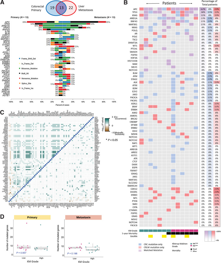
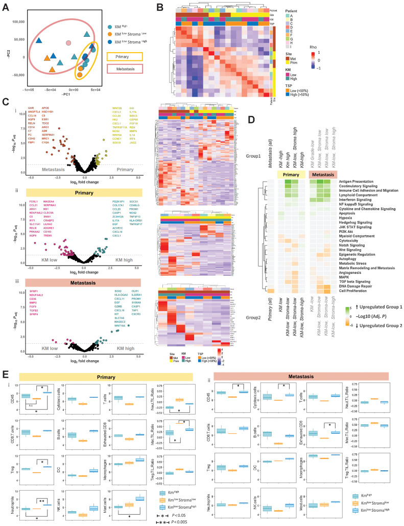
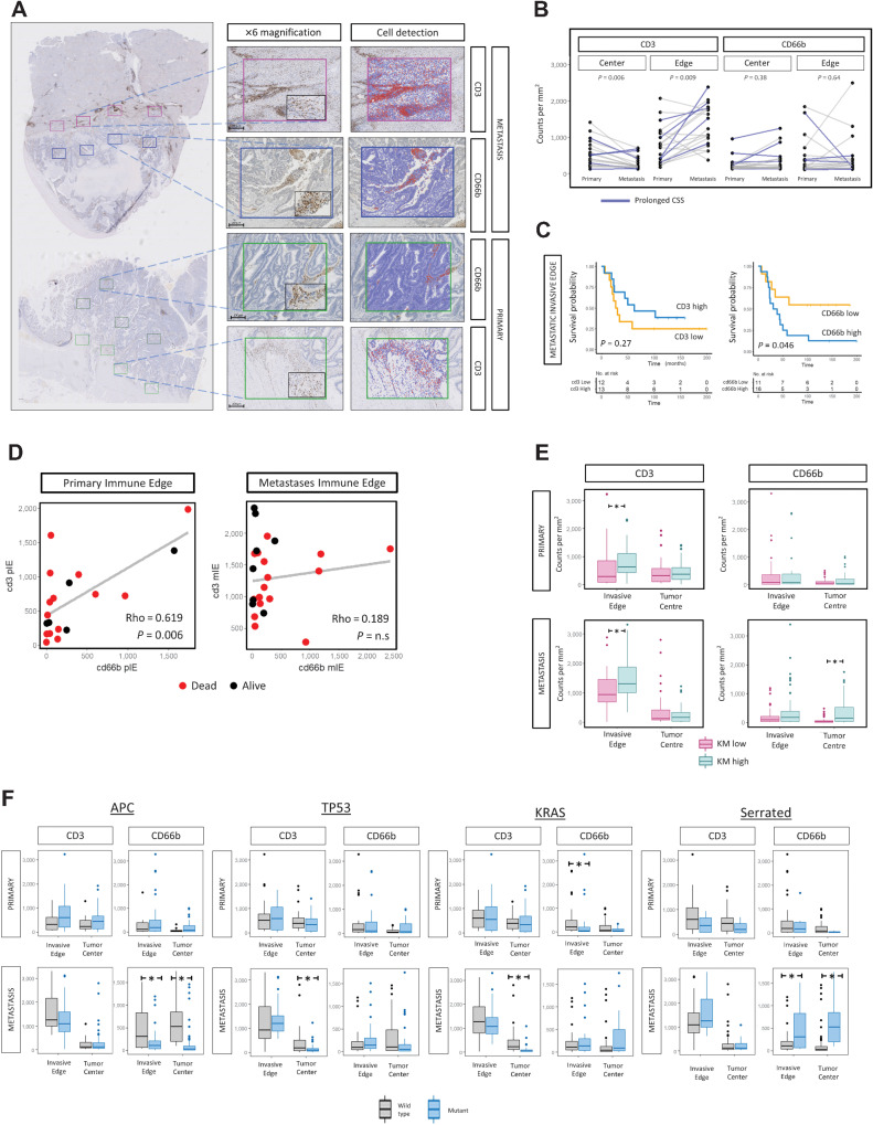
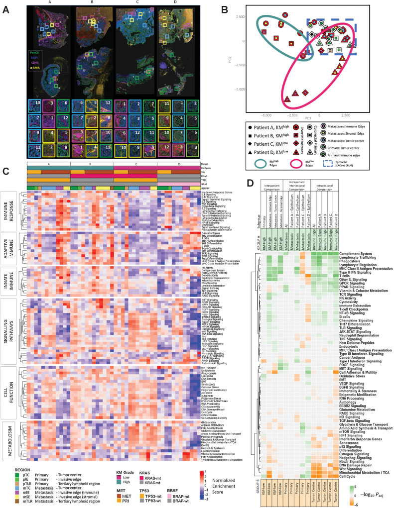
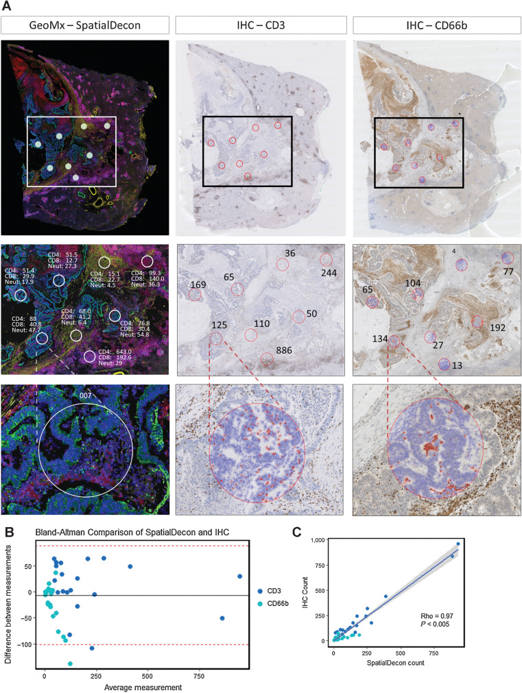
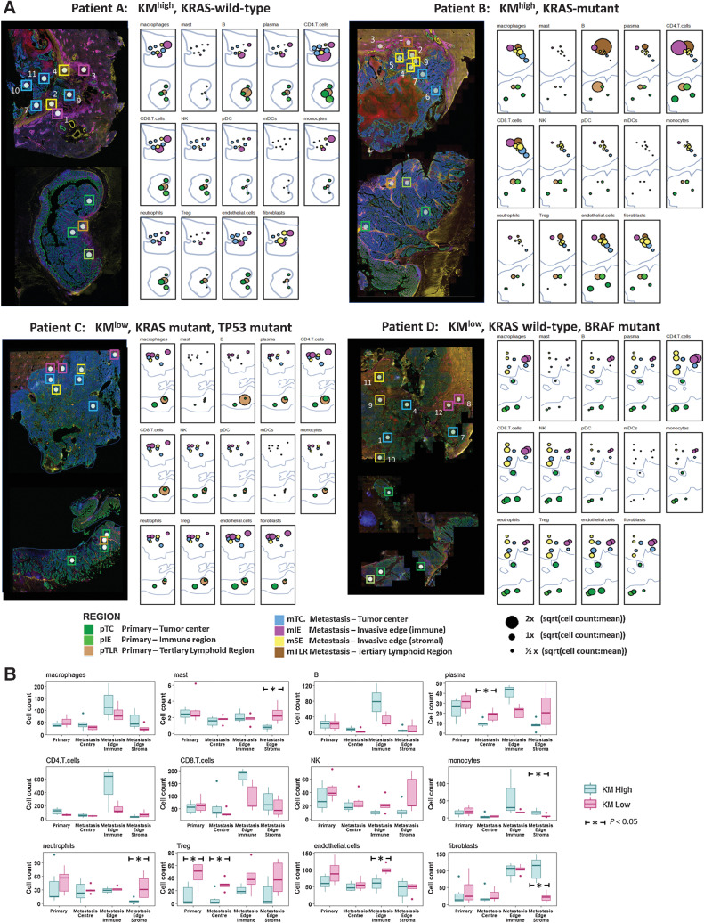
Strong immune responses in primary colorectal cancer correspond with better patient survival following surgery compared with tumors with predominantly stromal microenvironments. However, biomarkers to identify patients with colorectal cancer liver metastases (CRLM) with good prognosis following surgery for oligometastatic disease remain elusive. The aim of this study was to determine the practical application of a simple histological assessment of immune cell infiltration and stromal content in predicting outcome following synchronous resection of primary colorectal cancer and CRLM and to interrogate the underlying functional biology that drives disease progression. Samples from patients undergoing synchronous resection of primary colorectal cancer and CRLM were evaluated in detail through histological assessment, panel genomic and bulk transcriptomic assessment, IHC, and GeoMx spatial transcriptomics (ST) analysis. High immune infiltration of metastases was associated with improved cancer-specific survival. Bulk transcriptomic analysis was confounded by stromal content, but ST demonstrated that the invasive edge of the metastases of long-term survivors was characterized by adaptive immune cell populations enriched for type II IFN signaling and MHC-class II antigen presentation. In contrast, patients with poor prognosis demonstrated increased abundance of regulatory T cells and neutrophils with enrichment of Notch and TGFβ signaling pathways at the metastatic tumor center. In summary, histological assessment can stratify outcomes in patients undergoing synchronous resection of CRLM, suggesting that it has potential as a prognostic biomarker. Furthermore, ST analysis has revealed significant intratumoral and interlesional heterogeneity and identified the underlying transcriptomic programs driving each phenotype.

Significance: Spatial transcriptomics uncovers heterogeneity between patients, between matched lesions in the same patient, and within individual lesions and identifies drivers of metastatic progression in colorectal cancer with reactive and suppressed immune microenvironments.


Figures

Figure 1. Mutational Characterization of primary colorectal cancer and CRLM. A, Venn diagram demonstrating primary colorectal cancer and CRLM that underwent genomic analysis. Co-Barplot illustrating most frequently mutated genes across 13 matched primary colorectal cancer and CRLM, including mutation type. Genes are ordered by mutational frequency. Sections were sequenced using GPOL (Glasgow Precision Oncology Laboratory) mutational panel. B, Oncoplot demonstrating concurrent mutations in the 13 matched lesions. Patients are ranked according to co-mutational burden on the y-axis and ranked according to KM grade on the x-axis. Blue denotes gene mutated in primary only, red denotes gene mutated in metastasis only, purple denotes mutated in primary and metastasis. The right hand 3 columns denote the percentage of total patients with each mutation type. C, Correlation matrix demonstrating co-occurrence of mutations with left of the blue demarcation line representing primary colorectal cancer, right of the blue demarcation line representing CRLM (pair-wise Fisher's Exact test *, P < 0.05). Gene names are displayed along the x and y-axes ordered by mutational frequency. Dark green boxes represent significant co-occurrence. D, Box plot illustrating mutational burden in primary colorectal cancer and CRLM according to KM grade using the Mann–Whitney test to assess for statistically significant difference between KM groups.
Figure 1.
Mutational characterization of primary colorectal cancer and CRLM. A, Venn diagram demonstrating primary colorectal cancer and CRLM that underwent genomic analysis. Co-Barplot illustrating most frequently mutated genes across 13 matched primary colorectal cancer and CRLM, including mutation type. Genes are ordered by mutational frequency. Sections were sequenced using GPOL (Glasgow Precision Oncology Laboratory) mutational panel. B, Oncoplot demonstrating concurrent mutations in the 13 matched lesions. Patients are ranked according to co-mutational burden on the y-axis and ranked according to KM grade on the x-axis. Blue, gene mutated in primary only; red, gene mutated in metastasis only; purple, mutated in primary and metastasis. The right-hand three columns denote the percentage of total patients with each mutation type. C, Correlation matrix demonstrating co-occurrence of mutations, with left of the blue demarcation line representing primary colorectal cancer, right of the blue demarcation line representing CRLM (pair-wise Fisher exact test; *, P < 0.05). Gene names are displayed along the x- and y-axes ordered by mutational frequency. Dark green boxes represent significant co-occurrence. D, Box plot illustrating mutational burden in primary colorectal cancer and CRLM according to KM grade using the Mann–Whitney test to assess for statistically significant difference between KM groups.
Figure 2. BULK IO360 transcriptomic characterization of matched primary colorectal cancer and CRLM. A, PCA plot demonstrating 2 principal components of minimal variance for all samples. Primary colorectal cancer samples are demonstrated by circles and yellow outline. CRLM are demonstrated by triangular points and brown outline. KMhigh, KMlow Stromalow and KMlow Stromahigh samples are depicted by color. B, Unsupervised analysis using gene expression correlation matrix for all samples. Patient, site, KM grade, tumor stromal percentage (TSP) are depicted by key. Spearman correlation of all expressed genes performed between each sample sequenced and plotted on the heatmap. k-means clustering of heatmap to demonstrate correlated samples. Red represents strong correlation. Blue represents negative correlation. C, Volcano plots demonstrating differential gene expression results and clustered heatmap of significant genes for (i) all primary colorectal cancer versus CRLM; (ii) KM grade: KM high versus KM low primary colorectal cancer; (iii) KM grade: KM high versus KM low CRLM. The x-axis of volcano plot demonstrates log2fold change, y-axis demonstrates –log10P. Colored points demonstrate significant changes in gene expression between groups (P < 0.05 and logFC>1.5). Volcano plots demonstrate top 20 differentially expressed genes for each group. D, Heatmap demonstrating Gene Set Enrichment Analysis (GSEA) results comparing the different tumors grouped according with KM grade and TSP using io360-curated gene sets annotated on the right of the diagram. Heatmap squares represent Log10 Adjusted P value. Green demonstrates upregulation in group 1, orange represents upregulation in group 2. The heatmap is clustered by y-axis only to demonstrate frequently upregulated gene sets. E, Boxplot comparisons of immune cell populations and selected cell:cell ratios between KM grade and TSP segregated groups using deconvolution software included in the nCounter package. Annotated subgroups are: Kmhigh, Kmlow Stromalow, Kmlow Stromahigh. The y-axis represents log10 of estimated cell count. Primary colorectal cancer is represented in (i) and CRLM in (ii).
Figure 2.
BULK IO360 transcriptomic characterization of matched primary colorectal cancer and CRLM. A, PCA plot demonstrating two principal components of minimal variance for all samples. Primary colorectal cancer samples are demonstrated by circles and yellow outline. CRLM are demonstrated by triangular points and brown outline. KMhigh, KMlow Stromalow and KMlow Stromahigh samples are depicted by color. B, Unsupervised analysis using gene expression correlation matrix for all samples. Patient, site, KM grade, and TSP are depicted by key. Spearman correlation of all expressed genes performed between each sample sequenced and plotted on the heatmap. k-means clustering of heatmap to demonstrate correlated samples. Red, strong correlation. Blue, negative correlation. C, Volcano plots demonstrating differential gene expression results and clustered heatmap of significant genes for (i) all primary colorectal cancer versus CRLM; (ii) KM grade: KM high versus KM low primary colorectal cancer; (iii) KM grade: KM high versus KM low CRLM. The x-axis of volcano plot demonstrates log2-fold change; y-axis demonstrates –log10P. Colored points demonstrate significant changes in gene expression between groups (P < 0.05 and logFC > 1.5). Volcano plots demonstrate top 20 differentially expressed genes for each group. D, Heatmap demonstrating GSEA results comparing the different tumors grouped according with KM grade and TSP using io360-curated gene sets annotated on the right of the diagram. Heatmap squares represent log10-adjusted P value. Green, upregulation in group 1; orange, upregulation in group 2. The heatmap is clustered by y-axis only to demonstrate frequently upregulated gene sets. E, Box plot comparisons of immune cell populations and selected cell:cell ratios between KM grade and TSP segregated groups using deconvolution software included in the nCounter package. Annotated subgroups are: Kmhigh, Kmlow Stromalow, Kmlow Stromahigh. The y-axis represents log10 of estimated cell count. Primary colorectal cancer is represented in i and CRLM in ii.
Figure 3. IHC characterization of matched primary colorectal cancer and CRLM with integration of morphological and mutational features. A, Representative images of CD3 and CD66b immunohistochemical staining (Patient B). Whole section demonstrated at ×0.5 magnifications, ROIs corresponding to tumor center (TC) and invasive edge (IE) of primary and CRLM shown at ×6 and ×10 (black box); scale bar, 100 μm. B, Intra-patient comparison between primary and metastasis of CD3 and CD66b cell counts at tumor center and invasive edge. P values calculated using the Mann–Whitney test. C, Kaplan–Meier survival plots (log-rank test, P values displayed) demonstrating the prognostic impact of CD3 and CD66b cell density at CRLM IE identified by IHC for IE of CRLM. High and low values determined according to median expression. D, Comparison of CD3 and CD66b cell density at (i) primary IE (ii) CRLM IE (iii) CD3 primary IE and metastatic TC. Spearman's Rho analysis. E, Box plots illustrating the relationship between KM grade and CD3 and CD66b cell counts at the IE and TC of primary colorectal cancer and CRLM. The P value was calculated using the Mann–Whitney test. F, Box-plot representing relationship between mutational features (APC, TP53, KRAS, Serrated) and CD3 and CD66b cell density at TC and IE of primary colorectal cancer and CRLM. The P value was calculated using the Mann–Whitney test. Lesions of serrated origin were defined as APCwild + KRAS/BRAFmutation.
Figure 3.
IHC characterization of matched primary colorectal cancer and CRLM with integration of morphological and mutational features. A, Representative images of CD3 and CD66b immunohistochemical staining (Patient B). Whole section demonstrated at ×0.5 magnifications; ROIs corresponding to tumor center (TC) and invasive edge (IE) of primary and CRLM are shown at ×6 and ×10 (black box). Scale bar, 100 μm. B, Intrapatient comparison between primary and metastasis of CD3 and CD66b cell counts at tumor center and invasive edge. P values calculated using the Mann–Whitney test. C, Kaplan–Meier survival plots (log-rank test, P values displayed) demonstrating the prognostic impact of CD3 and CD66b cell density at CRLM IE identified by IHC for IE of CRLM. High and low values determined according to median expression. D, Comparison of CD3 and CD66b cell density at (i) primary IE, (ii) CRLM IE, and (iii) CD3 primary IE and metastatic TC. Spearman Rho analysis. E, Box plots illustrating the relationship between KM grade and CD3 and CD66b cell counts at the IE and TC of primary colorectal cancer and CRLM. The P value was calculated using the Mann–Whitney test. F, Box plot representing relationship between mutational features (APC, TP53, KRAS, Serrated) and CD3 and CD66b cell density at TC and IE of primary colorectal cancer and CRLM. The P value was calculated using the Mann–Whitney test. Lesions of serrated origin were defined as APCwild + KRAS/BRAFmutation.
Figure 4. Spatially resolved transcriptomic analysis using Nanostring Cancer Transcriptome Atlas gene sets. A, Representative images of multiplex immunofluorescence (mIF) staining of 4 matched primary colorectal cancer and CRLM. DAPI (blue), Pancytokeratin (green), CD45 (pink), and a-SMA (yellow). Topographic regions are annotated, each box represents hand-selected area of tumor. 8 regions were taken from CRLM and 4 from primary colorectal cancer per patient. Patient A: KMhigh KRAS-wt, good prognosis. Patient B: KMhigh, KRAS-mt, good prognosis. Patient C: KMlow, KRAS/TP53 co-mutation, poor prognosis. Patient D: KM-low, KRAS-wt, BRAF-mt high-mutational burden lesion. B, Principal Component Analysis plot of all regions of interest (ROI) selected. The patient from whom the lesion originated is represented by shape. A red border indicates region arises from CRLM and white border represents primary colorectal cancer. The topographical region within the lesion is illustrated by the innermost color of the shape. KM high metastatic edges are represented by a green circle, KMlow metastatic edges are represented by a red circle and a dashed blue line represents epithelial regions of primary colorectal cancer and CRLM. C, Heatmap demonstrating single sample GSEA for every ROI, ordered on the x-axis by Patient and ROI. Key presented to aid patient identification. The y-axis represents annotated gene sets from Cancer Transcriptome Atlas ordered by and clustered within modules of Immune Response, Adaptive Immune, Innate Immune, Signaling Pathways. Cell Function, Metabolism. Each cell represents the Normalized Enrichment Score scaled by pathway. D, Heatmap demonstrating GSEA providing inter-patient comparison of selected areas between KMhigh and KMlow patients and intra-patient comparison between primary and metastatic sites and intralesional comparison between tumor center and immune edge. Subset of regions filtered before GSEA is demonstrated in Subgroup. Subsequent groups compared in GSEA identified as Groups A and B. Group A annotated at top of diagram and represented by green. Group B annotated at bottom of diagram and represented by orange. Cells of heatmap represent −log10(Padj) for comparison, cell is tinted green if pathway is upregulated in group A and orange if upregulated in group B.
Figure 4.
Spatially resolved transcriptomic analysis using Nanostring Cancer Transcriptome Atlas gene sets. A, Representative images of mIF staining of four matched primary colorectal cancer and CRLM. DAPI, blue; Pancytokeratin, green; CD45, pink; αSMA, yellow. Topographic regions are annotated and each box represents hand-selected area of tumor. Eight regions were taken from CRLM and four from primary colorectal cancer per patient. Patient A: KMhigh, KRAS-wt, good prognosis. Patient B: KMhigh, KRAS-mt, good prognosis. Patient C: KMlow, KRAS/TP53 co-mutation, poor prognosis. Patient D: KM-low, KRAS-wt, BRAF-mt high-mutational burden lesion. B, PCA plot of all ROIs selected. The patient from whom the lesion originated is represented by shape. A red border indicates region arises from CRLM and white border represents primary colorectal cancer. The topographical region within the lesion is illustrated by the innermost color of the shape. KM high metastatic edges, green circle; KMlow metastatic edges, red circle; dashed blue line, epithelial regions of primary colorectal cancer and CRLM. C, Heatmap demonstrating single sample GSEA for every ROI, ordered on the x-axis by patient and ROI. Key presented to aid patient identification. The y-axis represents annotated gene sets from Cancer Transcriptome Atlas ordered by and clustered within modules of Immune Response, Adaptive Immune, Innate Immune, Signaling Pathways. Cell Function, Metabolism. Each cell represents the NES scaled by pathway. D, Heatmap demonstrating GSEA providing interpatient comparison of selected areas between KMhigh and KMlow patients and intrapatient comparison between primary and metastatic sites and intralesional comparison between tumor center and immune edge. Subset of regions filtered before GSEA is demonstrated in subgroup. Subsequent groups compared in GSEA identified as groups A and B. Group A annotated at top of diagram and represented by green. Group B annotated at bottom of diagram and represented by orange. Cells of heatmap represent −log10(Padj) for comparison; cell is tinted green if pathway is upregulated in group A and orange if upregulated in group B.
Figure 5. Immune cell spatial deconvolution. A, Representative images from 1 CRLM (Patient A, Fig. 4A) showing cell detection from the Qupath package used on a CD3 and CD66b IHC-stained liver metastasis to count number of CD3 and CD66b-positive cells from 21 regions from a total of 3 CRLM. This count was compared with the SpatialDecon-derived count that uses the transcriptomic data from the corresponding ROI in the GeoMx mIF-stained matched sample (See Supplementary Fig. S5). B, Bland Altman plot comparing transcriptome SpatialDecon-derived cell count versus the IHC-derived cell count. C, Correlation plot comparing transcriptome SpatialDecon-derived cell count versus the IHC-derived cell count.
Figure 5.
Immune cell spatial deconvolution. A, Representative images from 1 CRLM (Patient A, Fig. 4A) showing cell detection from the Qupath package used on a CD3 and CD66b IHC-stained liver metastasis to count number of CD3 and CD66b-positive cells from 21 regions from a total of three CRLM. This count was compared with the SpatialDecon-derived count that uses the transcriptomic data from the corresponding ROI in the GeoMx mIF-stained matched sample (See Supplementary Fig. S5). B, Bland Altman plot comparing transcriptome SpatialDecon-derived cell count versus the IHC-derived cell count. C, Correlation plot comparing transcriptome SpatialDecon-derived cell count versus the IHC-derived cell count.
Figure 6. Topographic immune cell deconvolution primary colorectal cancer and CRLM. A, Images (from Fig. 4A) of primary colorectal cancer (bottom) and CRLM (top) with 48 ROIs superimposed. Abundance estimates as determined from transcriptome by SpatialDecon for 14 cell populations illustrated for each ROI with color-coding detailing the annotated tumor region. Radius is proportional to the estimated cell counts within the ROI. The immune cell count per region was extracted and the square root of the ratio to the mean immune cell count per region (41.37) of all immune cells was calculated and displayed. The square root of the ratio was calculated to minimize the skew caused by variance of highly expressed cell types. B, Box plots demonstrating the median and interquartile range for each cell type analyzed organized by cell type and topographic region and grouped by KM grade. All ROI taken from primary colorectal cancer except TLR are grouped as Primary. Dendritic cells were removed due to insignificant counts. The Mann–Whitney test used to assess for statistical significance; *, P < 0.05.
Figure 6.
Topographic immune cell deconvolution primary colorectal cancer and CRLM. A, Images (from Fig. 4A) of primary colorectal cancer (bottom) and CRLM (top) with 48 ROIs superimposed. Abundance estimates as determined from transcriptome by SpatialDecon for 14 cell populations illustrated for each ROI with color coding detailing the annotated tumor region. Radius is proportional to the estimated cell counts within the ROI. The immune cell count per region was extracted and the square root of the ratio to the mean immune cell count per region (41.37) of all immune cells was calculated and is displayed. The square root of the ratio was calculated to minimize the skew caused by variance of highly expressed cell types. B, Box plots demonstrating the median and interquartile range for each cell type analyzed organized by cell type and topographic region and grouped by KM grade. All ROIs taken from primary colorectal cancer except TLR are grouped as primary. Dendritic cells were removed due to insignificant counts. The Mann–Whitney test was used to assess for statistical significance; *, P < 0.05.

References

    1. Dunne DF, Jones RP, Malik HZ, Fenwick SW, Poston GJ. Surgical management of colorectal liver metastases: a European perspective. Hepatic Oncol 2014;1:121–33. - PMC - PubMed
    1. Smith JJ, D'Angelica MI. Surgical management of hepatic metastases of colorectal cancer. Hematol Oncol Clin North Am 2015;29:61–84. - PubMed
    1. Dhir M, Sasson AR. Surgical management of liver metastases from colorectal cancer. J Oncol Pract 2016;12:33–9. - PubMed
    1. Fong Y, Fortner J, Sun RL, Brennan MF, Blumgart LH. Clinical score for predicting recurrence after hepatic resection for metastatic colorectal cancer: analysis of 1001 consecutive cases. Ann Surg 1999;230:309–18. - PMC - PubMed
    1. Steele CW, Whittle T, Smith JJ. Review: KRAS mutations are influential in driving hepatic metastases and predicting outcome in colorectal cancer. Chin Clin Oncol 2019;8:53. - PubMed

Publication types