Comment
. 2023 Aug 15;29(16):3051-3064.
doi: 10.1158/1078-0432.CCR-22-3790.

DNA-Methylome-Based Tumor Hypoxia Classifier Identifies HPV-Negative Head and Neck Cancer Patients at Risk for Locoregional Recurrence after Primary Radiochemotherapy

Affiliations
Comment

DNA-Methylome-Based Tumor Hypoxia Classifier Identifies HPV-Negative Head and Neck Cancer Patients at Risk for Locoregional Recurrence after Primary Radiochemotherapy

Bouchra Tawk et al. Clin Cancer Res. .

Abstract

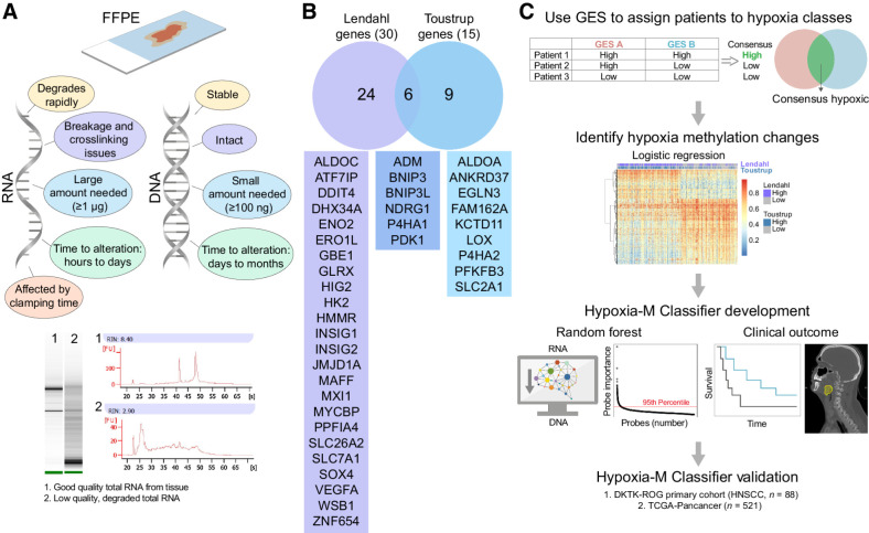
Purpose: Tumor hypoxia is a paradigmatic negative prognosticator of treatment resistance in head and neck squamous cell carcinoma (HNSCC). The lack of robust and reliable hypoxia classifiers limits the adaptation of stratified therapies. We hypothesized that the tumor DNA methylation landscape might indicate epigenetic reprogramming induced by chronic intratumoral hypoxia.

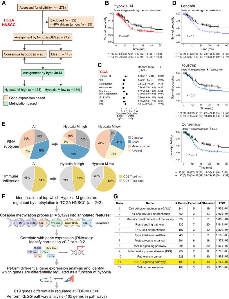
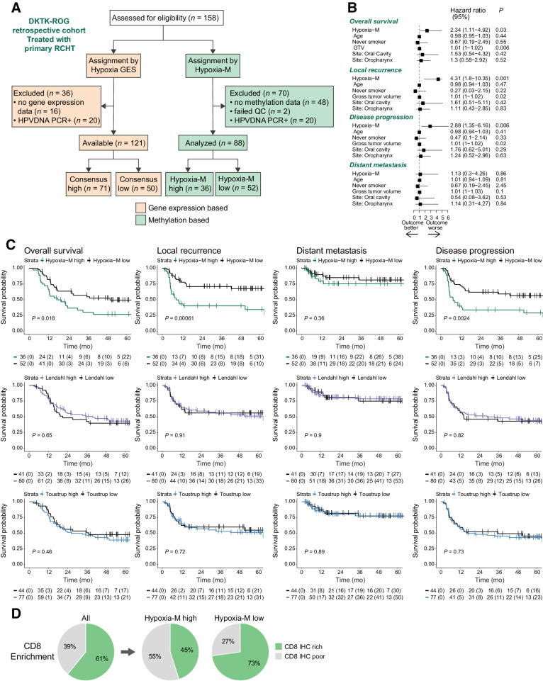
Experimental design: A DNA-methylome-based tumor hypoxia classifier (Hypoxia-M) was trained in the TCGA (The Cancer Genome Atlas)-HNSCC cohort based on matched assignments using gene expression-based signatures of hypoxia (Hypoxia-GES). Hypoxia-M was validated in a multicenter DKTK-ROG trial consisting of human papillomavirus (HPV)-negative patients with HNSCC treated with primary radiochemotherapy (RCHT).

Results: Although hypoxia-GES failed to stratify patients in the DKTK-ROG, Hypoxia-M was independently prognostic for local recurrence (HR, 4.3; P = 0.001) and overall survival (HR, 2.34; P = 0.03) but not distant metastasis after RCHT in both cohorts. Hypoxia-M status was inversely associated with CD8 T-cell infiltration in both cohorts. Hypoxia-M was further prognostic in the TCGA-PanCancer cohort (HR, 1.83; P = 0.04), underscoring the breadth of this classifier for predicting tumor hypoxia status.

Conclusions: Our findings highlight an unexplored avenue for DNA methylation-based classifiers as biomarkers of tumoral hypoxia for identifying high-risk features in patients with HNSCC tumors. See related commentary by Heft Neal and Brenner, p. 2954.


Figures

Figure 1. Development of Hypoxia-M Classifier. A, DNA methylation profiling offers technical advantages over gene expression, particularly for standard FFPE-derived biomaterial, where mRNA is prone to significant integrity and quality loss, for example, via degradation and crosslinking. B, To train for a DNA-methylation–based hypoxia classifier, well-established hypoxia gene expression signatures (GES) from Lendahl (n = 30) and Toustrup (n = 15) were used. C, To develop Hypoxia-M, a core of patients with consensus high tumor hypoxia GES was identified on the basis of the Toustrup and Lendahl GES in the TCGA-HNSCC (see Results). Methylation differences were identified using logistic regression between both groups. Hypoxia-M was built from a random forest classification using the 95th percentile most important probes and correlated with clinical outcome.
Figure 1.
Development of Hypoxia-M Classifier. A, DNA methylation profiling offers technical advantages over gene expression, particularly for standard FFPE-derived biomaterial, where mRNA is prone to significant integrity and quality loss, for example, via degradation and crosslinking. B, To train for a DNA-methylation–based hypoxia classifier, well-established hypoxia gene expression signatures (GES) from Lendahl (n = 30) and Toustrup (n = 15) were used. C, To develop Hypoxia-M, a core of patients with consensus high tumor hypoxia GES was identified on the basis of the Toustrup and Lendahl GES in the TCGA-HNSCC (see Results). Methylation differences were identified using logistic regression between both groups. Hypoxia-M was built from a random forest classification using the 95th percentile most important probes and correlated with clinical outcome.
Figure 2. Graphical representation of the clinical cohorts used in this study: The training cohort, TCGA-HNSCC (n = 242), and the validation cohort, DKTK-ROG (n = 88), of HPV-negative patients.
Figure 2.
Graphical representation of the clinical cohorts used in this study: The training cohort, TCGA-HNSCC (n = 242), and the validation cohort, DKTK-ROG (n = 88), of HPV-negative patients.
Figure 3. Evaluation of Hypoxia-M in the training cohort (TCGA-HNSCC). A, Consort chart of patients included in the study. B, Kaplan–Meier (KM) survival curves. Patients predicted to have Hypoxia-M high tumors had significantly increased rates of death (P = 0.015). C, On multivariate regression, Hypoxia-M retained its prognostic significance (HR, 1.66; P = 0.015). D, The GES from Toustrup and Lendahl used for derivation of hypoxia-M are also associated with significantly worsened OS in this cohort. E, Integration of Hypoxia-M with molecular biomarkers (top). Hypoxia-M is significantly enriched in the basal subtype (45%) and the classical subtype (30%; P < 0.05) and (bottom) there is a significant decrease in CD8 T cells immune cells in Hypoxia-M–high versus low tumors (P < 0.05). F, Integration of differentially methylated probes with differential gene expression in TCGA-HNSCC; 5,129 probes were differentially methylated between Consensus High versus low tumors. After collapsing methylation probes into methylation features, every feature was correlated with the expression of its corresponding gene (RNAseq). Only correlations >0.2 or < −2 were retained (n = 1,180). Differential gene expression was conducted for those genes between Hypoxia-M–high versus –low tumors, yielding 619 differentially regulated genes at FDR<0.05. G, Cell adhesion molecules (CAM), T-helper cell differentiation, and HIF-1alpha signaling pathways are differentially regulated among those 619 genes.
Figure 3.
Evaluation of Hypoxia-M in the training cohort (TCGA-HNSCC). A, Consort chart of patients included in the study. B, Kaplan–Meier (KM) survival curves. Patients predicted to have Hypoxia-M high tumors had significantly increased rates of death (P = 0.015). C, On multivariate regression, Hypoxia-M retained its prognostic significance (HR, 1.66; P = 0.015). D, The GES from Toustrup and Lendahl used for derivation of hypoxia-M are also associated with significantly worsened OS in this cohort. E, Integration of Hypoxia-M with molecular biomarkers (top). Hypoxia-M is significantly enriched in the basal subtype (45%) and the classical subtype (30%; P < 0.05) and (bottom) there is a significant decrease in CD8 T cells immune cells in Hypoxia-M–high versus low tumors (P < 0.05). F, Integration of differentially methylated probes with differential gene expression in TCGA-HNSCC; 5,129 probes were differentially methylated between Consensus High versus low tumors. After collapsing methylation probes into methylation features, every feature was correlated with the expression of its corresponding gene (RNAseq). Only correlations >0.2 or < −2 were retained (n = 1,180). Differential gene expression was conducted for those genes between Hypoxia-M–high versus –low tumors, yielding 619 differentially regulated genes at FDR<0.05. G, Cell adhesion molecules (CAM), T-helper cell differentiation, and HIF-1alpha signaling pathways are differentially regulated among those 619 genes.
Figure 4. Validation of Hypoxia‐M in the DKTK‐ROG cohort of patients with HPV-negative HNSCC treated with primary radiochemotherapy (RCHT). A, Consort chart of patients profiled in the study. B, On multivariate analysis, Hypoxia-M remained an independent inverse prognostic factor, particularly for LR (HR, 4.31; P = 0.001). C, Patients with Hypoxia-M tumors were at significantly higher risk of death (P = 0.018), disease progression (P = 0.0024), and local recurrence (LR; P < 0.0006) but not distant metastasis. D, Integration of Hypoxia-M with immune cell infiltration. Forty-five percent of Hypoxia-M high tumors had high CD8 T-cell IHC versus 73% of Hypoxia-M–Low tumors, representing a significant association between Hypoxia-M and distribution of immune cells (P < 0.05).
Figure 4.
Validation of Hypoxia‐M in the DKTK‐ROG cohort of patients with HPV-negative HNSCC treated with primary radiochemotherapy (RCHT). A, Consort chart of patients profiled in the study. B, On multivariate analysis, Hypoxia-M remained an independent inverse prognostic factor, particularly for LR (HR, 4.31; P = 0.001). C, Patients with Hypoxia-M tumors were at significantly higher risk of death (P = 0.018), disease progression (P = 0.0024), and local recurrence (LR; P < 0.0006) but not distant metastasis. D, Integration of Hypoxia-M with immune cell infiltration. Forty-five percent of Hypoxia-M high tumors had high CD8 T-cell IHC versus 73% of Hypoxia-M–Low tumors, representing a significant association between Hypoxia-M and distribution of immune cells (P < 0.05).
Figure 5. Validation of Hypoxia-M the TCGA-pancancer cohort. A, 521 patients with primary tumors were included in the validation of Hypoxia-M. B, Hypoxia-M tumors had significantly worsened outcomes in the TCGA-pancancer cohort (P < 0.0001). C, Hypoxia-M remained an independent prognostic factor after adjusting for age, race, tumor type, gender, stage, and treatment (HR, 1.83; P < 0.04). D, Prognostic utility of Hypoxia-M by anatomical site in TCGA-pancancer. Hypoxia-M was prognostic for squamous cell carcinomas (HNSC, lung squamous, and cervix) as well as bladder cancers. Buffa, Ragnum, and Winter GES were prognostic for glioblastoma and kidney. E, Hypoxia-M correlates with GES of hypoxia. Hypoxia-M correlates best with Lendahl, Toustrup, and Buffa signatures in the training cohort (TCGA-HNSCC). In the validation cohort (DKTK-ROG), it correlates best with Lendahl. Finally, in TCGA-pancancer, it correlates best with Winter, Ragnum, and Buffa GES.
Figure 5.
Validation of Hypoxia-M the TCGA-pancancer cohort. A, 521 patients with primary tumors were included in the validation of Hypoxia-M. B, Hypoxia-M tumors had significantly worsened outcomes in the TCGA-pancancer cohort (P < 0.0001). C, Hypoxia-M remained an independent prognostic factor after adjusting for age, race, tumor type, gender, stage, and treatment (HR, 1.83; P < 0.04). D, Prognostic utility of Hypoxia-M by anatomical site in TCGA-pancancer. Hypoxia-M was prognostic for squamous cell carcinomas (HNSC, lung squamous, and cervix) as well as bladder cancers. Buffa, Ragnum, and Winter GES were prognostic for glioblastoma and kidney. E, Hypoxia-M correlates with GES of hypoxia. Hypoxia-M correlates best with Lendahl, Toustrup, and Buffa signatures in the training cohort (TCGA-HNSCC). In the validation cohort (DKTK-ROG), it correlates best with Lendahl. Finally, in TCGA-pancancer, it correlates best with Winter, Ragnum, and Buffa GES.

Comment in

Comment on

Similar articles

Cited by

References

    1. Abdollahi A, Folkman J. Evading tumor evasion: current concepts and perspectives of anti-angiogenic cancer therapy. Drug Resist Updat 2010;13:16–28. - PubMed
    1. Salem A, Asselin M-C, Reymen B, Jackson A, Lambin P, West CML, et al. . Targeting hypoxia to improve non–small cell lung cancer outcome. JNCI J Natl Cancer Inst 2018;110:14–30. - PubMed
    1. Toustrup K, Sørensen BS, Metwally MAH, Tramm T, Mortensen LS, Overgaard J, et al. . Validation of a 15-gene hypoxia classifier in head and neck cancer for prospective use in clinical trials. Acta Oncol 2016;55:1091–8. - PubMed
    1. Cutsem EV, Lenz H-J, Furuse J, Tabernero J, Heinemann V, Ioka T, et al. . MAESTRO: a randomized, double-blind phase III study of evofosfamide (Evo) in combination with gemcitabine (Gem) in previously untreated patients (pts) with metastatic or locally advanced unresectable pancreatic ductal adenocarcinoma (PDAC). J Clin Oncol 2016;34:4007.
    1. Bhandari V, Li CH, Bristow RG, Boutros PC. Divergent mutational processes distinguish hypoxic and normoxic tumours. Nat Commun 2020;11:737. - PMC - PubMed

Publication types

MeSH terms