. 2023 Apr 25;42(4):112394.
doi: 10.1016/j.celrep.2023.112394. Epub 2023 Apr 13.

A plasma membrane-associated glycolytic metabolon is functionally coupled to KATP channels in pancreatic α and β cells from humans and mice

Affiliations

A plasma membrane-associated glycolytic metabolon is functionally coupled to KATP channels in pancreatic α and β cells from humans and mice

Thuong Ho et al. Cell Rep. .

Abstract

The ATP-sensitive K+ (KATP) channel is a key regulator of hormone secretion from pancreatic islet endocrine cells. Using direct measurements of KATP channel activity in pancreatic β cells and the lesser-studied α cells, from both humans and mice, we provide evidence that a glycolytic metabolon locally controls KATP channels on the plasma membrane. The two ATP-consuming enzymes of upper glycolysis, glucokinase and phosphofructokinase, generate ADP that activates KATP. Substrate channeling of fructose 1,6-bisphosphate through the enzymes of lower glycolysis fuels pyruvate kinase, which directly consumes the ADP made by phosphofructokinase to raise ATP/ADP and close the channel. We further show the presence of a plasma membrane-associated NAD+/NADH cycle whereby lactate dehydrogenase is functionally coupled to glyceraldehyde-3-phosphate dehydrogenase. These studies provide direct electrophysiological evidence of a KATP-controlling glycolytic signaling complex and demonstrate its relevance to islet glucose sensing and excitability.

Keywords: CP: Metabolism; K(ATP) channel; glycolysis; glycolytic metabolon; inside-out excised patch clamp; metabolic compartmentation; pyruvate kinase.


Conflict of interest statement

Declaration of interests The authors declare no competing interests.

Figures
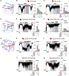
Figure 1.
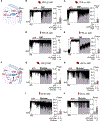
Figure 1.. Lower glycolytic enzymes raise ATP/ADP to close α and β cell KATP channels
(A–C) PEP (0.25, 1, and 5 mM) in the presence of high ADP (0.5 mM ADP, 0.1 mM ATP) led to the production of ATP by PK (A), thereby closing KATP channels in excised plasma membrane from mouse β cells (B) and mouse α cells (C). (D–F) In the high ADP condition, the chain reaction of PGM, ENO, and PK produced ATP and closed KATP channels (D) upon addition of 5 mM 3PG in mouse β cells (E) and 1 mM and 5 mM 3PG followed by 5 mM PEP in mouse α cells (F). (G–K) Lower glycolytic activity (from ALDO to PK) utilized 5 mM FBP as the substrate for ALDO and 5 mM NAD+ and Pi (in the form of KH2PO4) as the substrate for GAPDH in high ADP condition to produce ATP (G), closing KATP channels in mouse β cells (H), mouse α cells (I), human β cells (J), and human α cells (K). Example traces show closure of KATP channel under 1 mM ATP, reopening after switching to high ADP condition, followed by addition of glycolytic substrates. Graphs quantify channel activity (power) normalized to (B–C, E, and F) high ADP condition or (H–K) high ADP +5 mM FBP condition. Data are from at least 3 mice or 3 human donors and shown as mean ± SEM. #p < 0.1, *p < 0.05, **p < 0.01, ***p < 0.001, ****p < 0.0001 by ratio paired Student’s t test (E and H–K) or one-way ANOVA with post-hoc Sidak multiple comparisons (B, C, and F). Concentrations are in mM unless otherwise noted. See also Figure S2 and Table S1.
Figure 2.
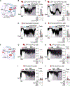
Figure 2.. Upper glycolytic enzymes produce ADP that activates KATP channels
(A–E) In the high ATP condition (1 mM ATP, 0.075 mM ADP), addition of 10 mM F6P led to production of ADP by PFK (A), thereby opening KATP channels in mouse β cells (B), mouse α cells (C), human β cells (D), and human α cells (E). (F–J) Addition of 10 mM glucose in high ATP (F) produced little effect in mouse β and α cells (G-H) but led to significant KATP activation through ADP production by GK in human β and α cells (I and J). Example recordings show KATP inhibition in the high ATP condition, followed by the addition of substrates for PFK and GK. Graphs quantify channel activity (power) normalized to high ATP condition. Data are from at least 3 mice or 3 human donors and shown as mean ± SEM. #p < 0.1, *p < 0.05, **p < 0.01, ***p < 0.001, ****p < 0.0001 by ratio paired Student’s t test. Concentrations are in mM unless otherwise noted. See also Table S1.
Figure 3.
Figure 3.. The ADP produced by PFK is used directly by PK, and NAD+ produced by plasma membrane-associated NADH oxidases is used directly by GAPDH to sustain ATP production
(A–E) Addition of 5 mM pyruvate (Pyr) and NADH allows NADH oxidation to provide NAD+ at the GAPDH step to metabolize 5 mM FBP (substrate for ALDO) to ATP that closes KATP channels in mouse and human β and α cells. (F–J) Addition of F6P (5 mM in mice, 1 mM in humans) under high ATP condition (1 mM ATP, 0.075 mM ADP) led to activation of KATP channels; the subsequent addition of PEP (5 mM in mice, 2 mM in humans) did not significantly alter KATP activity in mouse α cells (H) or human β cells (I) but led to KATP inhibition through PK-generated ATP in mouse β cells (G) and human α cells (J). Example traces show KATP inhibition in 1 mM ATP and reopening in the high ADP condition (0.5 mM ADP, 0.1 mM ATP) (B–E) or KATP inhibition in the high ATP condition (G–J), followed by the addition of test substrates. Graphs quantify channel activity (power) normalized to high ADP +5 mM FBP condition (B–E) or high ATP condition (G–J). Data are from at least 3 human donors and shown as mean ± SEM. #p < 0.1, *p < 0.05, **p < 0.01, ***p < 0.001, ****p < 0.0001 by ratio paired Student’s t test. Concentrations are in mM unless otherwise noted. See also Figures S3 and S4 and Table S1.

References

    1. Cook DL, and Hales CN (1984). Intracellular ATP directly blocks K+ channels in pancreatic B-cells. Nature 311, 271–273. 10.1038/311271a0. - DOI - PubMed
    1. Dunne MJ, and Petersen OH (1986). Intracellular ADP activates K+ channels that are inhibited by ATP in an insulin-secreting cell line. FEBS Lett 208, 59–62. 10.1016/0014-5793(86)81532-0. - DOI - PubMed
    1. Kakei M, Kelly RP, Ashcroft SJ, and Ashcroft FM (1986). The ATP-sensitivity of K+ channels in rat pancreatic B-cells is modulated by ADP. FEBS Lett 208, 63–66. 10.1016/0014-5793(86)81533-2. - DOI - PubMed
    1. Ashcroft FM, Harrison DE, and Ashcroft SJ (1984). Glucose induces closure of single potassium channels in isolated rat pancreatic β-cells. Nature 312, 446–448. 10.1038/312446a0. - DOI - PubMed
    1. Misler S, Falke LC, Gillis K, and McDaniel ML (1986). A metabolite-regulated potassium channel in rat pancreatic B cells. Proc. Natl. Acad. Sci. USA 83, 7119–7123. 10.1073/pnas.83.18.7119. - DOI - PMC - PubMed

Publication types