

Comparison of Novel Wide-Field In Vivo Corneal Confocal Microscopy With Skin Biopsy for Assessing Peripheral Neuropathy in Type 2 Diabetes
- PMID: 37058418
- PMCID: PMC10281223
- DOI: 10.2337/db22-0863
Comparison of Novel Wide-Field In Vivo Corneal Confocal Microscopy With Skin Biopsy for Assessing Peripheral Neuropathy in Type 2 Diabetes
Abstract
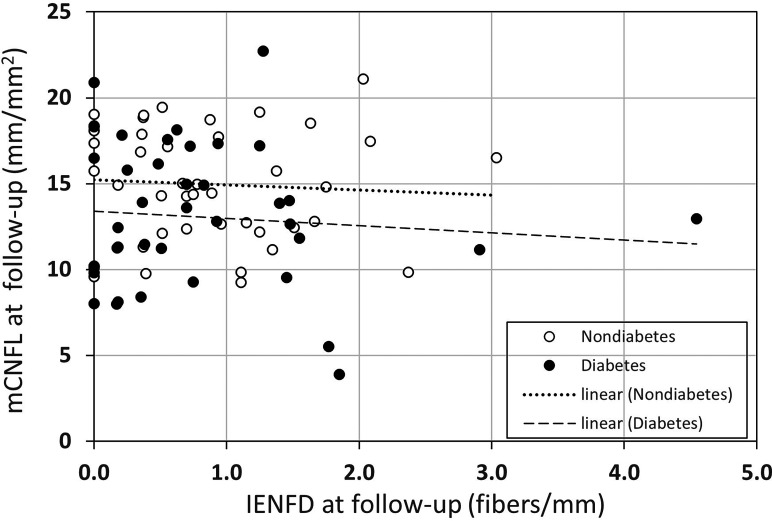
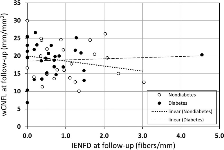
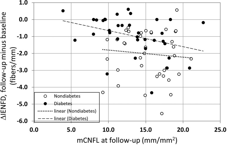
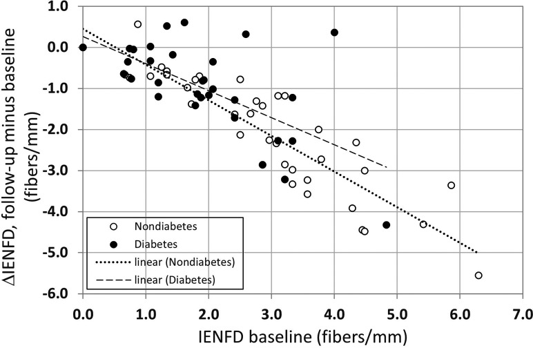
Diabetic peripheral neuropathy (DPN) is a serious complication of diabetes, where skin biopsy assessing intraepidermal nerve fiber density (IENFD) plays an important diagnostic role. In vivo confocal microscopy (IVCM) of the corneal subbasal nerve plexus has been proposed as a noninvasive diagnostic modality for DPN. Direct comparisons of skin biopsy and IVCM in controlled cohorts are lacking, as IVCM relies on subjective selection of images depicting only 0.2% of the nerve plexus. We compared these diagnostic modalities in a fixed-age cohort of 41 participants with type 2 diabetes and 36 healthy participants using machine algorithms to create wide-field image mosaics and quantify nerves in an area 37 times the size of prior studies to avoid human bias. In the same participants, and at the same time point, no correlation between IENFD and corneal nerve density was found. Corneal nerve density did not correlate with clinical measures of DPN, including neuropathy symptom and disability scores, nerve conduction studies, or quantitative sensory tests. Our findings indicate that corneal and intraepidermal nerves likely mirror different aspects of nerve degeneration, where only intraepidermal nerves appear to reflect the clinical status of DPN, suggesting that scrutiny is warranted concerning methodologies of studies using corneal nerves to assess DPN.
Article highlights: Comparison of intraepidermal nerve fiber density with automated wide-field corneal nerve fiber density in participants with type 2 diabetes revealed no correlation between these parameters. Intraepidermal and corneal nerve fibers both detected neurodegeneration in type 2 diabetes, but only intraepidermal nerve fibers were associated with clinical measures of diabetic peripheral neuropathy. A lack of association of corneal nerves with peripheral neuropathy measures suggests that corneal nerve fibers may be a poor biomarker for diabetic peripheral neuropathy.
© 2023 by the American Diabetes Association.
Conflict of interest statement
Figures

Similar articles
-
Assessing Corneal Confocal Microscopy and Other Small Fiber Measures in Diabetic Polyneuropathy.Neurology. 2023 Apr 18;100(16):e1680-e1690. doi: 10.1212/WNL.0000000000206902. Epub 2023 Feb 7. Neurology. 2023. PMID: 36750383 Free PMC article.
-
Utility of corneal confocal microscopy for assessing mild diabetic neuropathy: baseline findings of the LANDMark study.Clin Exp Optom. 2012 May;95(3):348-54. doi: 10.1111/j.1444-0938.2012.00740.x. Epub 2012 Apr 29. Clin Exp Optom. 2012. PMID: 22540156
-
Retinal and Corneal Neurodegeneration and Their Association with Systemic Signs of Peripheral Neuropathy in Type 2 Diabetes.Am J Ophthalmol. 2020 Jan;209:197-205. doi: 10.1016/j.ajo.2019.09.010. Epub 2019 Sep 19. Am J Ophthalmol. 2020. PMID: 31542341
-
Corneal nerves in diabetes-The role of the in vivo corneal confocal microscopy of the subbasal nerve plexus in the assessment of peripheral small fiber neuropathy.Surv Ophthalmol. 2021 May-Jun;66(3):493-513. doi: 10.1016/j.survophthal.2020.09.003. Epub 2020 Sep 19. Surv Ophthalmol. 2021. PMID: 32961210 Review.
-
Corneal confocal microscopy for the diagnosis of diabetic peripheral neuropathy: A systematic review and meta-analysis.J Diabetes Investig. 2022 Jan;13(1):134-147. doi: 10.1111/jdi.13643. Epub 2021 Aug 27. J Diabetes Investig. 2022. PMID: 34351711 Free PMC article.
Cited by
-
Research progress of corneal characteristics and changes in primary angle‑closure glaucoma (Review).Biomed Rep. 2025 Feb 5;22(4):59. doi: 10.3892/br.2025.1937. eCollection 2025 Apr. Biomed Rep. 2025. PMID: 39991004 Free PMC article. Review.
-
Advances in diagnostic methods for early-stage diabetic polyneuropathy.J Diabetes Investig. 2024 Jul;15(7):820-822. doi: 10.1111/jdi.14201. Epub 2024 Apr 6. J Diabetes Investig. 2024. PMID: 38581221 Free PMC article.
-
Peripheral Neuropathy Symptoms and Ocular Surface Lesions in Patients with Type 2 Diabetes Mellitus and Dry Eye: A Clinical Correlational Study.Ophthalmol Ther. 2025 Jul;14(7):1503-1519. doi: 10.1007/s40123-025-01150-x. Epub 2025 May 15. Ophthalmol Ther. 2025. PMID: 40372617 Free PMC article.
-
ConNeCT: weakly supervised corneal confocal microscopy image inpainting network based on a diffusion model.Biomed Opt Express. 2025 Jun 9;16(7):2615-2630. doi: 10.1364/BOE.562924. eCollection 2025 Jul 1. Biomed Opt Express. 2025. PMID: 40677830 Free PMC article.
-
Automated program using convolutional neural networks for objective and reproducible selection of corneal confocal microscopy images.Digit Health. 2025 Mar 17;11:20552076251326223. doi: 10.1177/20552076251326223. eCollection 2025 Jan-Dec. Digit Health. 2025. PMID: 40103638 Free PMC article.
References
-
- Saeedi P, Petersohn I, Salpea P, et al. .; IDF Diabetes Atlas Committee . Global and regional diabetes prevalence estimates for 2019 and projections for 2030 and 2045: results from the International Diabetes Federation Diabetes Atlas, 9th edition. Diabetes Res Clin Pract 2019;157:107843. - PubMed
-
- Neuropathy Spanish Study Group of the Spanish Diabetes Society (SDS) . The prevalence of clinical diabetic polyneuropathy in Spain: a study in primary care and hospital clinic groups. Diabetologia 1998;41:1263–1269 - PubMed
-
- Iqbal Z, Azmi S, Yadav R, et al. . Diabetic peripheral neuropathy: epidemiology, diagnosis, and pharmacotherapy. Clin Ther 2018;40:828–849 - PubMed
-
- Ziegler D, Tesfaye S, Spallone V, et al. . Screening, diagnosis and management of diabetic sensorimotor polyneuropathy in clinical practice: international expert consensus recommendations. Diabetes Res Clin Pract 2022;186:109063. - PubMed
